



ShK toxin
ShK toxin blocks voltage-dependent potassium channel (Kv1.3 channel). ShK toxin can be isolated from the whole body extract of the Caribbean sea anemone (Stichodactylu helianthus). ShK toxin competes with dendrotoxin I and α-dendrotoxin for binding to synaptosomal membranes of rat brain, facilitates acetylcholine release. ShK toxin suppresses K+ currents in cultured rat dorsal root ganglion neurons. ShK toxin also inhibits T lymphocyte proliferation.
For research use only. We do not sell to patients.
- CAS No.: 172450-46-3
- Formula: C169H274N54O48S7
- Molecular Weight:4054.80
-
Storage:
Please store the product under the recommended conditions in the Certificate of Analysis.

To place orders, for customer services and technical support, please contact: MedChemExpress USA
Tel: 609-228-6898 E-mail: [email protected] [email protected]


-
 Biological Activity
-
Chemical Information
- Protocols
- Purity & Documentation
- References
-
 Help & FAQs




Biological Activity
Description
IC50 & Target
Kv1.3 Channel[2]


Chemical Information
-
CAS No. 172450-46-3
-
Molecular Weight 4054.80
-
Formula C169H274N54O48S7
-
Synonyms
Stichodactyla helianthus neurotoxin
-
Sequence
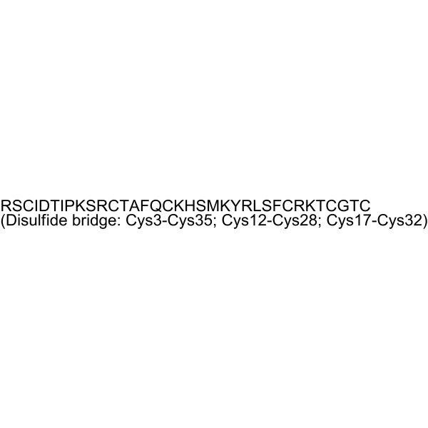
Arg-Ser-Cys-Ile-Asp-Thr-Ile-Pro-Lys-Ser-Arg-Cys-Thr-Ala-Phe-Gln-Cys-Lys-His-Ser-Met-Lys-Tyr-Arg-Leu-Ser-Phe-Cys-Arg-Lys-Thr-Cys-Gly-Thr-Cys (Disulfide bridge: Cys3-Cys35; Cys12-Cys28; Cys17-Cys32)
-
Sequence Shortening
RSCIDTIPKSRCTAFQCKHSMKYRLSFCRKTCGTC (Disulfide bridge: Cys3-Cys35; Cys12-Cys28; Cys17-Cys32)
-
Shipping
Room temperature in continental US; may vary elsewhere.
-
Storage
Please store the product under the recommended conditions in the Certificate of Analysis.


Purity & Documentation
References
[1]. Castañeda O, et al. Characterization of a potassium channel toxin from the Caribbean Sea anemone Stichodactyla helianthus. Toxicon. 1995 May;33(5):603-13. [Content Brief]
[2]. Beeton C, et al. The D-diastereomer of ShK toxin selectively blocks voltage-gated K+ channels and inhibits T lymphocyte proliferation. J Biol Chem. 2008 Jan 11;283(2):988-97. [Content Brief]

Calculators
Concentration (start) × Volume (start) = Concentration (final) × Volume (final)


ShK toxin Related Classifications
HY-P3071 Related Classifications
 Powered by Bioz