



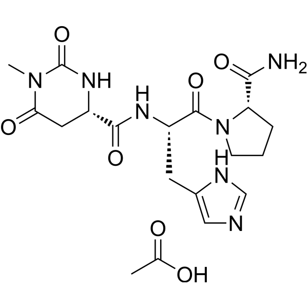
Taltirelin acetate
Based on 1 publication(s) in Google Scholar
Taltirelin acetate (TA-0910) is an acetate form of Taltirelin (TA-0910). Taltirelin (TA-0910) is an orally effective analogue of thyrotropin releasing hormone (TRH) and a TRH receptor (TRH-R) superagonist (IC50 at 910 nM). Taltirelin can cross the blood-brain barrier. Taltirelin stimulates an increase in cytosolic Ca2+ concentration (Ca2+ release) with an EC50 value of 36 nM. Taltirelin increases cell viability and reduces apoptosis in SH-SY5Y cells and primary rat mesencephalic neurons treated with MPP+ (HY-W008719) or Rotenone (HY-B1756). Taltirelin has neuroprotective effects in both cellular and animal models of Parkinson's disease. Taltirelin alleviates fatigue-like behavior in mouse models of cancer-related fatigue.
For research use only. We do not sell to patients.
- Purity : 99.75%
- CAS No.: 1549593-23-8
- Formula: C19H27N7O7
- Molecular Weight:465.46
-
Storage:
-20°C, sealed storage, away from moisture
* In solvent : -80°C, 6 months; -20°C, 1 month (sealed storage, away from moisture)

To place orders, for customer services and technical support, please contact: MedChemExpress USA
Tel: 609-228-6898 E-mail: [email protected] [email protected]


-
 Biological Activity
-
Chemical Information
-
Solvent & Solubility
- Protocols
- Purity & Documentation
- References
-
 Help & FAQs


-
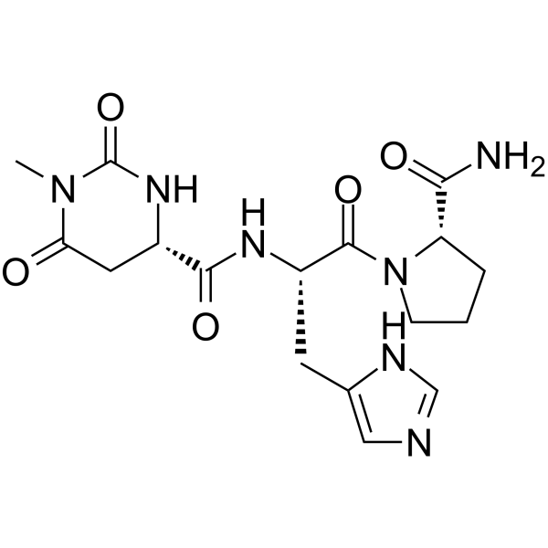
HY-B0596
TaltirelinThyroid Hormone Receptor Agonist -
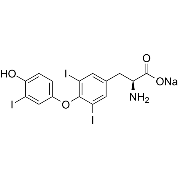
HY-A0070
Liothyronine sodiumTRα/TRβ Agonist -
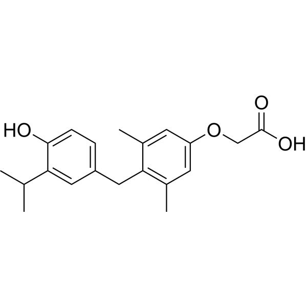
HY-14823
SobetiromeTRβ Agonist -

HY-A0070R
Liothyronine sodium (Standard)TRα/TRβ Agonist -
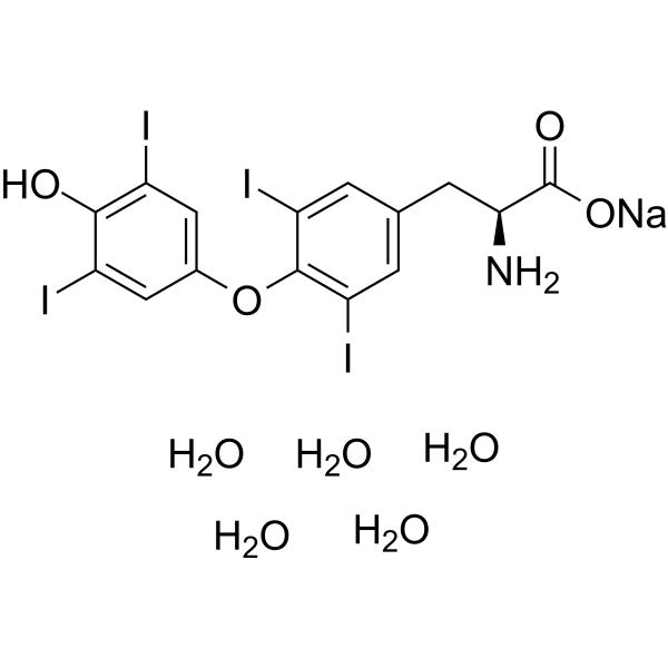
HY-18341A
L-Thyroxine sodium salt pentahydrateThyroid Hormone -
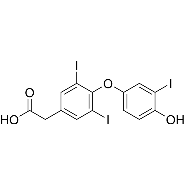
HY-B1201
TiratricolThyroid Hormone Receptor Inhibitor -
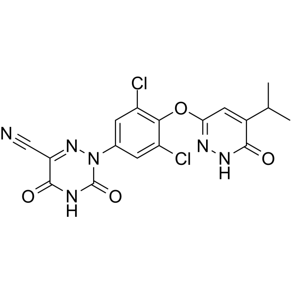
HY-12216
ResmetiromTHR-β Agonist -
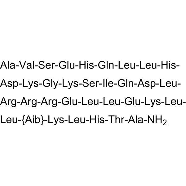
HY-108742
AbaloparatidePTHR1 Activator -
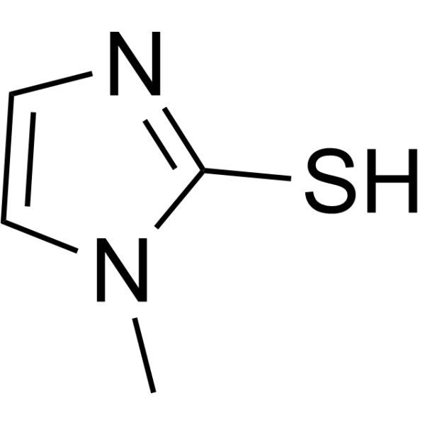
HY-B0208
MethimazoleAnti-thyroid Agent -
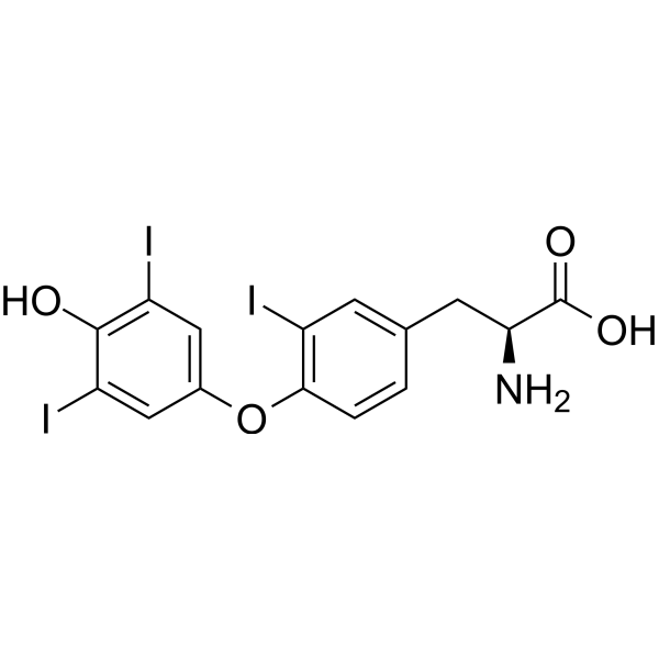
HY-W010696
Reverse T3Thyroid Hormone -
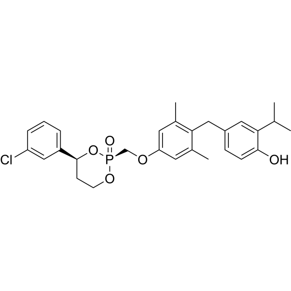
HY-111288
MB-07811
Publications Citing Use of MedChemExpress (MCE) Taltirelin acetate
More

Biological Activity
Description
IC50 & Target
IC50: 910 nM (Thyrotropin-releasing hormone receptor)[1]
In Vitro
Taltirelin (5 μM; 2 h) improves cell viability, reducess ROS production and apoptosis in MPP+ (HY-W008719) or Rotenone (HY-B1756) treated SH-SY5Y cells and primary rat midbrain neurons[2].
Taltirelin (5 μM; 2 h) exerts neuroprotective effects by inhibiting tau phosphorylation and AEP activation in SH-SY5Y cells treated with MPP+ (HY-W008719) or rotenone (HY-B1756)[2].
MedChemExpress (MCE) has not independently confirmed the accuracy of these methods. They are for reference only.
-
Cell Line:SH-SY5Y neuroblastoma cells and primary neonatal rat midbrain neurons
-
Concentration:5 μM
-
Incubation Time:2 h
-
Result:Increased cell viability.
-
Cell Line:SH-SY5Y neuroblastoma cells
-
Concentration:5 μM
-
Incubation Time:2 h
-
Result:Prevented the elevation of the cleaved caspase-3.
-
Cell Line:SH-SY5Y neuroblastoma cells
-
Concentration:5 μM
-
Incubation Time:2 h
-
Result:Reduced the levels of p-tau and AEP cleavage products: tau N368 and a-synuclein N103 fragments.
In Vivo
Taltirelin (0.2-5 mg/kg; Intraperitoneal injection; 14-56 days) has a neuroprotective effect in mouse models of MPTP (HY-15608) and chronic rotenone (HY-B1756) -induced PD, and improves locomotor function in mice [2].
Taltirelin (1 mg/kg Intraperitoneal injection; 10 mg/kg Oral administration; 1 day) alleviates the fatigue-like behavior caused by chemotherapy treatment, tumor burden and irradiation in mouse models [3].
MedChemExpress (MCE) has not independently confirmed the accuracy of these methods. They are for reference only.
-
Animal Model:MPTP (HY-15608) or Rotenone (HY-B1756) treated male C57/BL6 mice aged 8-9 weeks old[2]
-
Dosage:0.2 mg/kg, 1 mg/kg, 5 mg/kg 14 days; 0.2 mg/kg, 1 mg/kg 56 days
-
Administration:Intraperitoneal injection (i.p.)
-
Result:Improved the locomotor function and preserved dopaminergic neurons in the substantia nigra.
Down-regulated the levels of p-tau (S396), p-α-synuclein (S129) tau N368 and α-synuclein N103 fragments in substantia nigra and striatum.
-
Animal Model:5-FU (HY-90006) or radiation treated adult C57BL/6NCr mice aged 7-8 weeks;
Balb/cAnNCr mice aged 9 weeks old with colon 26 carcinoma[3] -
Dosage:1mg/kg;
10mg/kg -
Administration:Intraperitoneal injection (i.p.);
Oral administration (p.o.);
Twice a day before the test and once again 30 minutes prior to the test. -
Result:Reversed the fatigue-like effect, caused mice to run nearer to the top of the treadmill during the test.
- Detection of Apoptosis
- TUNEL staining for apoptotic DNA fragmentation
- Cell Cytotoxicity Assay
- Mammalian live/dead viability and cytotoxicity staining
- Annexin V plus membrane-impermeant dye apoptosis staining
- Research Protocol for Endocrine Diseases
- Human pluripotent stem cell midbrain dopaminergic neuron differentiation
- Apoptosis Solutions
- CCK-8/WST-8 Cell Proliferation Assay
- Ca2+ Staining Technique
- MTT Cell Proliferation Assay
- Cell Viability Determination by MTT Colorimetric Assay
Clinical Trial
| NCT Number | Sponsor | Condition | Start Date |
Phase

|
|---|---|---|---|---|
| NCT01329991 | 2011-05 | PHASE1 |


Chemical Information
-
CAS No. 1549593-23-8
-
Appearance Solid
-
Molecular Weight 465.46
-
Formula C19H27N7O7
-
Color White to off-white
-
SMILES
O=C(N(CCC1)[C@@H]1C(N)=O)[C@@H](NC([C@@H](NC2=O)CC(N2C)=O)=O)CC3=CN=CN3.CC(O)=O
-
Synonyms
TA-0910 acetate
-
Shipping
Room temperature in continental US; may vary elsewhere.
-
Storage
-20°C, sealed storage, away from moisture
* In solvent : -80°C, 6 months; -20°C, 1 month (sealed storage, away from moisture)


Publications (1)
-
Journal Impact Factor
-
Most Recent
-
J Mol Neurosci
A Ketogenic Diet Affects Gut Microbiota by Regulating Gut Microbiota and Promoting Hippocampal TRHR Expression to Combat Seizures. [Abstract]2024 Nov 3;74(4):104. PMID: 39489848


Solvent & Solubility
In Vitro:
DMSO : 125 mg/mL (268.55 mM; Need ultrasonic; Hygroscopic DMSO has a significant impact on the solubility of product, please use newly opened DMSO)
Please refer to the solubility information to select the appropriate solvent. Once prepared, please aliquot and store the solution to prevent product inactivation from repeated freeze-thaw cycles.
Storage method and period of stock solution: -80°C, 6 months; -20°C, 1 month (sealed storage, away from moisture). When stored at -80°C, please use it within 6 months. When stored at -20°C, please use it within 1 month.
Please refer to the solubility information to select the appropriate solvent. Once prepared, please aliquot and store the solution to prevent product inactivation from repeated freeze-thaw cycles.
Storage method and period of stock solution: -80°C, 6 months; -20°C, 1 month (sealed storage, away from moisture). When stored at -80°C, please use it within 6 months. When stored at -20°C, please use it within 1 month.
Concentration (start) × Volume (start) = Concentration (final) × Volume (final)
In Vivo:
Select the appropriate dissolution method based on your experimental animal and administration route.
- For the following dissolution methods, please ensure to first prepare a clear stock solution using an In Vitro approach and then sequentially add co-solvents:
- To ensure reliable experimental results, the clarified stock solution can be appropriately stored based on storage conditions. As for the working solution for In Vivo experiments, it is recommended to prepare freshly and use it on the same day.
- The percentages shown for the solvents indicate their volumetric ratio in the final prepared solution. If precipitation or phase separation occurs during preparation, heat and/or sonication can be used to aid dissolution.
Add each solvent one by one: 10% DMSO 40% PEG300 5% Tween-80 45% Saline
Solubility: ≥ 2.08 mg/mL (4.47 mM); Clear solution
This protocol yields a clear solution of ≥ 2.08 mg/mL (saturation unknown).
Taking 1 mL working solution as an example, add 100 μL DMSO stock solution (20.8 mg/mL) to 400 μL PEG300, and mix evenly; then add 50 μL Tween-80 and mix evenly; then add 450 μL Saline to adjust the volume to 1 mL.
Preparation of Saline: Dissolve 0.9 g sodium chloride in ddH₂O and dilute to 100 mL to obtain a clear Saline solution.
Add each solvent one by one: 10% DMSO 90% (20% SBE-β-CD in Saline)
Solubility: ≥ 2.08 mg/mL (4.47 mM); Clear solution
This protocol yields a clear solution of ≥ 2.08 mg/mL (saturation unknown).
Taking 1 mL working solution as an example, add 100 μL DMSO stock solution (20.8 mg/mL) to 900 μL 20% SBE-β-CD in Saline, and mix evenly.
Preparation of 20% SBE-β-CD in Saline (4°C, storage for one week): 2 g SBE-β-CD powder is dissolved in 10 mL Saline, completely dissolve until clear.
In Vivo Dissolution Calculator
Please enter the basic information of animal experiments:
-
-
-
-
Recommended: Prepare an additional quantity of animals to account for potential losses during experiments.
Please enter your animal formula composition:
-
%DMSO +
Recommended: Keep the proportion of DMSO in working solution below 2% if your animal is weak.
-
%+
-
+%Tween-80 + +
-
%Saline +
The co-solvents required include: DMSO, . All of co-solvents are available by MedChemExpress (MCE). , Tween 80. All of co-solvents are available by MedChemExpress (MCE).
Working solution concentration: 0.22 mg/mL
Method for preparing stock solution: mg drug dissolved in μL DMSO. Stock solution concentration: mg/mL. * In solvent : -80°C, 6 months; -20°C, 1 month (sealed storage, away from moisture)
1. Take μL DMSO stock solution;
2. Add μL .
μL , mix evenly;
3. Then add μL Tween 80, mix evenly;
4. Then add μL
Please ensure that the stock solution in the first step is dissolved to a clear state, and add co-solvents in sequence. You can use ultrasonic heating (ultrasonic cleaner, recommended frequency 20-40 kHz), vortexing, etc. to assist dissolution.


Purity & Documentation
-
Data Sheet (284 KB)
-
SDS (252 KB)
- English - EN (252 KB)
- Français - FR (252 KB)
- Deutsch - DE (252 KB)
- Norwegian - NO (252 KB)
- Español - ES (252 KB)
- Swedish - SV (252 KB)
- Italian - IT (252 KB)
- Korean - KR (252 KB)
- Portuguese - PT (252 KB)
-
Handling Instructions (2659 KB)
References
[1]. Thirunarayanan N, et al. Taltirelin is a superagonist at the human thyrotropin-releasing hormone receptor. Front Endocrinol (Lausanne). 2012 Oct 9;3:120. [Content Brief]
[2]. Zheng C, et al. TRH Analog, Taltirelin Protects Dopaminergic Neurons From Neurotoxicity of MPTP and Rotenone. Front Cell Neurosci. 2018 Dec 20;12:485. [Content Brief]
[3]. Dougherty JP, et al. Taltirelin alleviates fatigue-like behavior in mouse models of cancer-related fatigue. Pharmacol Res. 2017 Oct;124:1-8. [Content Brief]


Complete Stock Solution Preparation Table
Please refer to the solubility information to select the appropriate solvent. Once prepared, please aliquot and store the solution to prevent product inactivation from repeated freeze-thaw cycles.
Storage method and period of stock solution: -80°C, 6 months; -20°C, 1 month (sealed storage, away from moisture). When stored at -80°C, please use it within 6 months. When stored at -20°C, please use it within 1 month.
| Optional Solvent | Concentration Solvent Mass | 1 mg | 5 mg | 10 mg | 25 mg |
|---|---|---|---|---|---|
| DMSO | 1 mM | 2.1484 mL | 10.7421 mL | 21.4841 mL | 53.7103 mL |
| 5 mM | 0.4297 mL | 2.1484 mL | 4.2968 mL | 10.7421 mL | |
| 10 mM | 0.2148 mL | 1.0742 mL | 2.1484 mL | 5.3710 mL | |
| 15 mM | 0.1432 mL | 0.7161 mL | 1.4323 mL | 3.5807 mL | |
| 20 mM | 0.1074 mL | 0.5371 mL | 1.0742 mL | 2.6855 mL | |
| 25 mM | 0.0859 mL | 0.4297 mL | 0.8594 mL | 2.1484 mL | |
| 30 mM | 0.0716 mL | 0.3581 mL | 0.7161 mL | 1.7903 mL | |
| 40 mM | 0.0537 mL | 0.2686 mL | 0.5371 mL | 1.3428 mL | |
| 50 mM | 0.0430 mL | 0.2148 mL | 0.4297 mL | 1.0742 mL | |
| 60 mM | 0.0358 mL | 0.1790 mL | 0.3581 mL | 0.8952 mL | |
| 80 mM | 0.0269 mL | 0.1343 mL | 0.2686 mL | 0.6714 mL | |
| 100 mM | 0.0215 mL | 0.1074 mL | 0.2148 mL | 0.5371 mL |


Taltirelin acetate Related Classifications
HY-B0596A Related Classifications
 Powered by Bioz