



THPN

Based on 1 Customer Validation
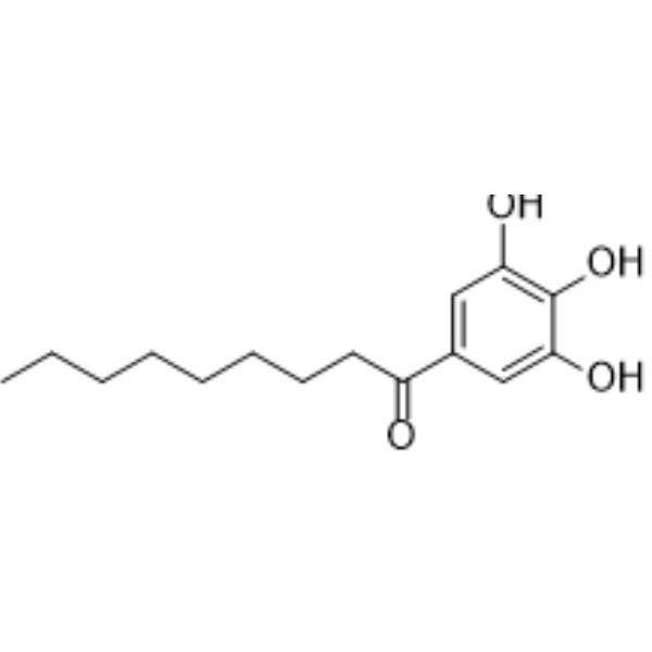
THPN is a potent Nur77 agonist. THPN specifically binds the LBD of Nur77 (TR3) but not that of retinoic acid receptor α and PPARγ with a Kd of 270 nM. THPN leads to Nur77 translocation to the mitochondria to induce autophagic cell death in melanoma.
For research use only. We do not sell to patients.
- Purity : 98.00%
- CAS No.: 100079-26-3
- Formula: C15H22O4
- Molecular Weight:266.33
-
Storage:Powder -20°C, 3 years , 4°C, 2 years ; In solvent -80°C, 6 months , -20°C, 1 month

To place orders, for customer services and technical support, please contact: MedChemExpress USA
Tel: 609-228-6898 E-mail: [email protected] [email protected]


-
 Biological Activity
-
Chemical Information
- Protocols
- Purity & Documentation
- References
-
 Help & FAQs


All Nuclear Hormone Receptor 4A/NR4A Isoforms
More

Biological Activity
Description
IC50 & Target
|
Nur77/NR4A1 |
Cellular Effect
|
Cell Line

|
Type | Value | Description | References |
|---|---|---|---|---|
| CCRF-CEM | IC50 |
43 μM
Compound: 8
|
Cytotoxicity against human CEM cells after 72 hrs by MTT assay
Cytotoxicity against human CEM cells after 72 hrs by MTT assay
|
[PMID: 18295493] |
| CCRF-CEM | IC50 |
50 μM
Compound: 8
|
Cytotoxicity against human CEM cells after 24 hrs by MTT assay
Cytotoxicity against human CEM cells after 24 hrs by MTT assay
|
[PMID: 18295493] |
| L1210 | IC50 |
22 μM
Compound: 8
|
Cytotoxicity against mouse L1210 cells after 48 hrs by MTT assay
Cytotoxicity against mouse L1210 cells after 48 hrs by MTT assay
|
[PMID: 18295493] |
| L1210 | IC50 |
22 μM
Compound: 8
|
Cytotoxicity against mouse L1210 cells after 72 hrs by MTT assay
Cytotoxicity against mouse L1210 cells after 72 hrs by MTT assay
|
[PMID: 18295493] |
| Vero | IC50 |
>1 μM
Compound: 8
|
Cytotoxicity against african green monkey VERO cells
Cytotoxicity against african green monkey VERO cells
|
[PMID: 18295493] |
| Vero | IC50 |
>299 μM
Compound: 8
|
Genotoxicity against african green monkey VERO cells
Genotoxicity against african green monkey VERO cells
|
[PMID: 18295493] |


Chemical Information
-
CAS No. 100079-26-3
-
Appearance Solid
-
Molecular Weight 266.33
-
Formula C15H22O4
-
Color White to off-white
-
SMILES
CCCCCCCCC(C1=CC(O)=C(O)C(O)=C1)=O
-
Shipping
Room temperature in continental US; may vary elsewhere.
-
Storage
Powder -20°C 3 years 4°C 2 years In solvent -80°C 6 months -20°C 1 month


Purity & Documentation
-
Data Sheet (265 KB)
-
SDS (392 KB)
- English - EN (392 KB)
- Français - FR (392 KB)
- Deutsch - DE (392 KB)
- Norwegian - NO (392 KB)
- Español - ES (392 KB)
- Swedish - SV (392 KB)
- Italian - IT (392 KB)
- Korean - KR (392 KB)
- Portuguese - PT (392 KB)
-
Handling Instructions (2659 KB)
References
[1]. Wei-jia Wang, et al. Orphan nuclear receptor TR3 acts in autophagic cell death via mitochondrial signaling pathway. Nat Chem Biol. 2014 Feb;10(2):133-40. [Content Brief]
[2]. Li Li, et al. Impeding the interaction between Nur77 and p38 reduces LPS-induced inflammation. Nat Chem Biol. 2015 May;11(5):339-46. [Content Brief]

Calculators
Concentration (start) × Volume (start) = Concentration (final) × Volume (final)


THPN Related Classifications
HY-113827 Related Classifications
 Powered by Bioz