



ULK1-IN-2

Based on 1 Customer Validation
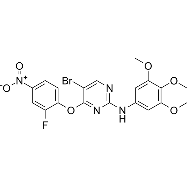
ULK1-IN-2 (compound 3s) is a potent ULK1 inhibitor. ULK1-IN-2 shows highest cytotoxic effect against cancer cell lines, with IC50 of 1.94 μM in A549. ULK1-IN-2 can induce apoptosis and simultaneously block autophagy, and can be used to study NSCLC (Non-small cell lung cancer).
Nos produits utilisent uniquement pour la recherche. Nous ne vendons pas aux patients.
- Pureté : 98.34%
- CAS No.: 2497409-01-3
- Formule: C19H16BrFN4O6
- Masse moléculaire:495.26
-
Stockage:
4°C, sealed storage, away from moisture and light
* In solvent : -80°C, 6 months; -20°C, 1 month (sealed storage, away from moisture and light)

To place orders, for customer services and technical support, please contact: MedChemExpress USA
Tel: 609-228-6898 E-mail: [email protected] [email protected]


-
 Activité biologique
-
Chemical Information
-
Solvant et solubilité
- Protocole
- Pureté et documentation
- Références
-
 Help & FAQs


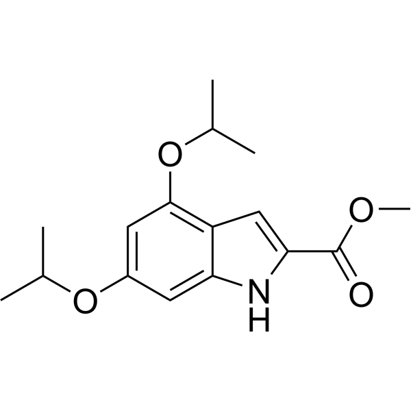
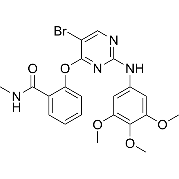
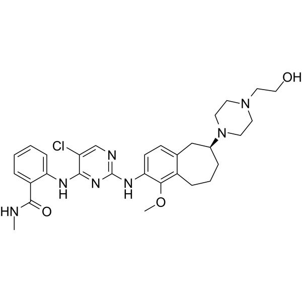
Voir tous les produits spécifiques à Isoform AMPK
More

Activité biologique
Description
IC50 & Target
|
ULK1 |
Cellular Effect
|
Cell Line

|
Type | Value | Description | References |
|---|---|---|---|---|
| A549 | IC50 |
1.94 μM
Compound: 3s
|
Antiproliferative activity against human A549 cells assessed as reduction in cell viability after 24 hrs by MTT assay
Antiproliferative activity against human A549 cells assessed as reduction in cell viability after 24 hrs by MTT assay
|
[PMID: 32961380] |
| HL-60 | IC50 |
10.89 μM
Compound: 3s
|
Antiproliferative activity against human HL60 cells assessed as reduction in cell viability after 24 hrs by MTT assay
Antiproliferative activity against human HL60 cells assessed as reduction in cell viability after 24 hrs by MTT assay
|
[PMID: 32961380] |
| MCF7 | IC50 |
19.6 μM
Compound: 3s
|
Antiproliferative activity against human MCF-7 cells assessed as reduction in cell viability after 24 hrs by MTT assay
Antiproliferative activity against human MCF-7 cells assessed as reduction in cell viability after 24 hrs by MTT assay
|
[PMID: 32961380] |
| MDA-MB-468 | IC50 |
16.83 μM
Compound: 3s
|
Antiproliferative activity against human MDA-MB-468 cells assessed as reduction in cell viability after 24 hrs by MTT assay
Antiproliferative activity against human MDA-MB-468 cells assessed as reduction in cell viability after 24 hrs by MTT assay
|
[PMID: 32961380] |
| U-937 | IC50 |
12.92 μM
Compound: 3s
|
Antiproliferative activity against human U937 cells assessed as reduction in cell viability after 24 hrs by MTT assay
Antiproliferative activity against human U937 cells assessed as reduction in cell viability after 24 hrs by MTT assay
|
[PMID: 32961380] |
In Vitro
ULK1-IN-2 (compound 3s) (10 μM, 24 h) shows strong anti-proliferative activity against A549, U937, HL60, MDA-MB-468 and MCF-7[1].
ULK1-IN-2 (0-8 μM, 24 h) blocks autophagy via inhibiting ULK1 in A549 cells[1].
ULK1-IN-2 (0-8 μM, 24 h) induces apoptosis via the mitochondrial pathways in A549 cells in dose department manner[1].
ULK1-IN-2 (0-8 μM, 24 h) inhibits ULK1 and p-ULK1ser317 expression in a concentration-dependent manner, remarkably decreases Bcl-2 expression, increases Bax and the active form of Caspase-3 expression.[1].
MedChemExpress (MCE) has not independently confirmed the accuracy of these methods. They are for reference only.
-
Cell Line:Human cancer cell lines A549, U937, HL60, MDA-MB-468 and MCF-7[1]
-
Concentration:10 μM
-
Incubation Time:24 h
-
Result:Significantly improved anti-proliferative activity against A549, U937, HL60, MDA-MB-468 and MCF-7, with kinase inhibitory activity of 99.15% and IC50 values of 1.94, 12.92, 10.89, 16.83, and 19.60 μM, respectively.
-
Cell Line:A549 cells[1]
-
Concentration:0, 2, 4, 8 μM
-
Incubation Time:24 h
-
Result:Blocked autophagy of A549 cells via inhibiting ULK.
-
Cell Line:A549 cells[1]
-
Concentration:0, 2, 4, 8 μM
-
Incubation Time:24 h
-
Result:Inhibited expression of ULK1 and p-ULK1ser317 in a concentration-dependent manner. Increased the autophagy substrate P62, reduced LC3-I conversion to LC3-II, and decreased the levels of Beclin1. Remarkably decreased Bcl-2 expression, increased Bax and the active form of Caspase-3 expression.


Chemical Information
-
CAS No. 2497409-01-3
-
Appearance Solid
-
Masse moléculaire 495.26
-
Formule C19H16BrFN4O6
-
Color Light yellow to yellow
-
SMILES
COC1=C(OC)C(OC)=CC(NC2=NC=C(Br)C(OC3=CC=C([N+]([O-])=O)C=C3F)=N2)=C1
-
Livraison
Room temperature in continental US; may vary elsewhere.
-
Stockage
4°C, sealed storage, away from moisture and light
* In solvent : -80°C, 6 months; -20°C, 1 month (sealed storage, away from moisture and light)


Solvant et solubilité
In Vitro:
DMSO : 33.33 mg/mL (67.30 mM; Need ultrasonic; Hygroscopic DMSO has a significant impact on the solubility of product, please use newly opened DMSO)
Please refer to the solubility information to select the appropriate solvent. Once prepared, please aliquot and store the solution to prevent product inactivation from repeated freeze-thaw cycles.
Storage method and period of stock solution: -80°C, 6 months; -20°C, 1 month (sealed storage, away from moisture and light). When stored at -80°C, please use it within 6 months. When stored at -20°C, please use it within 1 month.
Please refer to the solubility information to select the appropriate solvent. Once prepared, please aliquot and store the solution to prevent product inactivation from repeated freeze-thaw cycles.
Storage method and period of stock solution: -80°C, 6 months; -20°C, 1 month (sealed storage, away from moisture and light). When stored at -80°C, please use it within 6 months. When stored at -20°C, please use it within 1 month.
Concentration (start) × Volume (start) = Concentration (final) × Volume (final)


Pureté et documentation
-
Fiche technique (273 KB)
-
SDS (252 KB)
- English - EN (252 KB)
- Français - FR (252 KB)
- Deutsch - DE (252 KB)
- Norwegian - NO (252 KB)
- Español - ES (252 KB)
- Swedish - SV (252 KB)
- Italian - IT (252 KB)
- Korean - KR (252 KB)
- Portuguese - PT (252 KB)
-
Instruction de manipulation (2659 KB)
Références


Complete Stock Solution Preparation Table
Please refer to the solubility information to select the appropriate solvent. Once prepared, please aliquot and store the solution to prevent product inactivation from repeated freeze-thaw cycles.
Storage method and period of stock solution: -80°C, 6 months; -20°C, 1 month (sealed storage, away from moisture and light). When stored at -80°C, please use it within 6 months. When stored at -20°C, please use it within 1 month.
| Optional Solvent | Concentration Solvent Mass | 1 mg | 5 mg | 10 mg | 25 mg |
|---|---|---|---|---|---|
| DMSO | 1 mM | 2.0191 mL | 10.0957 mL | 20.1914 mL | 50.4785 mL |
| 5 mM | 0.4038 mL | 2.0191 mL | 4.0383 mL | 10.0957 mL | |
| 10 mM | 0.2019 mL | 1.0096 mL | 2.0191 mL | 5.0479 mL | |
| 15 mM | 0.1346 mL | 0.6730 mL | 1.3461 mL | 3.3652 mL | |
| 20 mM | 0.1010 mL | 0.5048 mL | 1.0096 mL | 2.5239 mL | |
| 25 mM | 0.0808 mL | 0.4038 mL | 0.8077 mL | 2.0191 mL | |
| 30 mM | 0.0673 mL | 0.3365 mL | 0.6730 mL | 1.6826 mL | |
| 40 mM | 0.0505 mL | 0.2524 mL | 0.5048 mL | 1.2620 mL | |
| 50 mM | 0.0404 mL | 0.2019 mL | 0.4038 mL | 1.0096 mL | |
| 60 mM | 0.0337 mL | 0.1683 mL | 0.3365 mL | 0.8413 mL |


ULK1-IN-2 Related Classifications
HY-143466 Related Classifications
 Powered by Bioz