



Urolithin C
Based on 5 publication(s) in Google Scholar
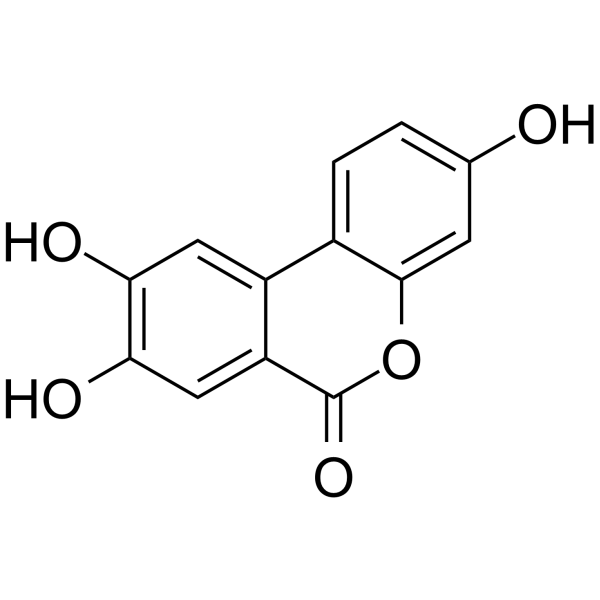
Urolithin C, a gut-microbial metabolite of Ellagic acid, is a glucose-dependent activator of insulin secretion. Urolithin C is a L-type Ca2+ channel opener and enhances Ca2+ influx. Urolithin C induces cell apoptosis through a mitochondria-mediated pathway and also stimulates reactive oxygen species (ROS) formation.
For research use only. We do not sell to patients.
- Purity : 98.0%
- CAS No.: 165393-06-6
- Formula: C13H8O5
- Molecular Weight:244.20
-
Storage:Powder -20°C, 3 years , 4°C, 2 years ; In solvent -80°C, 6 months , -20°C, 1 month

To place orders, for customer services and technical support, please contact: MedChemExpress USA
Tel: 609-228-6898 E-mail: [email protected] [email protected]


-
 Biological Activity
-
Chemical Information
-
Solvent & Solubility
- Protocols
- Purity & Documentation
- References
-
 Help & FAQs


-
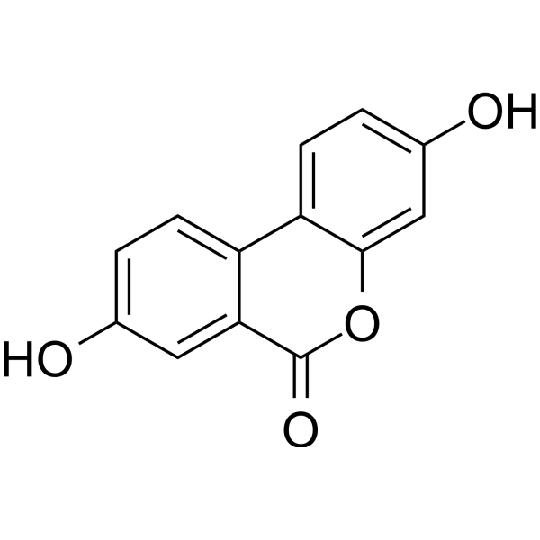
HY-100599
Urolithin AMitophagy Activator -
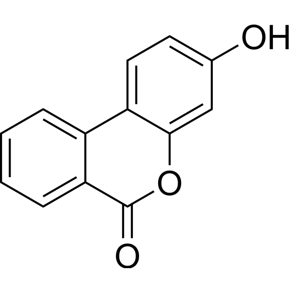
HY-126307
Urolithin BNF-κB Inhibitor -
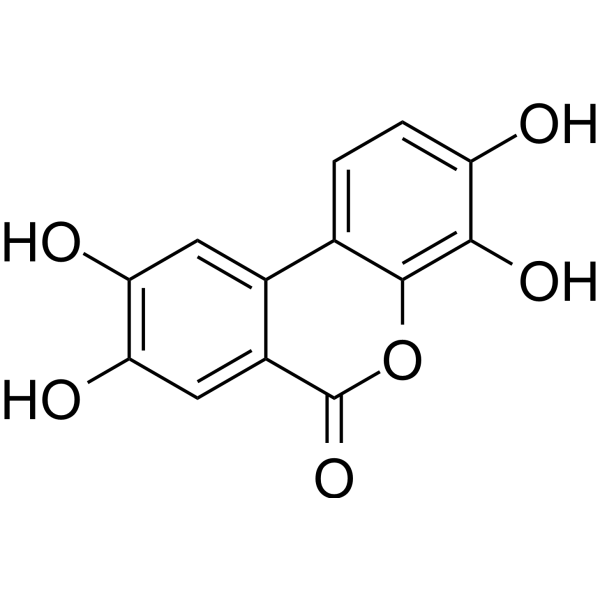
HY-133178
Urolithin DEphA Receptor Antagonist -
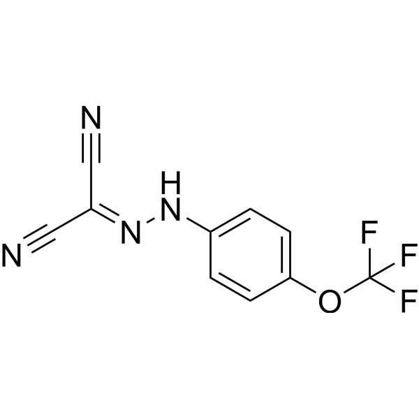
HY-100410
FCCPOXPHOS Uncoupler -
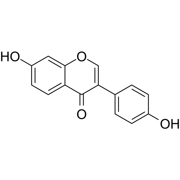
HY-N0019
DaidzeinEndogenous Metabolite -
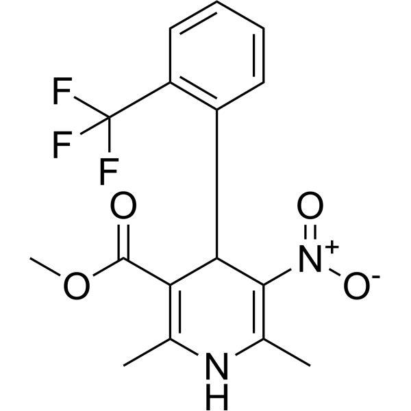
HY-10588
Bay K 8644L-type Ca2+ Channel Agonist -
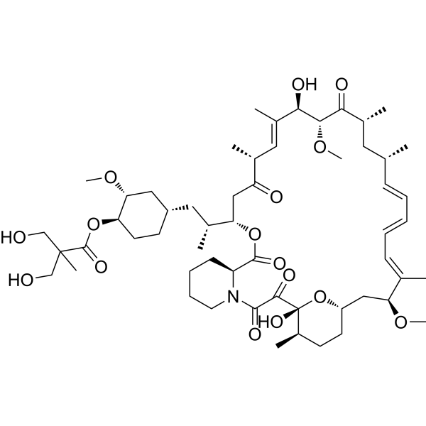
HY-50910
TemsirolimusmTOR Inhibitor -
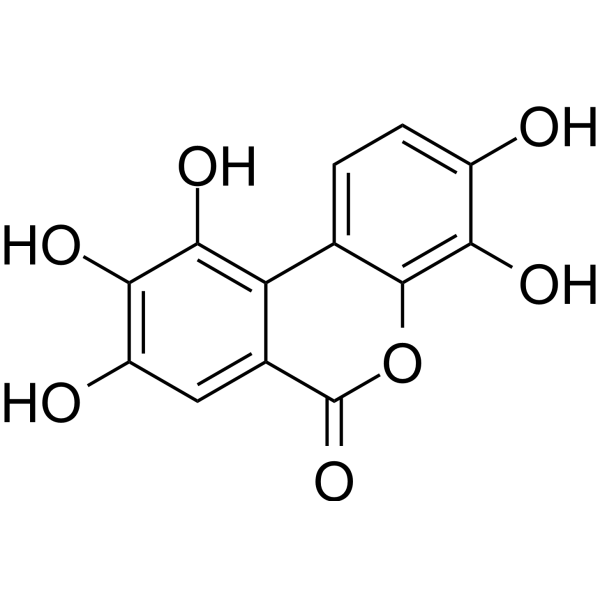
HY-N7922
Urolithin M5NA Inhibitor -
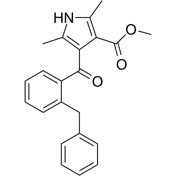
HY-103307
FPL64176L-type Ca2+ Channels Agonist -

HY-135897R
Urolithin C (Standard)Insulin Secretion Activator -
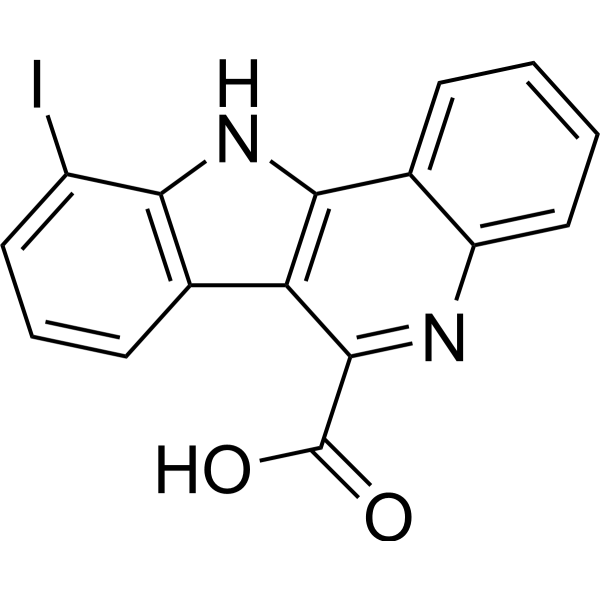
HY-146221
Dyrk1A-IN-5DYRK1A Inhibitor
Publications Citing Use of MedChemExpress (MCE) Urolithin C
MoreAll Calcium Channel Isoforms
MoreAll Endogenous Metabolite Isoforms
More

Biological Activity
Description
IC50 & Target
|
L-type calcium channel |
In Vitro
Urolithin C (20-100 μM; 1 hour; INS-1 cells) treatment enhances glucose-induced extracellular signal-regulated kinases 1/2 (ERK1/2) activation in INS-1 β-cells[1].
?
Urolithin C significantly inhibits the proliferation of PC12 cells. Urolithin C treatment actively increases the lactate dehydrogenase (LDH) release and lipid peroxidation malondialdehyde (MDA), stimulates reactive oxygen species (ROS) formation and mitochondrial membrane depolarization, and caused calcium dyshomeostasis[2].
?
Urolithin C treatment induces apoptosis and S phase cell cycle arrest[2].
MedChemExpress (MCE) has not independently confirmed the accuracy of these methods. They are for reference only. Further protocols information, click here.
-
Cell Line:INS-1 cells
-
Concentration:20 μM, 100 μM
-
Incubation Time:1 hour
-
Result:Enhanced glucose-induced extracellular signal-regulated kinases 1/2 (ERK1/2) activation.
In Vivo
MedChemExpress (MCE) has not independently confirmed the accuracy of these methods. They are for reference only.


Chemical Information
-
CAS No. 165393-06-6
-
Appearance Solid
-
Molecular Weight 244.20
-
Formula C13H8O5
-
Color Light brown to brown
-
SMILES
O=C1C2=CC(O)=C(O)C=C2C3=CC=C(O)C=C3O1
-
Structure Classification
-
Initial Source
-
Shipping
Room temperature in continental US; may vary elsewhere.
-
Storage
Powder -20°C 3 years 4°C 2 years In solvent -80°C 6 months -20°C 1 month


Publications (5)
-
Journal Impact Factor
-
Most Recent
-
Cell Discov
Cancer cells sense solid stress to enhance metastasis by CKAP4 phase separation-mediated microtubule branching. [Abstract]2024 Nov 12;10(1):114. PMID: 39528501 -
Ecotoxicol Environ Saf
CDK5-USP30 signaling pathway regulates MAVS-mediated inflammation via suppressing mitophagy in MPTP/MPP+ PD model. [Abstract]2024 Jul 1:279:116446. PMID: 38772138 -
Int Immunopharmacol
The ellagitannin metabolite urolithin C attenuated cognitive impairment by inhibiting neuroinflammation via downregulation of MAPK/NF-kB signaling pathways in aging mice. [Abstract]2024 Sep 19;142(Pt B):113151. PMID: 39303538 -
Naunyn Schmiedebergs Arch Pharmacol
Urolithin C reveals anti-NAFLD potential via AMPK-ferroptosis axis and modulating gut microbiota. [Abstract]2023 Oct;396(10):2687-2699. PMID: 37126194 -


Solvent & Solubility
In Vitro:
DMSO : 50 mg/mL (204.75 mM; Need ultrasonic; Hygroscopic DMSO has a significant impact on the solubility of product, please use newly opened DMSO)
Please refer to the solubility information to select the appropriate solvent. Once prepared, please aliquot and store the solution to prevent product inactivation from repeated freeze-thaw cycles.
Storage method and period of stock solution: -80°C, 6 months; -20°C, 1 month. When stored at -80°C, please use it within 6 months. When stored at -20°C, please use it within 1 month.
Please refer to the solubility information to select the appropriate solvent. Once prepared, please aliquot and store the solution to prevent product inactivation from repeated freeze-thaw cycles.
Storage method and period of stock solution: -80°C, 6 months; -20°C, 1 month. When stored at -80°C, please use it within 6 months. When stored at -20°C, please use it within 1 month.
Concentration (start) × Volume (start) = Concentration (final) × Volume (final)
In Vivo:
Select the appropriate dissolution method based on your experimental animal and administration route.
- For the following dissolution methods, please ensure to first prepare a clear stock solution using an In Vitro approach and then sequentially add co-solvents:
- To ensure reliable experimental results, the clarified stock solution can be appropriately stored based on storage conditions. As for the working solution for In Vivo experiments, it is recommended to prepare freshly and use it on the same day.
- The percentages shown for the solvents indicate their volumetric ratio in the final prepared solution. If precipitation or phase separation occurs during preparation, heat and/or sonication can be used to aid dissolution.
Add each solvent one by one: 10% DMSO 40% PEG300 5% Tween-80 45% Saline
Solubility: ≥ 2.08 mg/mL (8.52 mM); Clear solution
This protocol yields a clear solution of ≥ 2.08 mg/mL (saturation unknown).
Taking 1 mL working solution as an example, add 100 μL DMSO stock solution (20.8 mg/mL) to 400 μL PEG300, and mix evenly; then add 50 μL Tween-80 and mix evenly; then add 450 μL Saline to adjust the volume to 1 mL.
Preparation of Saline: Dissolve 0.9 g sodium chloride in ddH₂O and dilute to 100 mL to obtain a clear Saline solution.
Add each solvent one by one: 10% DMSO 90% (20% SBE-β-CD in Saline)
Solubility: ≥ 2.08 mg/mL (8.52 mM); Clear solution
This protocol yields a clear solution of ≥ 2.08 mg/mL (saturation unknown).
Taking 1 mL working solution as an example, add 100 μL DMSO stock solution (20.8 mg/mL) to 900 μL 20% SBE-β-CD in Saline, and mix evenly.
Preparation of 20% SBE-β-CD in Saline (4°C, storage for one week): 2 g SBE-β-CD powder is dissolved in 10 mL Saline, completely dissolve until clear.
In Vivo Dissolution Calculator
Please enter the basic information of animal experiments:
-
-
-
-
Recommended: Prepare an additional quantity of animals to account for potential losses during experiments.
Please enter your animal formula composition:
-
%DMSO +
Recommended: Keep the proportion of DMSO in working solution below 2% if your animal is weak.
-
%+
-
+%Tween-80 + +
-
%Saline +
The co-solvents required include: DMSO, . All of co-solvents are available by MedChemExpress (MCE). , Tween 80. All of co-solvents are available by MedChemExpress (MCE).
Working solution concentration: 0.22 mg/mL
Method for preparing stock solution: mg drug dissolved in μL DMSO. Stock solution concentration: mg/mL.
1. Take μL DMSO stock solution;
2. Add μL .
μL , mix evenly;
3. Then add μL Tween 80, mix evenly;
4. Then add μL
Please ensure that the stock solution in the first step is dissolved to a clear state, and add co-solvents in sequence. You can use ultrasonic heating (ultrasonic cleaner, recommended frequency 20-40 kHz), vortexing, etc. to assist dissolution.

Protocols
-
Apoptosis
Apoptosis, also called programmed cell death, is generally characterized by distinct morphological characteristics.
-
TUNEL staining for apoptotic DNA fragmentation
TUNEL staining detects DNA strand breaks by using terminal deoxynucleotidyl transferase to add labeled nucleotides to exposed 3′-OH DNA termini, generating either microscopic staining in fixed cells or tissue sections, or fluorescence/cytometric signal in cell suspensions. TUNEL positivity reflects DNA fragmentation but should not be interpreted alone as definitive apoptosis, because TUNEL can also label necrotic, autolytic, mechanically damaged, or DNA-repair-associated DNA breaks.
-
Annexin V plus membrane-impermeant dye apoptosis staining
Annexin V-based apoptosis assays rely on the detection of phosphatidylserine (PS) externalization from the inner leaflet of the plasma membrane to the outer leaflet, an early biochemical hallmark of apoptosis. Fluorescently labeled Annexin V binds PS in a calcium-dependent manner, enabling identification of early apoptotic cells by flow cytometry or fluorescence microscopy. When combined with a membrane-impermeant DNA-binding dye (e. g. , propidium iodide), this approach allows discrimination between viable (Annexin V−/dye−), early apoptotic (Annexin V+/dye−), and late apoptotic or necrotic (Annexin V+/dye+) cell populations by assessing membrane integrity and PS exposure.
-
ROS/oxidative-stress fluorescent staining
ROS/oxidative-stress fluorescent staining uses cell-permeant fluorogenic probes that become fluorescent after oxidation inside cells or tissues; commonly used examples include DCFH-DA/DCFDA for broad cellular oxidant detection, DHE for superoxide-related signal detection, MitoSOX for mitochondrial superoxide-related signal detection, and CellROX probes for oxidative-stress-associated fluorescence readouts. The assay detects probe oxidation rather than a single ROS species unless the probe and analysis method have been chemically validated for that species. DCFH-DA enters cells, is deacetylated by intracellular esterases to DCFH, and produces fluorescent DCF after oxidation, so the readout is used as an operational measure of total cellular oxidative stress rather than a species-specific ROS measurement. DHE and MitoSOX can report superoxide-related oxidation, but red fluorescence alone can include non-specific ethidium-like oxidation products; HPLC or optimized spectral approaches are
-
Research Protocol for Endocrine Diseases
Endocrine diseases often arise from disrupted hormone production, hormone signaling, or target-tissue responsiveness; for diabetes-focused endocrine disease models, insulin signaling regulates glucose uptake, hepatic glucose output, lipid metabolism, and β-cell compensation. Type 2 diabetes develops through interacting defects in insulin resistance, β-cell dysfunction, adipose inflammation, hepatic glucose overproduction, altered incretin signaling, and ectopic lipid metabolism. A major unresolved question is whether endocrine dysfunction is driven primarily by target-tissue insulin resistance, intrinsic β-cell failure, immune/inflammatory stress, or combined multi-organ failure that differs by disease stage.
-
Human Islet Cell Culture
The method of preserving islets in vitro, with purified reduced immunogenicity. The steps are islet isolation, islet cell purification, in vitro determination of islet function and islet cell culture.
-
Apoptosis Solutions
Apoptosis is a regulated, generally non-lytic cell-death pathway that removes unwanted, damaged, infected, or abnormal cells through coordinated morphological changes, caspase activation, DNA fragmentation, and membrane remodeling. The intrinsic apoptosis pathway is controlled mainly by mitochondrial outer membrane permeabilization, BCL-2 family proteins, cytochrome c release, apoptosome formation, caspase-9 activation, and downstream executioner caspase-3/7 activation. The extrinsic apoptosis pathway is initiated by death receptors such as Fas, TNFR, and TRAIL receptors, which recruit adaptor proteins and activate caspase-8 before engaging executioner caspases or mitochondrial amplification through BID cleavage. Apoptosis is linked to many phenotypes, including cancer cell killing, tissue homeostasis, immune regulation, neurodegeneration, infection response, and treatment-induced cytotoxicity; unresolved questions include how apoptosis interacts with necroptosis, pyroptosis, ferroptos
-
Ca2+ Staining Technique
Ca2+ staining is an experimental technique that utilizes specific fluorescent probes (such as Fluo-4 AM, Fura-2, etc.) to qualitatively or quantitatively detect dynamic changes in intracellular Ca2+ concentrations; this is achieved by monitoring the changes in fluorescent signals generated when these probes bind to free intracellular calcium ions. The underlying principle relies primarily on the presence of chelating groups within the probe's molecular structure that possess high affinity for calcium ions.


Purity & Documentation
-
Data Sheet (278 KB)
-
SDS (393 KB)
- English - EN (393 KB)
- Français - FR (393 KB)
- Deutsch - DE (393 KB)
- Norwegian - NO (393 KB)
- Español - ES (393 KB)
- Swedish - SV (393 KB)
- Italian - IT (393 KB)
- Korean - KR (393 KB)
- Portuguese - PT (393 KB)
-
Handling Instructions (2659 KB)
References
[1]. Slimane Toubal, et al. Urolithin C Increases Glucose-Induced ERK Activation Which Contributes to Insulin Secretion. Fundam Clin Pharmacol. 2020 Feb 21. [Content Brief]
[3]. Morgane Bayle, et al. Development and Validation of a Liquid Chromatography-Electrospray Ionization-Tandem Mass Spectrometry Method for the Determination of Urolithin C in Rat Plasma and Its Application to a Pharmacokinetic Study. J Pharm Biomed Anal. 2016 Nov 30;131:33-39. [Content Brief]


Complete Stock Solution Preparation Table
Please refer to the solubility information to select the appropriate solvent. Once prepared, please aliquot and store the solution to prevent product inactivation from repeated freeze-thaw cycles.
Storage method and period of stock solution: -80°C, 6 months; -20°C, 1 month. When stored at -80°C, please use it within 6 months. When stored at -20°C, please use it within 1 month.
| Optional Solvent | Concentration Solvent Mass | 1 mg | 5 mg | 10 mg | 25 mg |
|---|---|---|---|---|---|
| DMSO | 1 mM | 4.0950 mL | 20.4750 mL | 40.9500 mL | 102.3751 mL |
| 5 mM | 0.8190 mL | 4.0950 mL | 8.1900 mL | 20.4750 mL | |
| 10 mM | 0.4095 mL | 2.0475 mL | 4.0950 mL | 10.2375 mL | |
| 15 mM | 0.2730 mL | 1.3650 mL | 2.7300 mL | 6.8250 mL | |
| 20 mM | 0.2048 mL | 1.0238 mL | 2.0475 mL | 5.1188 mL | |
| 25 mM | 0.1638 mL | 0.8190 mL | 1.6380 mL | 4.0950 mL | |
| 30 mM | 0.1365 mL | 0.6825 mL | 1.3650 mL | 3.4125 mL | |
| 40 mM | 0.1024 mL | 0.5119 mL | 1.0238 mL | 2.5594 mL | |
| 50 mM | 0.0819 mL | 0.4095 mL | 0.8190 mL | 2.0475 mL | |
| 60 mM | 0.0683 mL | 0.3413 mL | 0.6825 mL | 1.7063 mL | |
| 80 mM | 0.0512 mL | 0.2559 mL | 0.5119 mL | 1.2797 mL | |
| 100 mM | 0.0410 mL | 0.2048 mL | 0.4095 mL | 1.0238 mL |


Urolithin C Related Classifications
HY-135897 Related Classifications
 Powered by Bioz