



Sabizabulin
Based on 4 publication(s) in Google Scholar
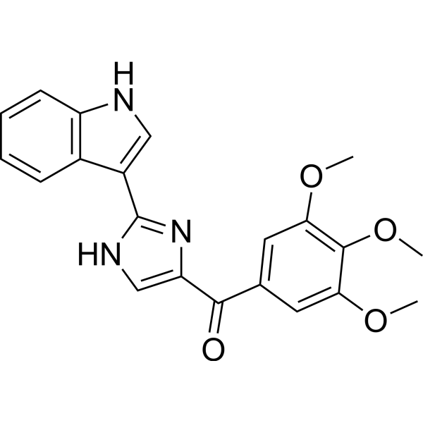
VERU-111 (ABI-231) is a potent and orally active α and β tubulin inhibitor, which displays strong antiproliferative activity, with an average IC50 of 5.2 nM against panels of melanoma and prostate cancer cell lines. VERU-111 (ABI-231) suppresses tumor growth and metastatic phenotypes of cervical cancer cells via targeting HPV E6 and E7, and has potential for the treatment of prostate cancer.
For research use only. We do not sell to patients.
- Purity : 99.00%
- CAS No.: 1332881-26-1
- Formula: C21H19N3O4
- Molecular Weight:377.39
-
Storage:
4°C, sealed storage, away from moisture and light
* In solvent : -80°C, 6 months; -20°C, 1 month (sealed storage, away from moisture and light)

To place orders, for customer services and technical support, please contact: MedChemExpress USA
Tel: 609-228-6898 E-mail: [email protected] [email protected]


-
 Biological Activity
-
Chemical Information
-
Solvent & Solubility
- Protocols
- Purity & Documentation
- References
-
 Help & FAQs


-
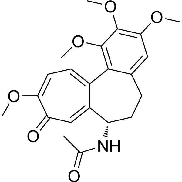
HY-16569
ColchicineTubulin Inhibitor -

HY-B0015
PaclitaxelAntitumor Agent -
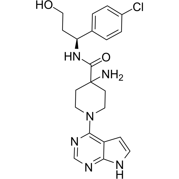
HY-15431
CapivasertibAkt Inhibitor -
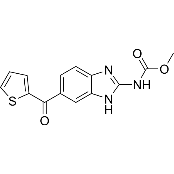
HY-13520
NocodazoleMicrotubule Inhibitor -
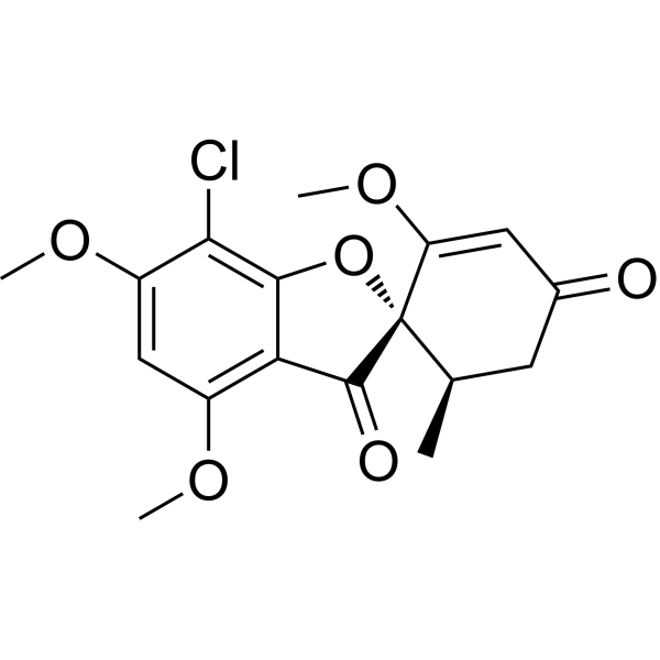
HY-17583
GriseofulvinAntifungal Antibiotic -

HY-16569R
Colchicine (Standard)Tubulin Inhibitor -
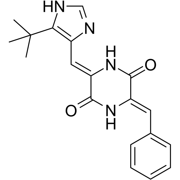
HY-14444
PlinabulinVascular Disrupting Agent -
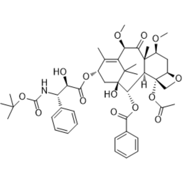
HY-15459
Cabazitaxel -
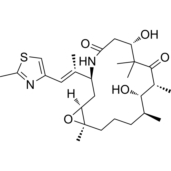
HY-10222
IxabepiloneMicrotubule Inhibitor -
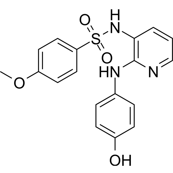
HY-13270
ABT-751Sulfonamides Antimitotic Compounds/Tubulin Binders Agent -
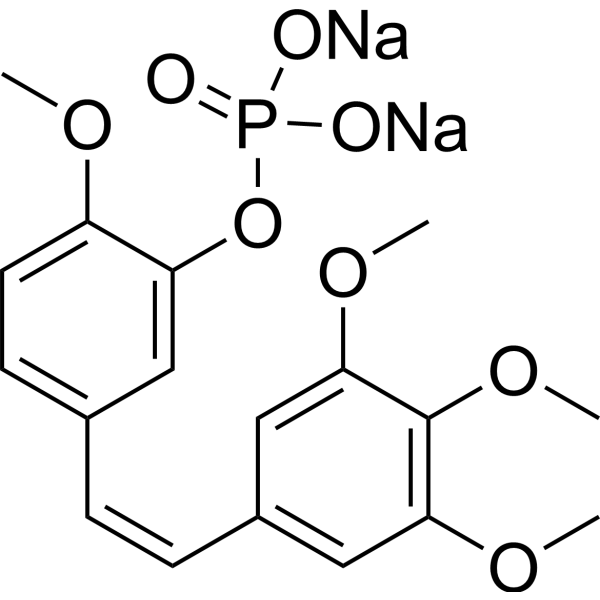
HY-17449
Fosbretabulin disodiumTubulin Destabilizing Agent -
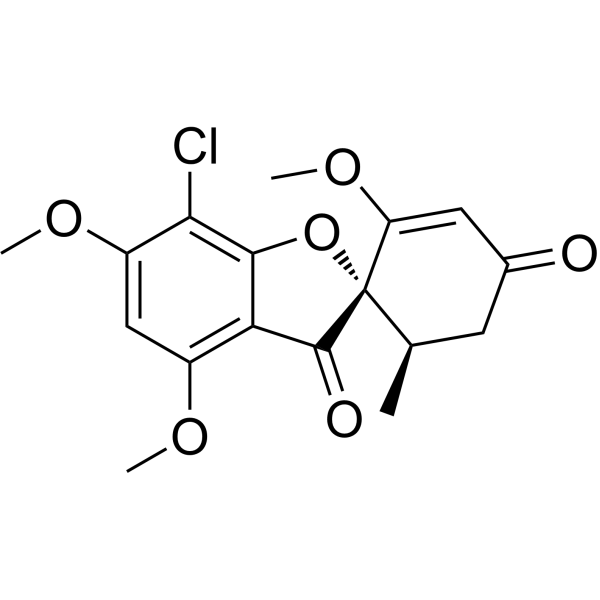
HY-17583R
Griseofulvin (Standard)
Publications Citing Use of MedChemExpress (MCE) Sabizabulin
More

Biological Activity
Description
IC50 & Target
tubulin[1]
Cellular Effect
|
Cell Line

|
Type | Value | Description | References |
|---|---|---|---|---|
| A-375 | IC50 |
10.4 nM
Compound: ABI-231
|
Antiproliferative activity against human A375 cells measured after 72 hrs by MTS assay
Antiproliferative activity against human A375 cells measured after 72 hrs by MTS assay
|
[PMID: 31860298] |
| A-375 | IC50 |
3.2 nM
Compound: 6a
|
Antiproliferative activity against human A375 cells after 48 hrs by SRB assay
Antiproliferative activity against human A375 cells after 48 hrs by SRB assay
|
[PMID: 22783954] |
| A-375 | IC50 |
8.1 nM
Compound: ABI-231
|
Antiproliferative activity against human A375 cells after 72 hrs by MTT assay
Antiproliferative activity against human A375 cells after 72 hrs by MTT assay
|
[PMID: 31251599] |
| A-375 | IC50 |
8.1 nM
Compound: VERU-111
|
Cytotoxicity in human A375 cells assessed as reduction in cell viability incubated for 72 hrs by MTS assay
Cytotoxicity in human A375 cells assessed as reduction in cell viability incubated for 72 hrs by MTS assay
|
[PMID: 30122035] |
| DU-145 | IC50 |
3.9 nM
Compound: 6a
|
Antiproliferative activity against human DU145 cells after 48 hrs by SRB assay
Antiproliferative activity against human DU145 cells after 48 hrs by SRB assay
|
[PMID: 22783954] |
| LNCaP | IC50 |
2.8 nM
Compound: 6a
|
Antiproliferative activity against human LNCAP cells after 48 hrs by SRB assay
Antiproliferative activity against human LNCAP cells after 48 hrs by SRB assay
|
[PMID: 22783954] |
| M14 | IC50 |
5.6 nM
Compound: ABI-231
|
Antiproliferative activity against human M14 cells after 72 hrs by MTT assay
Antiproliferative activity against human M14 cells after 72 hrs by MTT assay
|
[PMID: 31251599] |
| M14 | IC50 |
5.6 nM
Compound: VERU-111
|
Cytotoxicity in human M14 cells assessed as reduction in cell viability incubated for 72 hrs by MTS assay
Cytotoxicity in human M14 cells assessed as reduction in cell viability incubated for 72 hrs by MTS assay
|
[PMID: 30122035] |
| M14 | IC50 |
8.9 nM
Compound: ABI-231
|
Antiproliferative activity against human M14 cells measured after 72 hrs by MTS assay
Antiproliferative activity against human M14 cells measured after 72 hrs by MTS assay
|
[PMID: 31860298] |
| MDA-MB-231 | IC50 |
8.2 nM
Compound: ABI-231
|
Antiproliferative activity against human MDA-MB-231 cells measured after 72 hrs by MTS assay
Antiproliferative activity against human MDA-MB-231 cells measured after 72 hrs by MTS assay
|
[PMID: 31860298] |
| MDA-MB-453 | IC50 |
13.6 nM
Compound: ABI-231
|
Antiproliferative activity against human MDA-MB-453 cells measured after 72 hrs by MTS assay
Antiproliferative activity against human MDA-MB-453 cells measured after 72 hrs by MTS assay
|
[PMID: 31860298] |
| PC-3 | IC50 |
3.7 nM
Compound: 6a
|
Antiproliferative activity against human PC3 cells after 48 hrs by SRB assay
Antiproliferative activity against human PC3 cells after 48 hrs by SRB assay
|
[PMID: 22783954] |
| RPMI-7951 | IC50 |
7.2 nM
Compound: VERU-111
|
Cytotoxicity in human RPMI7951 cells assessed as reduction in cell viability incubated for 72 hrs by MTS assay
Cytotoxicity in human RPMI7951 cells assessed as reduction in cell viability incubated for 72 hrs by MTS assay
|
[PMID: 30122035] |
| RPMI-7951 | IC50 |
8.8 nM
Compound: ABI-231
|
Antiproliferative activity against human RPMI7951 cells measured after 72 hrs by MTS assay
Antiproliferative activity against human RPMI7951 cells measured after 72 hrs by MTS assay
|
[PMID: 31860298] |
| SK-BR-3 | IC50 |
13.3 nM
Compound: ABI-231
|
Antiproliferative activity against human SK-BR-3 cells measured after 72 hrs by MTS assay
Antiproliferative activity against human SK-BR-3 cells measured after 72 hrs by MTS assay
|
[PMID: 31860298] |
| WM164 | IC50 |
5.3 nM
Compound: 6a
|
Antiproliferative activity against human WM164 cells after 48 hrs by SRB assay
Antiproliferative activity against human WM164 cells after 48 hrs by SRB assay
|
[PMID: 22783954] |
| WM164 | IC50 |
7.2 nM
Compound: ABI-231
|
Antiproliferative activity against human WM164 cells after 72 hrs by MTT assay
Antiproliferative activity against human WM164 cells after 72 hrs by MTT assay
|
[PMID: 31251599] |
In Vitro
VERU-111 (2.5-80 nM; 24-48 hours) inhibits Panc-1, AsPC-1 and HPAF-II cells growth in a dose and time-dependent manner (24 hours: IC50s of 25, 35 and 35 nM, respectively; 48 hours: IC50s of 11.8, 15.5, and 25 nM, respectively)[4].
VERU-111 (5-20 nM; 24 hours) arrests Panc-1 and AsPC-1 cells in G2/M phase in a dose-dependent manner[4].
VERU-111 (5-20 nM; 24 hours) shows dose-dependent inhibition of pro-Caspase 3 and 9 and activation of Caspase-3 and 9, induces the expression of Bax and Bad, and inhibits the expression of Bcl-2 and Bcl-xl proteins in both AsPC-1 and Panc-1 cells[4].
MedChemExpress (MCE) has not independently confirmed the accuracy of these methods. They are for reference only.
-
Cell Line:Panc-1, AsPC-1, HPAF-II cells
-
Concentration:2.5, 5, 10, 20, 40, 80 nM
-
Incubation Time:24, 48 hours
-
Result:Inhibited the growth of PanCa cells in a dose and time-dependent manner. The IC50 of VERU-111 was 25, 35 and 35 nM in Panc-1, AsPC-1 and HPAF-II, respectively after 24 h treatment, while 48 h post-treatment it was 11.8, 15.5, and 25 nM.
-
Cell Line:Panc-1, AsPC-1 cells
-
Concentration:5, 10, 20 nM
-
Incubation Time:24 hours
-
Result:Arrested Panc-1 and AsPC-1 cells in G2/M phase in a dose-dependent manner.
-
Cell Line:AsPC-1 and Panc-1 cells
-
Concentration:5, 10, 20 nM
-
Incubation Time:24 hours
-
Result:Dose-dependent inhibition of pro-Caspase 3 and 9 and activation of Caspase-3 and 9 in both AsPC-1 and Panc-1 cells. Induces the expression of Bax and Bad and inhibited the expression of Bcl-2 and Bcl-xl proteins.
In Vivo
MedChemExpress (MCE) has not independently confirmed the accuracy of these methods. They are for reference only.
-
Animal Model:Six-week-old female athymic nude mice (bearing AsPC-1 cells)
-
Dosage:50 μg/mouse
-
Administration:Intra-tumorally; 3 times per week for 3 weeks
-
Result:Effectively inhibited tumor growth.
Clinical Trial
| NCT Number | Sponsor | Condition | Start Date |
Phase
|
|---|---|---|---|---|
| NCT01329991 | 2011-05 | PHASE1 |


Chemical Information
-
CAS No. 1332881-26-1
-
Appearance Solid
-
Molecular Weight 377.39
-
Formula C21H19N3O4
-
Color Light yellow to yellow
-
SMILES
O=C(C1=CNC(C2=CNC3=C2C=CC=C3)=N1)C4=CC(OC)=C(OC)C(OC)=C4
-
Synonyms
VERU-111; ABI-231
-
Shipping
Room temperature in continental US; may vary elsewhere.
-
Storage
4°C, sealed storage, away from moisture and light
* In solvent : -80°C, 6 months; -20°C, 1 month (sealed storage, away from moisture and light)


Publications (4)
-
Journal Impact Factor
-
Most Recent
-
Mol Cancer Ther
RGN6024 is a brain-penetrant, small molecule tubulin destabilizer for the treatment of glioblastoma. [Abstract]2025 Aug 1;24(8):1129-1144. PMID: 40397474 -
Biomedicines
Novel Brain-Penetrant, Small-Molecule Tubulin Destabilizers for the Treatment of Glioblastoma. [Abstract]2024 Feb 9;12(2):406. PMID: 38398008 -
-


Solvent & Solubility
In Vitro:
DMSO : 20 mg/mL (53.00 mM; Need ultrasonic; Hygroscopic DMSO has a significant impact on the solubility of product, please use newly opened DMSO)
Please refer to the solubility information to select the appropriate solvent. Once prepared, please aliquot and store the solution to prevent product inactivation from repeated freeze-thaw cycles.
Storage method and period of stock solution: -80°C, 6 months; -20°C, 1 month (sealed storage, away from moisture and light). When stored at -80°C, please use it within 6 months. When stored at -20°C, please use it within 1 month.
Please refer to the solubility information to select the appropriate solvent. Once prepared, please aliquot and store the solution to prevent product inactivation from repeated freeze-thaw cycles.
Storage method and period of stock solution: -80°C, 6 months; -20°C, 1 month (sealed storage, away from moisture and light). When stored at -80°C, please use it within 6 months. When stored at -20°C, please use it within 1 month.
Concentration (start) × Volume (start) = Concentration (final) × Volume (final)
In Vivo:
Select the appropriate dissolution method based on your experimental animal and administration route.
- For the following dissolution methods, please ensure to first prepare a clear stock solution using an In Vitro approach and then sequentially add co-solvents:
- To ensure reliable experimental results, the clarified stock solution can be appropriately stored based on storage conditions. As for the working solution for In Vivo experiments, it is recommended to prepare freshly and use it on the same day.
- The percentages shown for the solvents indicate their volumetric ratio in the final prepared solution. If precipitation or phase separation occurs during preparation, heat and/or sonication can be used to aid dissolution.
Add each solvent one by one: 10% DMSO 40% PEG300 5% Tween-80 45% Saline
Solubility: ≥ 2 mg/mL (5.30 mM); Clear solution
This protocol yields a clear solution of ≥ 2 mg/mL (saturation unknown).
Taking 1 mL working solution as an example, add 100 μL DMSO stock solution (20.0 mg/mL) to 400 μL PEG300, and mix evenly; then add 50 μL Tween-80 and mix evenly; then add 450 μL Saline to adjust the volume to 1 mL.
Preparation of Saline: Dissolve 0.9 g sodium chloride in ddH₂O and dilute to 100 mL to obtain a clear Saline solution.
Add each solvent one by one: 10% DMSO 90% (20% SBE-β-CD in Saline)
Solubility: 2 mg/mL (5.30 mM); Suspended solution; Need ultrasonic
This protocol yields a suspended solution of 2 mg/mL. Suspended solution can be used for oral and intraperitoneal injection.
Taking 1 mL working solution as an example, add 100 μL DMSO stock solution (20.0 mg/mL) to 900 μL 20% SBE-β-CD in Saline, and mix evenly.
Preparation of 20% SBE-β-CD in Saline (4°C, storage for one week): 2 g SBE-β-CD powder is dissolved in 10 mL Saline, completely dissolve until clear.
In Vivo Dissolution Calculator
Please enter the basic information of animal experiments:
-
-
-
-
Recommended: Prepare an additional quantity of animals to account for potential losses during experiments.
Please enter your animal formula composition:
-
%DMSO +
Recommended: Keep the proportion of DMSO in working solution below 2% if your animal is weak.
-
%+
-
+%Tween-80 + +
-
%Saline +
The co-solvents required include: DMSO, . All of co-solvents are available by MedChemExpress (MCE). , Tween 80. All of co-solvents are available by MedChemExpress (MCE).
Working solution concentration: 0.22 mg/mL
Method for preparing stock solution: mg drug dissolved in μL DMSO. Stock solution concentration: mg/mL. * In solvent : -80°C, 6 months; -20°C, 1 month (sealed storage, away from moisture and light)
1. Take μL DMSO stock solution;
2. Add μL .
μL , mix evenly;
3. Then add μL Tween 80, mix evenly;
4. Then add μL
Please ensure that the stock solution in the first step is dissolved to a clear state, and add co-solvents in sequence. You can use ultrasonic heating (ultrasonic cleaner, recommended frequency 20-40 kHz), vortexing, etc. to assist dissolution.


Purity & Documentation
-
Data Sheet (284 KB)
-
SDS (252 KB)
- English - EN (252 KB)
- Français - FR (252 KB)
- Deutsch - DE (252 KB)
- Norwegian - NO (252 KB)
- Español - ES (252 KB)
- Swedish - SV (252 KB)
- Italian - IT (252 KB)
- Korean - KR (252 KB)
- Portuguese - PT (252 KB)
-
Handling Instructions (2659 KB)
References
[1]. Wang Q, et al. Structure-Guided Design, Synthesis, and Biological Evaluation of (2-(1H-Indol-3-yl)-1H-imidazol-4-yl)(3,4,5-trimethoxyphenyl) Methanone (ABI-231) Analogues Targeting the Colchicine Binding Site in Tubulin. J Med Chem. 2019 Jul 12. [Content Brief]
[4]. Kashyap VK, et al. Therapeutic efficacy of a novel βIII/βIV-tubulin inhibitor (VERU-111) in pancreatic cancer. J Exp Clin Cancer Res. 2019 Jan 23;38(1):29. [Content Brief]


Complete Stock Solution Preparation Table
Please refer to the solubility information to select the appropriate solvent. Once prepared, please aliquot and store the solution to prevent product inactivation from repeated freeze-thaw cycles.
Storage method and period of stock solution: -80°C, 6 months; -20°C, 1 month (sealed storage, away from moisture and light). When stored at -80°C, please use it within 6 months. When stored at -20°C, please use it within 1 month.
| Optional Solvent | Concentration Solvent Mass | 1 mg | 5 mg | 10 mg | 25 mg |
|---|---|---|---|---|---|
| DMSO | 1 mM | 2.6498 mL | 13.2489 mL | 26.4978 mL | 66.2445 mL |
| 5 mM | 0.5300 mL | 2.6498 mL | 5.2996 mL | 13.2489 mL | |
| 10 mM | 0.2650 mL | 1.3249 mL | 2.6498 mL | 6.6244 mL | |
| 15 mM | 0.1767 mL | 0.8833 mL | 1.7665 mL | 4.4163 mL | |
| 20 mM | 0.1325 mL | 0.6624 mL | 1.3249 mL | 3.3122 mL | |
| 25 mM | 0.1060 mL | 0.5300 mL | 1.0599 mL | 2.6498 mL | |
| 30 mM | 0.0883 mL | 0.4416 mL | 0.8833 mL | 2.2081 mL | |
| 40 mM | 0.0662 mL | 0.3312 mL | 0.6624 mL | 1.6561 mL | |
| 50 mM | 0.0530 mL | 0.2650 mL | 0.5300 mL | 1.3249 mL |


Sabizabulin Related Classifications
HY-120599 Related Classifications
 Powered by Bioz