



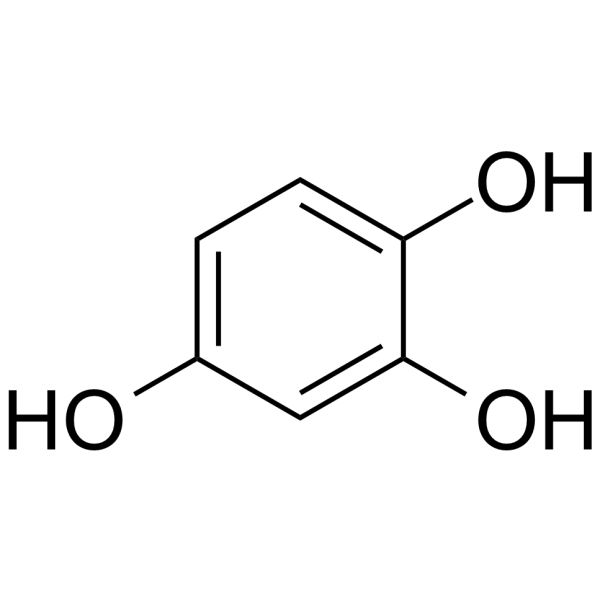
1,2,4-Trihydroxybenzene

Based on 1 Customer Validation
1,2,4-Trihydroxybenzene (Hydroxyhydroquinone) is an ER stress inducer that targets proteins such as PKR-like ER kinase PERK to induce cytotoxicity. 1,2,4-Trihydroxybenzene selectively activates eIF2α phosphorylation, activates the PERK-eIF2α signaling pathway and induces stress granule formation. 1,2,4-Trihydroxybenzene subsequently exacerbates oxidative stress and causes DNA double-strand breaks, destroying organelles such as mitochondria and ER, and inducing cell death. 1,2,4-Trihydroxybenzene also has the potential to exhibit anti-tumor effect, increase blood pressure, and relieve spasm.
For research use only. We do not sell to patients.
- Purity : 99.56%
- CAS No.: 533-73-3
- Formula: C6H6O3
- Molecular Weight:126.11
-
Storage:Powder -20°C, 3 years , 4°C, 2 years ; In solvent -80°C, 6 months , -20°C, 1 month

To place orders, for customer services and technical support, please contact: MedChemExpress USA
Tel: 609-228-6898 E-mail: [email protected] [email protected]


-
 Biological Activity
-
Chemical Information
-
Solvent & Solubility
- Protocols
- Purity & Documentation
- References
-
 Help & FAQs





Biological Activity
Description
IC50 & Target
|
eIF2-α |
In Vitro
1,2,4-Trihydroxybenzene (HHQ) (10-100 μM; 2-4 h) increases intracellular Ca2+ concentration, induces membrane potential hyperpolarization and activates Ca2+-dependent K+ channels in rat thymic lymphocyte experiments[1].
1,2,4-Trihydroxybenzene (THB) (50-800 μM; 24 h) dose-dependently reduces the cell viability of human keratinocytes (HaCaT) and induces stress granule formation and eIF2α/PERK phosphorylation. Continued treatment with 1,2,4-Trihydroxybenzene (THB) (50-800 μM; 1 h, followed by 24 h recovery) leads to DNA damage and apoptosis[2].
1,2,4-Trihydroxybenzene (20-40 μg/mL; 6-24 h) significantly inhibits the cell viability of HeLa [Chang liver] (human Chang liver cells), induces G2/M phase arrest, and non-apoptotic cell death, accompanied by mitochondrial structural destruction and increasing ROS levels[3].
MedChemExpress (MCE) has not independently confirmed the accuracy of these methods. They are for reference only. Further protocols information, click here.
-
Cell Line:HeLa Chang liver cells
-
Concentration:20, 40 μg/mL
-
Incubation Time:6, 12, 24 h
-
Result:Resulted G2/M phase arrest (40% cell accumulation) at 40 μg/mL.
Showed mitochondrial swelling, cristae disruption, and ER volume reduction.
In Vivo
MedChemExpress (MCE) has not independently confirmed the accuracy of these methods. They are for reference only.


Chemical Information
-
CAS No. 533-73-3
-
Appearance Solid
-
Molecular Weight 126.11
-
Formula C6H6O3
-
Color Brown to gray
-
SMILES
OC1=CC=C(O)C=C1O
-
Synonyms
Hydroxyhydroquinone
-
Structure Classification
-
Initial Source
Phanerochaete chrysosporium
-
Shipping
Room temperature in continental US; may vary elsewhere.
-
Storage
Powder -20°C 3 years 4°C 2 years In solvent -80°C 6 months -20°C 1 month


Solvent & Solubility
In Vitro:
DMSO : 50 mg/mL (396.48 mM; Need ultrasonic; Hygroscopic DMSO has a significant impact on the solubility of product, please use newly opened DMSO)
Please refer to the solubility information to select the appropriate solvent. Once prepared, please aliquot and store the solution to prevent product inactivation from repeated freeze-thaw cycles.
Storage method and period of stock solution: -80°C, 6 months; -20°C, 1 month. When stored at -80°C, please use it within 6 months. When stored at -20°C, please use it within 1 month.
Please refer to the solubility information to select the appropriate solvent. Once prepared, please aliquot and store the solution to prevent product inactivation from repeated freeze-thaw cycles.
Storage method and period of stock solution: -80°C, 6 months; -20°C, 1 month. When stored at -80°C, please use it within 6 months. When stored at -20°C, please use it within 1 month.
Concentration (start) × Volume (start) = Concentration (final) × Volume (final)
In Vivo:
Select the appropriate dissolution method based on your experimental animal and administration route.
- For the following dissolution methods, please ensure to first prepare a clear stock solution using an In Vitro approach and then sequentially add co-solvents:
- To ensure reliable experimental results, the clarified stock solution can be appropriately stored based on storage conditions. As for the working solution for In Vivo experiments, it is recommended to prepare freshly and use it on the same day.
- The percentages shown for the solvents indicate their volumetric ratio in the final prepared solution. If precipitation or phase separation occurs during preparation, heat and/or sonication can be used to aid dissolution.
Add each solvent one by one: 10% DMSO 40% PEG300 5% Tween-80 45% Saline
Solubility: 2.5 mg/mL (19.82 mM); Suspended solution; Need ultrasonic
This protocol yields a suspended solution of 2.5 mg/mL. Suspended solution can be used for oral and intraperitoneal injection.
Taking 1 mL working solution as an example, add 100 μL DMSO stock solution (25.0 mg/mL) to 400 μL PEG300, and mix evenly; then add 50 μL Tween-80 and mix evenly; then add 450 μL Saline to adjust the volume to 1 mL.
Preparation of Saline: Dissolve 0.9 g sodium chloride in ddH₂O and dilute to 100 mL to obtain a clear Saline solution.
Add each solvent one by one: 10% DMSO 90% (20% SBE-β-CD in Saline)
Solubility: ≥ 2.5 mg/mL (19.82 mM); Clear solution
This protocol yields a clear solution of ≥ 2.5 mg/mL (saturation unknown).
Taking 1 mL working solution as an example, add 100 μL DMSO stock solution (25.0 mg/mL) to 900 μL 20% SBE-β-CD in Saline, and mix evenly.
Preparation of 20% SBE-β-CD in Saline (4°C, storage for one week): 2 g SBE-β-CD powder is dissolved in 10 mL Saline, completely dissolve until clear.
In Vivo Dissolution Calculator
Please enter the basic information of animal experiments:
-
-
-
-
Recommended: Prepare an additional quantity of animals to account for potential losses during experiments.
Please enter your animal formula composition:
-
%DMSO +
Recommended: Keep the proportion of DMSO in working solution below 2% if your animal is weak.
-
%+
-
+%Tween-80 + +
-
%Saline +
The co-solvents required include: DMSO, . All of co-solvents are available by MedChemExpress (MCE). , Tween 80. All of co-solvents are available by MedChemExpress (MCE).
Working solution concentration: 0.22 mg/mL
Method for preparing stock solution: mg drug dissolved in μL DMSO. Stock solution concentration: mg/mL.
1. Take μL DMSO stock solution;
2. Add μL .
μL , mix evenly;
3. Then add μL Tween 80, mix evenly;
4. Then add μL
Please ensure that the stock solution in the first step is dissolved to a clear state, and add co-solvents in sequence. You can use ultrasonic heating (ultrasonic cleaner, recommended frequency 20-40 kHz), vortexing, etc. to assist dissolution.

Protocols
-
Kinase activity and phosphorylation assays
Kinase activity assays measure the ability of kinases to transfer phosphate groups from ATP to specific substrates, while phosphorylation assays detect the presence and levels of phosphorylated proteins. Common methods include radiolabeled ATP incorporation (e. g. ,), ADP release detection via bioluminescence (e. g. ,[3]), enzyme-linked immunosorbent assays (ELISA) for phospho-specific epitopes (e. g. ,[6]), and microtiter-based formats for high-throughput screening (e. g. ,[8]). The ADP-Glo assay quantifies kinase activity by measuring ADP produced during phosphorylation using a luciferase-based system. Radiometric assays involve autoradiography or scintillation counting after incorporation of 32P-labeled ATP into substrate proteins. ELISA-based approaches rely on phospho-specific antibodies to detect activated kinases in cell lysates or purified samples.
-
Western Blot
Western blotting (WB) is a commonly used experimental method in molecular biology, biochemistry, and immunogenetics for identifying and quantifying target proteins. It combines gel electrophoresis with immunoassay, enabling researchers to analyze protein expression, post-translational modifications, and molecular weight.
-
Cell Cytotoxicity Assay
Cytotoxicity assays are usually based on the assessment of cell membrane damage, which can also be indirectly detected by measuring cell viability. Detection methods include MTT assay, CKK-8 assay, LDH assay and ATP assay, etc.
-
Mammalian live/dead viability and cytotoxicity staining
Live/dead viability and cytotoxicity staining assays are based on the simultaneous detection of intracellular esterase activity in metabolically active (viable) cells and membrane integrity loss in non-viable cells. In commonly used dual-staining approaches, membrane-permeant fluorogenic substrates are converted by intracellular esterases into fluorescent products in live cells, while impermeant DNA-binding dyes selectively enter cells with compromised plasma membranes and label nucleic acids in dead or dying cells, enabling discrimination between viable and non-viable populations by fluorescence microscopy or flow cytometry.
-
ROS/oxidative-stress fluorescent staining
ROS/oxidative-stress fluorescent staining uses cell-permeant fluorogenic probes that become fluorescent after oxidation inside cells or tissues; commonly used examples include DCFH-DA/DCFDA for broad cellular oxidant detection, DHE for superoxide-related signal detection, MitoSOX for mitochondrial superoxide-related signal detection, and CellROX probes for oxidative-stress-associated fluorescence readouts. The assay detects probe oxidation rather than a single ROS species unless the probe and analysis method have been chemically validated for that species. DCFH-DA enters cells, is deacetylated by intracellular esterases to DCFH, and produces fluorescent DCF after oxidation, so the readout is used as an operational measure of total cellular oxidative stress rather than a species-specific ROS measurement. DHE and MitoSOX can report superoxide-related oxidation, but red fluorescence alone can include non-specific ethidium-like oxidation products; HPLC or optimized spectral approaches are
-
Apoptosis Solutions
Apoptosis is a regulated, generally non-lytic cell-death pathway that removes unwanted, damaged, infected, or abnormal cells through coordinated morphological changes, caspase activation, DNA fragmentation, and membrane remodeling. The intrinsic apoptosis pathway is controlled mainly by mitochondrial outer membrane permeabilization, BCL-2 family proteins, cytochrome c release, apoptosome formation, caspase-9 activation, and downstream executioner caspase-3/7 activation. The extrinsic apoptosis pathway is initiated by death receptors such as Fas, TNFR, and TRAIL receptors, which recruit adaptor proteins and activate caspase-8 before engaging executioner caspases or mitochondrial amplification through BID cleavage. Apoptosis is linked to many phenotypes, including cancer cell killing, tissue homeostasis, immune regulation, neurodegeneration, infection response, and treatment-induced cytotoxicity; unresolved questions include how apoptosis interacts with necroptosis, pyroptosis, ferroptos
-
Protocol for Kinase activity and phosphorylation assays
Kinase activity assays measure transfer of phosphate from ATP to a protein or peptide substrate, generating phosphorylated substrate, ADP, or incorporated radiolabeled phosphate as the readout; phosphorylation assays measure site-specific phosphorylation in cells or tissues as a proxy for kinase-pathway activation, inhibition, or substrate regulation. Phosphorylation can be detected by phospho-specific Western blot, immunoprecipitation kinase assay, phospho-immunofluorescence, phospho-flow cytometry, luminescent ADP detection, radiolabeled ATP incorporation, or reporter-based pathway assays, and these readouts can be applied to cancer cells, primary neurons, mouse tumors, organoids, inflammatory macrophages, ferroptosis studies, and mitophagy studies when the kinase target is biologically relevant.
-
Cell Viability Determination by MTT Colorimetric Assay
The following protocol uses the MTT colorimetric assay as a classic literature-established method for assessing cell viability/metabolic activity in cultured mammalian cells. MTT[3-(4,5-dimethylthiazol-2-yl)-2,5-diphenyltetrazolium bromide] is reduced by metabolically active cells to a colored formazan product; the amount of formazan is quantified spectrophotometrically and provides an indirect measure of metabolically active viable cells. Importantly, MTT reduction reflects cellular oxidoreductase/metabolic activity rather than an absolute direct count of living cells, so changes in cellular metabolism can alter the signal independently of cell number.


Purity & Documentation
-
Data Sheet (276 KB)
-
SDS (557 KB)
- English - EN (557 KB)
- Français - FR (557 KB)
- Deutsch - DE (557 KB)
- Norwegian - NO (557 KB)
- Español - ES (557 KB)
- Swedish - SV (557 KB)
- Italian - IT (557 KB)
- Korean - KR (557 KB)
- Portuguese - PT (557 KB)
-
Handling Instructions (2659 KB)
References
[1]. Risa Kamae, et al. Hydroxyhydroquinone, a by-product of coffee bean roasting, increases intracellular Ca2+ concentration in rat thymic lymphocytes. Food Chem Toxicol. 2017 Apr;102:39-45. [Content Brief]
[2]. Jung DM, et al. 1,2,4-trihydroxybenzene induces stress granule formation and causes DNA damage in human keratinocytes. Toxicol In Vitro. 2023 Oct;92:105638. [Content Brief]
[3]. Park EJ, et al. 1,2,4-trihydroxybenzene induces non-apoptotic cell death via the structural damage of intracellular organelles. Toxicol Appl Pharmacol. 2024 Nov;492:117096. [Content Brief]
[4]. Inoue S. Effects of 1,3,5-trihydroxybenzene and 2,4,6-trihydroxy-1-propiophenone on the smooth muscle organs. Jpn J Pharmacol. 1969 Jun;19(2):224-33. [Content Brief]


Complete Stock Solution Preparation Table
Please refer to the solubility information to select the appropriate solvent. Once prepared, please aliquot and store the solution to prevent product inactivation from repeated freeze-thaw cycles.
Storage method and period of stock solution: -80°C, 6 months; -20°C, 1 month. When stored at -80°C, please use it within 6 months. When stored at -20°C, please use it within 1 month.
| Optional Solvent | Concentration Solvent Mass | 1 mg | 5 mg | 10 mg | 25 mg |
|---|---|---|---|---|---|
| DMSO | 1 mM | 7.9296 mL | 39.6479 mL | 79.2959 mL | 198.2396 mL |
| 5 mM | 1.5859 mL | 7.9296 mL | 15.8592 mL | 39.6479 mL | |
| 10 mM | 0.7930 mL | 3.9648 mL | 7.9296 mL | 19.8240 mL | |
| 15 mM | 0.5286 mL | 2.6432 mL | 5.2864 mL | 13.2160 mL | |
| 20 mM | 0.3965 mL | 1.9824 mL | 3.9648 mL | 9.9120 mL | |
| 25 mM | 0.3172 mL | 1.5859 mL | 3.1718 mL | 7.9296 mL | |
| 30 mM | 0.2643 mL | 1.3216 mL | 2.6432 mL | 6.6080 mL | |
| 40 mM | 0.1982 mL | 0.9912 mL | 1.9824 mL | 4.9560 mL | |
| 50 mM | 0.1586 mL | 0.7930 mL | 1.5859 mL | 3.9648 mL | |
| 60 mM | 0.1322 mL | 0.6608 mL | 1.3216 mL | 3.3040 mL | |
| 80 mM | 0.0991 mL | 0.4956 mL | 0.9912 mL | 2.4780 mL | |
| 100 mM | 0.0793 mL | 0.3965 mL | 0.7930 mL | 1.9824 mL |


1,2,4-Trihydroxybenzene Related Classifications
HY-W010451 Related Classifications
 Powered by Bioz