



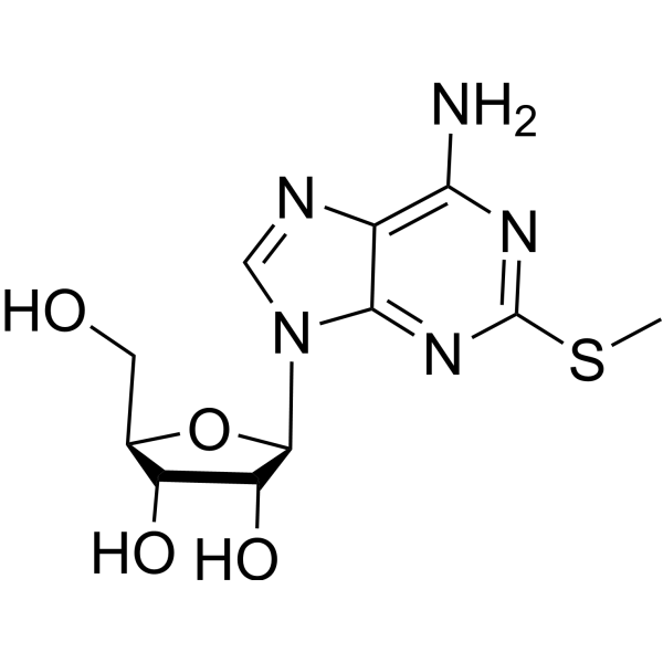
2-Methylthioadenosine
Based on 1 publication(s) in Google Scholar
2-Methylthioadenosine is a purine nucleoside analog. Purine nucleoside analogs have broad antitumor activity targeting indolent lymphoid malignancies. Anticancer mechanisms in this process rely on inhibition of DNA synthesis, induction of apoptosis, etc.
For research use only. We do not sell to patients.
- Purity : 98.82%
- CAS No.: 4105-39-9
- Formula: C11H15N5O4S
- Molecular Weight:313.33
-
Storage:Powder -20°C, 3 years , 4°C, 2 years ; In solvent -80°C, 6 months , -20°C, 1 month

To place orders, for customer services and technical support, please contact: MedChemExpress USA
Tel: 609-228-6898 E-mail: [email protected] [email protected]


-
 Biological Activity
-
Chemical Information
-
Solvent & Solubility
- Protocols
- Purity & Documentation
- References
-
 Help & FAQs


Publications Citing Use of MedChemExpress (MCE) 2-Methylthioadenosine
More

Biological Activity
Description


Chemical Information
-
CAS No. 4105-39-9
-
Appearance Solid
-
Molecular Weight 313.33
-
Formula C11H15N5O4S
-
Color White to off-white
-
SMILES
NC1=C2N=CN([C@H]3[C@@H](O)[C@@H](O)[C@@H](CO)O3)C2=NC(SC)=N1
-
Shipping
Room temperature in continental US; may vary elsewhere.
-
Storage
Powder -20°C 3 years 4°C 2 years In solvent -80°C 6 months -20°C 1 month


Publications (1)
-
Journal Impact Factor
-
Most Recent
-
Cell
Adenosine kinase and ADAL coordinate detoxification of modified adenosines to safeguard metabolism. [Abstract]2025 Oct 30;188(22):6151-6169.e24. PMID: 40840445


Solvent & Solubility
In Vitro:
DMSO : 25 mg/mL (79.79 mM; Need ultrasonic; Hygroscopic DMSO has a significant impact on the solubility of product, please use newly opened DMSO)
Please refer to the solubility information to select the appropriate solvent. Once prepared, please aliquot and store the solution to prevent product inactivation from repeated freeze-thaw cycles.
Storage method and period of stock solution: -80°C, 6 months; -20°C, 1 month. When stored at -80°C, please use it within 6 months. When stored at -20°C, please use it within 1 month.
Please refer to the solubility information to select the appropriate solvent. Once prepared, please aliquot and store the solution to prevent product inactivation from repeated freeze-thaw cycles.
Storage method and period of stock solution: -80°C, 6 months; -20°C, 1 month. When stored at -80°C, please use it within 6 months. When stored at -20°C, please use it within 1 month.
Concentration (start) × Volume (start) = Concentration (final) × Volume (final)


Purity & Documentation
-
Data Sheet (276 KB)
-
SDS (393 KB)
- English - EN (393 KB)
- Français - FR (393 KB)
- Deutsch - DE (393 KB)
- Norwegian - NO (393 KB)
- Español - ES (393 KB)
- Swedish - SV (393 KB)
- Italian - IT (393 KB)
- Korean - KR (393 KB)
- Portuguese - PT (393 KB)
-
Handling Instructions (2659 KB)
References
[1]. Man S, et al. Potential and promising anticancer drugs from adenosine and its analogs. Drug Discov Today. 2021 Jun;26(6):1490-1500. [Content Brief]
[2]. Robak T, Robak P. Purine nucleoside analogs in the treatment of rarer chronic lymphoid leukemias. Curr Pharm Des. 2012;18(23):3373-88. [Content Brief]


Complete Stock Solution Preparation Table
Please refer to the solubility information to select the appropriate solvent. Once prepared, please aliquot and store the solution to prevent product inactivation from repeated freeze-thaw cycles.
Storage method and period of stock solution: -80°C, 6 months; -20°C, 1 month. When stored at -80°C, please use it within 6 months. When stored at -20°C, please use it within 1 month.
| Optional Solvent | Concentration Solvent Mass | 1 mg | 5 mg | 10 mg | 25 mg |
|---|---|---|---|---|---|
| DMSO | 1 mM | 3.1915 mL | 15.9576 mL | 31.9152 mL | 79.7881 mL |
| 5 mM | 0.6383 mL | 3.1915 mL | 6.3830 mL | 15.9576 mL | |
| 10 mM | 0.3192 mL | 1.5958 mL | 3.1915 mL | 7.9788 mL | |
| 15 mM | 0.2128 mL | 1.0638 mL | 2.1277 mL | 5.3192 mL | |
| 20 mM | 0.1596 mL | 0.7979 mL | 1.5958 mL | 3.9894 mL | |
| 25 mM | 0.1277 mL | 0.6383 mL | 1.2766 mL | 3.1915 mL | |
| 30 mM | 0.1064 mL | 0.5319 mL | 1.0638 mL | 2.6596 mL | |
| 40 mM | 0.0798 mL | 0.3989 mL | 0.7979 mL | 1.9947 mL | |
| 50 mM | 0.0638 mL | 0.3192 mL | 0.6383 mL | 1.5958 mL | |
| 60 mM | 0.0532 mL | 0.2660 mL | 0.5319 mL | 1.3298 mL |


2-Methylthioadenosine Related Classifications
HY-131795 Related Classifications
 Powered by Bioz