



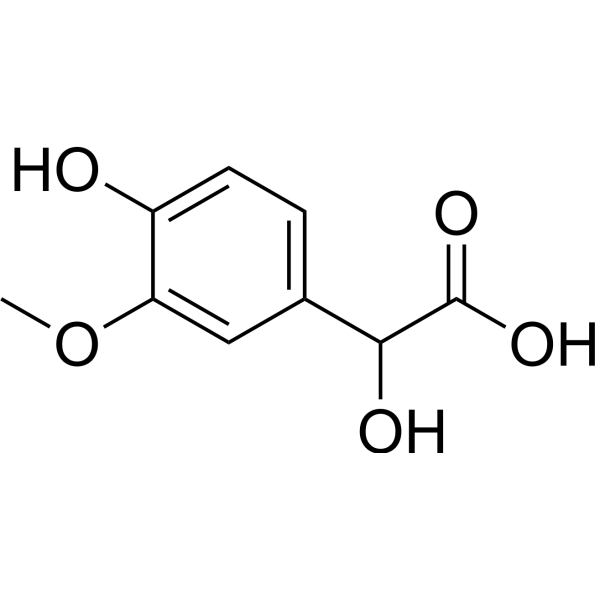
3,4-Dihydroxymandelic acid
Based on 1 publication(s) in Google Scholar
3,4‑Dihydroxymandelic acid is a metabolite of Norepinephrine (HY-13715) with free radical scavenging and antioxidant activities. 3,4-Dihydroxymandelic acid acts as a chemoattractant for enterohaemorrhagic Escherichia coli (EHEC), regulates virulence gene expression and bacterial adhesion to intestinal epithelial cells in a QseC-dependent manner, and targets EHEC to preferred infection sites. 3,4‑Dihydroxymandelic acid can be used in studies related to oxidative stress and enterohaemorrhagic Escherichia coli infection.
For research use only. We do not sell to patients.
- Purity : 98.0%
- CAS No.: 775-01-9
- Formula: C8H8O5
- Molecular Weight:184.15
-
Storage:Powder -20°C, 3 years , 4°C, 2 years ; In solvent -80°C, 6 months , -20°C, 1 month

To place orders, for customer services and technical support, please contact: MedChemExpress USA
Tel: 609-228-6898 E-mail: [email protected] [email protected]


-
 Biological Activity
-
Chemical Information
-
Solvent & Solubility
- Protocols
- Purity & Documentation
- References
-
 Help & FAQs


-
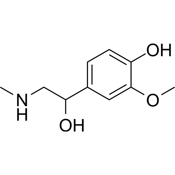
HY-113299
MetanephrineEpinephrine Metabolite -
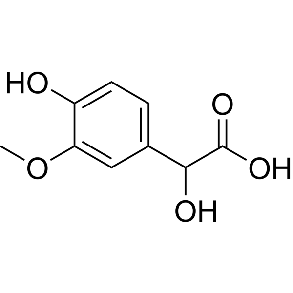
HY-113121
Vanillylmandelic acidAntioxidant Agent -

HY-113474R
3,4-Dihydroxymandelic acid (Standard)Reference Standard -
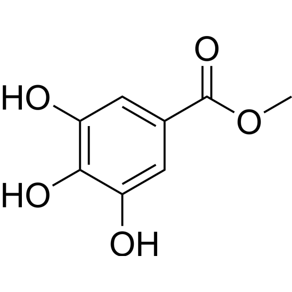
HY-N2010
Methyl gallateAntioxidant -
HY-113121R
Vanillylmandelic acid (Standard)Antioxidant Agent -

HY-N2010R
Methyl gallate (Standard)Antioxidant
Publications Citing Use of MedChemExpress (MCE) 3,4-Dihydroxymandelic acid
MoreAll Endogenous Metabolite Isoforms
More

Biological Activity
Description
In Vitro
3,4-Dihydroxymandelic acid (DHMA) acts as a chemoattractant for the EHEC O157:H7 strain 86-24, which is evidenced by the cell aggregation observed at the edge of agarose plugs containing 500 μM DHMA after 30 min of incubation; the MMC value increases at concentrations ranging from 50 to 5000 μM[1].
3,4-Dihydroxymandelic acid (50 μM) induces QseC-dependent upregulation of virulence genes in EHEC O157:H7 strain 86-24; it also increases the adhesion of the strain to HeLa S3 cells in a QseC-dependent manner[1].
3,4-Dihydroxymandelic acid (DHMA) potently scavenges DPPH free radicals in cell-free assays, with an EC50 of 0.062 mol/mol and an ARP of 16; it also efficiently scavenges superoxide anion radicals in cell-free chemiluminescence assays, with an IC50 of 130 nM[2].
3,4-Dihydroxymandelic acid (0.0001-0.001%; 48-hour incubation) enhances the antioxidant capacity of human skin fibroblasts in a dose-dependent manner[2].
3,4-Dihydroxymandelic acid (0.005-0.05%) potently inhibits accelerated lipid autoxidation in degummed soybean oil (AOI 9.5), degummed squalene (AOI 34) and evening primrose oil (AOI 6.7), and exhibits synergistic activity with α-tocopherol in soybean oil and squalene[2].
3,4-Dihydroxymandelic acid (0.0001-0.01%; 24-72 h incubations) exhibits acute cytotoxicity only at the concentration of 0.01% after 24 h of incubation[2].
MedChemExpress (MCE) has not independently confirmed the accuracy of these methods. They are for reference only. Further protocols information, click here.
-
Cell Line:Human dermal fibroblasts
-
Concentration:0.0001, 0.001, 0.01%
-
Incubation Time:24 h, 48 h, 72 h
-
Result:Showed no cytotoxic effect at concentrations of 0.001% and 0.0001%.
Caused a 2.45-fold increase in extracellular LDH at the 0.01% concentration after 24 h, with MTT conversion initially increasing then decreasing at 48 h and 72 h.
In Vivo
3,4-Dihydroxymandelic acid (DHMA) accumulates at high levels in the myocardium of aged rats compared with other tested tissues[2].
MedChemExpress (MCE) has not independently confirmed the accuracy of these methods. They are for reference only.


Chemical Information
-
CAS No. 775-01-9
-
Appearance Solid
-
Molecular Weight 184.15
-
Formula C8H8O5
-
Color White to off-white
-
SMILES
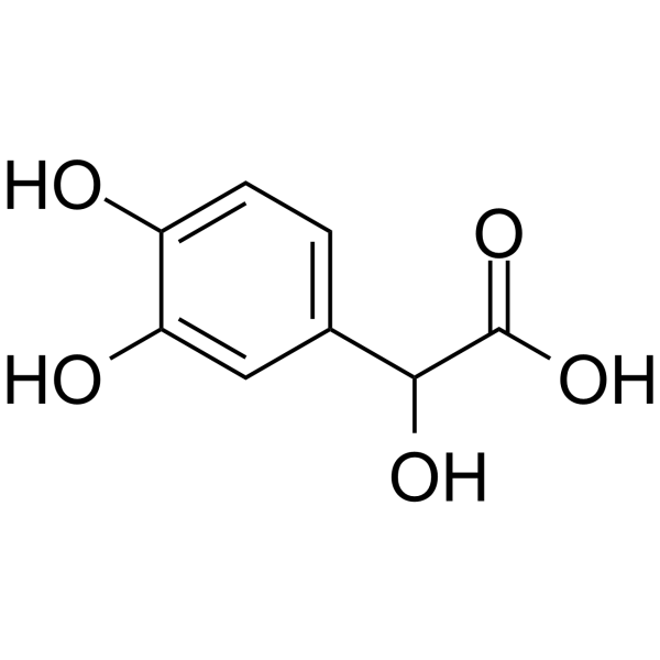
O=C(O)C(O)C1=CC=C(O)C(O)=C1
-
Structure Classification
-
Initial Source
-
Shipping
Room temperature in continental US; may vary elsewhere.
-
Storage
Powder -20°C 3 years 4°C 2 years In solvent -80°C 6 months -20°C 1 month


Publications (1)
-
Journal Impact Factor
-
Most Recent
-
Anal Chem
High-Throughput Reactive Desorption Electrospray Ionization Mass Spectrometry for Targeted Derivatization and Analysis of Poorly Ionized Small Molecules. [Abstract]2026 Jun 16;98(23):16962-16970. PMID: 42223360


Solvent & Solubility
In Vitro:
H2O : 100 mg/mL (543.04 mM; Need ultrasonic)
DMSO : 83.33 mg/mL (452.51 mM; Need ultrasonic; Hygroscopic DMSO has a significant impact on the solubility of product, please use newly opened DMSO)
Please refer to the solubility information to select the appropriate solvent. Once prepared, please aliquot and store the solution to prevent product inactivation from repeated freeze-thaw cycles.
Storage method and period of stock solution: -80°C, 6 months; -20°C, 1 month. When stored at -80°C, please use it within 6 months. When stored at -20°C, please use it within 1 month.
* Note: If you choose water as the stock solution, please dilute it to the working solution, then filter and sterilize it with a 0.22 μm filter before use.
Please refer to the solubility information to select the appropriate solvent. Once prepared, please aliquot and store the solution to prevent product inactivation from repeated freeze-thaw cycles.
Storage method and period of stock solution: -80°C, 6 months; -20°C, 1 month. When stored at -80°C, please use it within 6 months. When stored at -20°C, please use it within 1 month.
* Note: If you choose water as the stock solution, please dilute it to the working solution, then filter and sterilize it with a 0.22 μm filter before use.
Concentration (start) × Volume (start) = Concentration (final) × Volume (final)
In Vivo:
Select the appropriate dissolution method based on your experimental animal and administration route.
- For the following dissolution methods, please ensure to first prepare a clear stock solution using an In Vitro approach and then sequentially add co-solvents:
- To ensure reliable experimental results, the clarified stock solution can be appropriately stored based on storage conditions. As for the working solution for In Vivo experiments, it is recommended to prepare freshly and use it on the same day.
- The percentages shown for the solvents indicate their volumetric ratio in the final prepared solution. If precipitation or phase separation occurs during preparation, heat and/or sonication can be used to aid dissolution.
Add each solvent one by one: 10% DMSO 40% PEG300 5% Tween-80 45% Saline
Solubility: ≥ 2.08 mg/mL (11.30 mM); Clear solution
This protocol yields a clear solution of ≥ 2.08 mg/mL (saturation unknown).
Taking 1 mL working solution as an example, add 100 μL DMSO stock solution (20.8 mg/mL) to 400 μL PEG300, and mix evenly; then add 50 μL Tween-80 and mix evenly; then add 450 μL Saline to adjust the volume to 1 mL.
Preparation of Saline: Dissolve 0.9 g sodium chloride in ddH₂O and dilute to 100 mL to obtain a clear Saline solution.
Add each solvent one by one: 10% DMSO 90% (20% SBE-β-CD in Saline)
Solubility: ≥ 2.08 mg/mL (11.30 mM); Clear solution
This protocol yields a clear solution of ≥ 2.08 mg/mL (saturation unknown).
Taking 1 mL working solution as an example, add 100 μL DMSO stock solution (20.8 mg/mL) to 900 μL 20% SBE-β-CD in Saline, and mix evenly.
Preparation of 20% SBE-β-CD in Saline (4°C, storage for one week): 2 g SBE-β-CD powder is dissolved in 10 mL Saline, completely dissolve until clear.
In Vivo Dissolution Calculator
Please enter the basic information of animal experiments:
-
-
-
-
Recommended: Prepare an additional quantity of animals to account for potential losses during experiments.
Working solution concentration: 0.22 mg/mL
This product has good water solubility, please refer to the measured solubility data in water/PBS/Saline for details.

Protocols
-
RT-PCR
Reverse transcription technology uses RNA as a template to synthesize DNA. RT-PCR is simple, specific and sensitive, and can be used to detect gene expression levels and expression differences in cells; detect RNA virus content; clone cDNA sequences of specific genes.
-
RNA extraction experimental
By lysing cells, releasing RNA, and removing impurities such as proteins and DNA, high-purity RNA products are finally obtained. The commonly used traditional method is the guanidine isothiocyanate/phenol/chloroform method (Trizol), which is suitable for a variety of animal materials including animal tissues, microorganisms, cultured cells, etc., and most plant materials.
-
Research Protocol for Infectious Diseases
Infectious-disease experiments test how pathogens interact with host barriers, innate immune receptors, inflammatory signaling, pathogen replication, and tissue injury; pattern-recognition receptors such as TLRs, RIG-I-like receptors, NOD-like receptors, and inflammasomes detect microbial molecules and activate NF-κB, interferon, and cytokine responses. The central hypothesis is that infection severity reflects the balance between pathogen burden and host response: protective inflammation restricts pathogen growth, whereas excessive or mislocalized inflammation contributes to tissue damage and disease phenotype. Unresolved questions include which host pathways are protective versus pathogenic, why some infection models fail to translate to human disease, and which combined readouts best predict clinically relevant infection outcomes.
-
ROS/oxidative-stress fluorescent staining
ROS/oxidative-stress fluorescent staining uses cell-permeant fluorogenic probes that become fluorescent after oxidation inside cells or tissues; commonly used examples include DCFH-DA/DCFDA for broad cellular oxidant detection, DHE for superoxide-related signal detection, MitoSOX for mitochondrial superoxide-related signal detection, and CellROX probes for oxidative-stress-associated fluorescence readouts. The assay detects probe oxidation rather than a single ROS species unless the probe and analysis method have been chemically validated for that species. DCFH-DA enters cells, is deacetylated by intracellular esterases to DCFH, and produces fluorescent DCF after oxidation, so the readout is used as an operational measure of total cellular oxidative stress rather than a species-specific ROS measurement. DHE and MitoSOX can report superoxide-related oxidation, but red fluorescence alone can include non-specific ethidium-like oxidation products; HPLC or optimized spectral approaches are
-
Real Time qPCR (Q-PCR)
Real-time quantitative PCR (qPCR) quantifies an amplifiable nucleic-acid target by monitoring fluorescence during PCR cycling rather than measuring product only after amplification. The increase in fluorescence tracks accumulation of PCR product, and the quantification cycle (Cq; historically also Ct/CP) is related to the initial amount of target: samples containing more starting target generally reach the defined fluorescence threshold in fewer cycles.


Purity & Documentation
-
Data Sheet (287 KB)
-
SDS (393 KB)
- English - EN (393 KB)
- Français - FR (393 KB)
- Deutsch - DE (393 KB)
- Norwegian - NO (393 KB)
- Español - ES (393 KB)
- Swedish - SV (393 KB)
- Italian - IT (393 KB)
- Korean - KR (393 KB)
- Portuguese - PT (393 KB)
-
Handling Instructions (2659 KB)
References


Complete Stock Solution Preparation Table
Please refer to the solubility information to select the appropriate solvent. Once prepared, please aliquot and store the solution to prevent product inactivation from repeated freeze-thaw cycles.
Storage method and period of stock solution: -80°C, 6 months; -20°C, 1 month. When stored at -80°C, please use it within 6 months. When stored at -20°C, please use it within 1 month.
| Optional Solvent | Concentration Solvent Mass | 1 mg | 5 mg | 10 mg | 25 mg |
|---|---|---|---|---|---|
| DMSO / H2O | 1 mM | 5.4304 mL | 27.1518 mL | 54.3036 mL | 135.7589 mL |
| 5 mM | 1.0861 mL | 5.4304 mL | 10.8607 mL | 27.1518 mL | |
| 10 mM | 0.5430 mL | 2.7152 mL | 5.4304 mL | 13.5759 mL | |
| 15 mM | 0.3620 mL | 1.8101 mL | 3.6202 mL | 9.0506 mL | |
| 20 mM | 0.2715 mL | 1.3576 mL | 2.7152 mL | 6.7879 mL | |
| 25 mM | 0.2172 mL | 1.0861 mL | 2.1721 mL | 5.4304 mL | |
| 30 mM | 0.1810 mL | 0.9051 mL | 1.8101 mL | 4.5253 mL | |
| 40 mM | 0.1358 mL | 0.6788 mL | 1.3576 mL | 3.3940 mL | |
| 50 mM | 0.1086 mL | 0.5430 mL | 1.0861 mL | 2.7152 mL | |
| 60 mM | 0.0905 mL | 0.4525 mL | 0.9051 mL | 2.2626 mL | |
| 80 mM | 0.0679 mL | 0.3394 mL | 0.6788 mL | 1.6970 mL | |
| 100 mM | 0.0543 mL | 0.2715 mL | 0.5430 mL | 1.3576 mL |
* Note: If you choose water as the stock solution, please dilute it to the working solution, then filter and sterilize it with a 0.22 μm filter before use.


3,4-Dihydroxymandelic acid Related Classifications
HY-113474 Related Classifications
 Powered by Bioz