



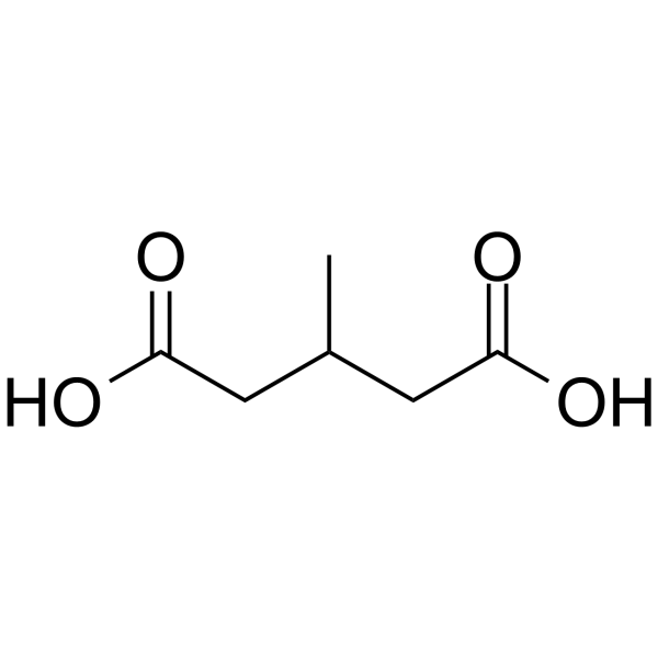
3-Methylglutaric acid

Based on 1 Customer Validation
3-Methylglutaric acid is a non-selective inhibitor of mitochondrial function and Na+, K+-ATPase, with an inhibition rate of 30% on rat cortical synaptosomal Na+, K+-ATPase. 3-Methylglutaric acid can induce reactive oxygen species (ROS) generation, thereby causing oxidative damage and inhibiting mitochondrial redox potential and ion pump function of cell membranes. 3-Methylglutaric acid can be used to study the neuropathological mechanisms of metabolic diseases and the role of oxidative stress-mediated neuronal damage in neurodegeneration.
For research use only. We do not sell to patients.
- Purity : 98.0%
- CAS No.: 626-51-7
- Formula: C6H10O4
- Molecular Weight:146.14
-
Storage:Powder -20°C, 3 years , 4°C, 2 years ; In solvent -80°C, 6 months , -20°C, 1 month

To place orders, for customer services and technical support, please contact: MedChemExpress USA
Tel: 609-228-6898 E-mail: [email protected] [email protected]


-
 Biological Activity
-
Chemical Information
-
Solvent & Solubility
- Protocols
- Purity & Documentation
- References
-
 Help & FAQs


-
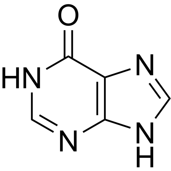
HY-N0091
HypoxanthineEndogenous Metabolite -
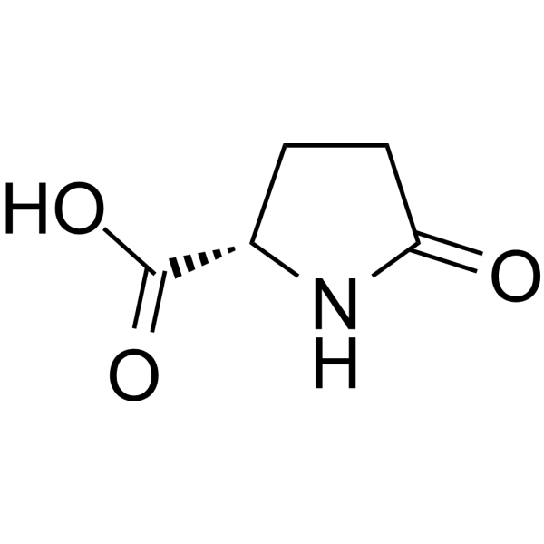
HY-76082
L-Pyroglutamic acidEndogenous Metabolite -
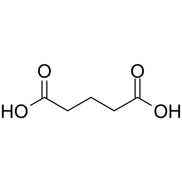
HY-W008820
Glutaric acid -
HY-Y1139
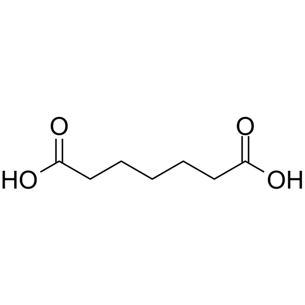
Pimelic acidRetinopathy Marker -
HY-N0666
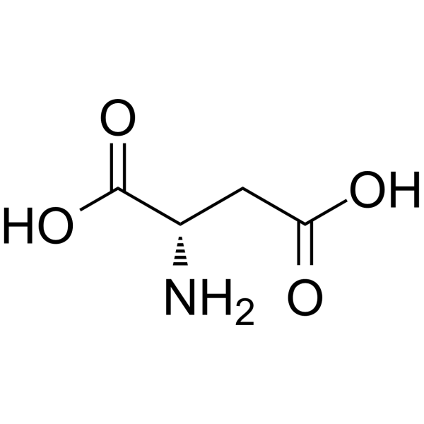
L-Aspartic acidNMDA Receptor Agonist -
HY-W017522
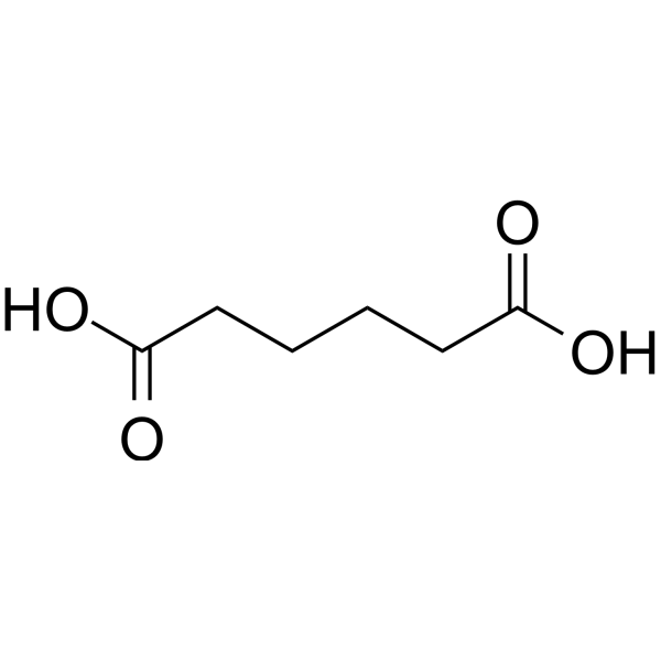
Adipic acidAnti-HSV Agent -
HY-W015300
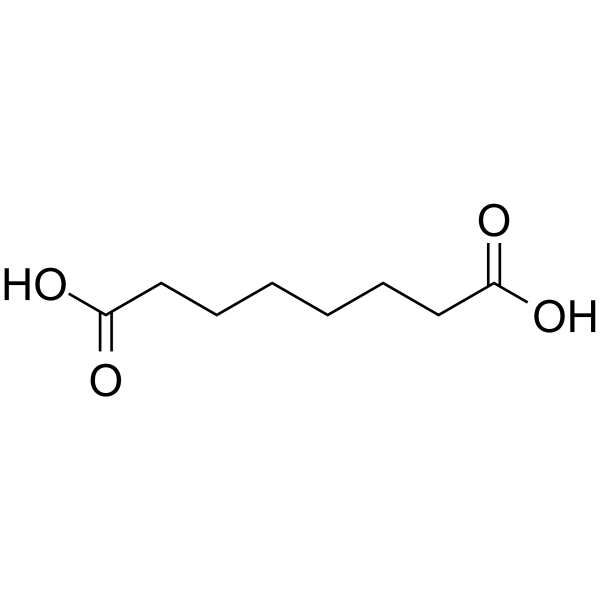
Suberic acidCrystalline Dibasic Acid -
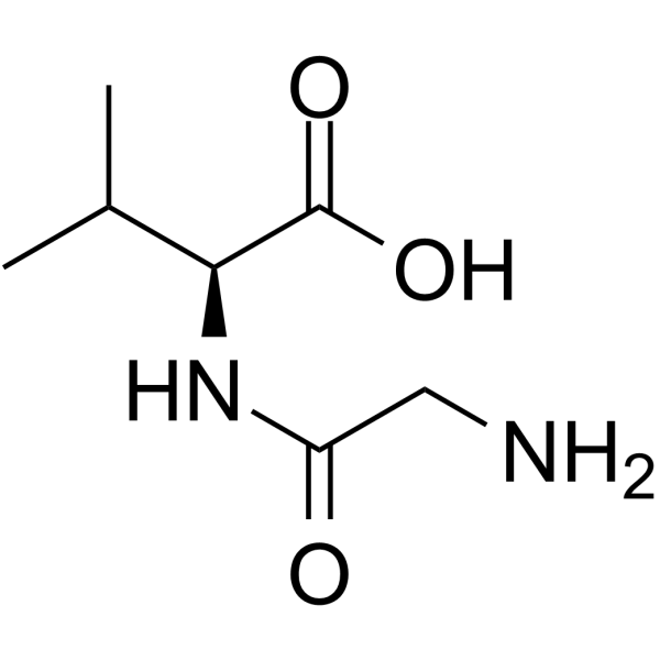
HY-W016785
Glycyl-L-valineDipeptide -

HY-113410R
3-Methylglutaric acid (Standard)Na+/K+ ATPase Inhibitor -
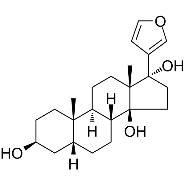
HY-12283
RostafuroxinATP1A1 Antognist -
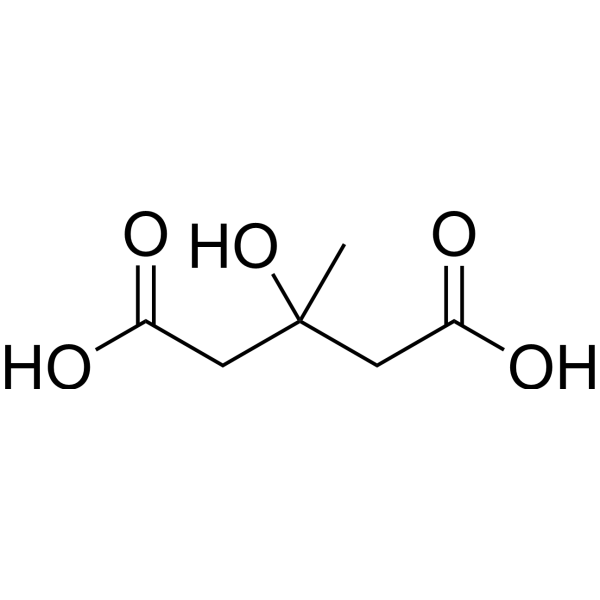
HY-B1189
MeglutolEndogenous Metabolite -
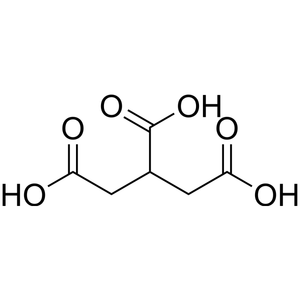
HY-W020215
Tricarballylic acidAconitate Hydratase Inhibitor


Biological Activity
Description
IC50 & Target
|
Human Endogenous Metabolite |
In Vitro
3-Methylglutaric acid (0.1-5.0 mM; 1 h) inhibits the mitochondrial redox potential of rat cortical synaptosomes, and the reduction rate of Resazurin (HY-118540) is reduced by 25%[1].
3-Methylglutaric acid (0.1-5.0 mM; 1 h) significantly inhibits the synaptosomal Na+,K+-ATPase activity by 30%, and this inhibitory effect can be completely reversed by the antioxidants creatine and melatonin[1].
3-Methylglutaric acid (0.1-5.0 mM; 1 h) induces synaptosomal ROS generation and increases the DCFH oxidation level by 30%[1].
MedChemExpress (MCE) has not independently confirmed the accuracy of these methods. They are for reference only. Further protocols information, click here.
-
Cell Line:Rat cerebral cortex synaptosomes (viability assessed by LDH release)
-
Concentration:0.1 mM, 1.0 mM, 5.0 mM
-
Incubation Time:1 h, 2 h
-
Result:Unchanged LDH leakage compared to control (≤10% of total release), indicating no significant membrane damage or cell death at tested concentrations.
In Vivo
MedChemExpress (MCE) has not independently confirmed the accuracy of these methods. They are for reference only.
-
Animal Model:
-
Dosage:10 μmol/g (first injection), 5 μmol/g (subsequent two injections); with or without Bezafibrate (30 or 100 mg/kg/day, dissolved in corn oil)
-
Administration:Intraperitoneal injection, three doses with 90-min intervals on the same day;
while Bezafibrate administratied via Oral gavage, once daily for 7 consecutive days before 3-Methylglutaric acid administration. -
Result:Significantly increased malondialdehyde (MDA) levels (lipid peroxidation) in liver and cerebral cortex, decreased reduced glutathione (GSH) content, and altered activities of antioxidant enzymes (catalase ↑, glutathione peroxidase ↓, glucose-6-phosphate dehydrogenase ↓).
Bezafibrate (100 mg/kg/day) completely prevented MDA increase and GSH depletion, and partially normalized enzyme activities.


Chemical Information
-
CAS No. 626-51-7
-
Appearance Solid
-
Molecular Weight 146.14
-
Formula C6H10O4
-
Color White to off-white
-
SMILES
O=C(O)CC(C)CC(O)=O
-
Structure Classification
-
Shipping
Room temperature in continental US; may vary elsewhere.
-
Storage
Powder -20°C 3 years 4°C 2 years In solvent -80°C 6 months -20°C 1 month


Solvent & Solubility
In Vitro:
H2O : ≥ 100 mg/mL (684.28 mM)
DMSO : 100 mg/mL (684.28 mM; Need ultrasonic; Hygroscopic DMSO has a significant impact on the solubility of product, please use newly opened DMSO)
* "≥" means soluble, but saturation unknown.
Please refer to the solubility information to select the appropriate solvent. Once prepared, please aliquot and store the solution to prevent product inactivation from repeated freeze-thaw cycles.
Storage method and period of stock solution: -80°C, 6 months; -20°C, 1 month. When stored at -80°C, please use it within 6 months. When stored at -20°C, please use it within 1 month.
* Note: If you choose water as the stock solution, please dilute it to the working solution, then filter and sterilize it with a 0.22 μm filter before use.
Please refer to the solubility information to select the appropriate solvent. Once prepared, please aliquot and store the solution to prevent product inactivation from repeated freeze-thaw cycles.
Storage method and period of stock solution: -80°C, 6 months; -20°C, 1 month. When stored at -80°C, please use it within 6 months. When stored at -20°C, please use it within 1 month.
* Note: If you choose water as the stock solution, please dilute it to the working solution, then filter and sterilize it with a 0.22 μm filter before use.
Concentration (start) × Volume (start) = Concentration (final) × Volume (final)
In Vivo:
Select the appropriate dissolution method based on your experimental animal and administration route.
- For the following dissolution methods, please ensure to first prepare a clear stock solution using an In Vitro approach and then sequentially add co-solvents:
- To ensure reliable experimental results, the clarified stock solution can be appropriately stored based on storage conditions. As for the working solution for In Vivo experiments, it is recommended to prepare freshly and use it on the same day.
- The percentages shown for the solvents indicate their volumetric ratio in the final prepared solution. If precipitation or phase separation occurs during preparation, heat and/or sonication can be used to aid dissolution.
Add each solvent one by one: 10% DMSO 40% PEG300 5% Tween-80 45% Saline
Solubility: ≥ 2.5 mg/mL (17.11 mM); Clear solution
This protocol yields a clear solution of ≥ 2.5 mg/mL (saturation unknown).
Taking 1 mL working solution as an example, add 100 μL DMSO stock solution (25.0 mg/mL) to 400 μL PEG300, and mix evenly; then add 50 μL Tween-80 and mix evenly; then add 450 μL Saline to adjust the volume to 1 mL.
Preparation of Saline: Dissolve 0.9 g sodium chloride in ddH₂O and dilute to 100 mL to obtain a clear Saline solution.
Add each solvent one by one: 10% DMSO 90% (20% SBE-β-CD in Saline)
Solubility: ≥ 2.5 mg/mL (17.11 mM); Clear solution
This protocol yields a clear solution of ≥ 2.5 mg/mL (saturation unknown).
Taking 1 mL working solution as an example, add 100 μL DMSO stock solution (25.0 mg/mL) to 900 μL 20% SBE-β-CD in Saline, and mix evenly.
Preparation of 20% SBE-β-CD in Saline (4°C, storage for one week): 2 g SBE-β-CD powder is dissolved in 10 mL Saline, completely dissolve until clear.
In Vivo Dissolution Calculator
Please enter the basic information of animal experiments:
-
-
-
-
Recommended: Prepare an additional quantity of animals to account for potential losses during experiments.
Working solution concentration: 0.22 mg/mL
This product has good water solubility, please refer to the measured solubility data in water/PBS/Saline for details.

Protocols
-
Neurotoxicity Study
This protocol assesses in vitro neurotoxicity by combining neuronal viability, mitochondrial/metabolic activity, neurite outgrowth, and optional neuronal network function readouts. Calcein-AM or resazurin/PrestoBlue readouts estimate viable or metabolically active cells; βIII-tubulin immunofluorescence detects neuronal morphology and neurite networks; TMRE detects mitochondrial membrane potential; and MEA recordings detect functional changes in neuronal network activity.
-
Mitochondrial membrane-potential fluorescent assay
Mitochondrial membrane potential fluorescent assays estimate ΔΨm in living cells using lipophilic cationic dyes such as TMRM, TMRE, rhodamine 123, and JC-1, which accumulate in mitochondria according to membrane polarization; loss of signal after FCCP or CCCP treatment is interpreted as mitochondrial depolarization. TMRM/TMRE and rhodamine 123 are commonly used for semi-quantitative live-cell microscopy or flow cytometry, while JC-1 can report a shift from red aggregate fluorescence to green monomer fluorescence during depolarization; interpretation requires controls because dye concentration, quenching mode, cell type, dye efflux, and mitochondrial mass can affect fluorescence independently of ΔΨm.
-
ROS/oxidative-stress fluorescent staining
ROS/oxidative-stress fluorescent staining uses cell-permeant fluorogenic probes that become fluorescent after oxidation inside cells or tissues; commonly used examples include DCFH-DA/DCFDA for broad cellular oxidant detection, DHE for superoxide-related signal detection, MitoSOX for mitochondrial superoxide-related signal detection, and CellROX probes for oxidative-stress-associated fluorescence readouts. The assay detects probe oxidation rather than a single ROS species unless the probe and analysis method have been chemically validated for that species. DCFH-DA enters cells, is deacetylated by intracellular esterases to DCFH, and produces fluorescent DCF after oxidation, so the readout is used as an operational measure of total cellular oxidative stress rather than a species-specific ROS measurement. DHE and MitoSOX can report superoxide-related oxidation, but red fluorescence alone can include non-specific ethidium-like oxidation products; HPLC or optimized spectral approaches are
-
Mitochondrial membrane-potential and mitochondrial mass staining
Mitochondrial membrane potential staining measures the electrochemical polarization across the mitochondrial inner membrane in live cells using lipophilic cationic fluorescent probes; early rhodamine-based work showed that selective mitochondrial dye accumulation is lost when the mitochondrial transmembrane potential is dissipated. JC-1 reports mitochondrial polarization by shifting from green monomer fluorescence to red J-aggregate fluorescence as dye concentration increases within energized mitochondria; therefore, the red/green fluorescence ratio is used as a relative readout of mitochondrial membrane potential. TMRE or TMRM staining provides a single-channel relative readout because these cationic rhodamine esters accumulate in polarized mitochondria, and lower fluorescence indicates reduced mitochondrial polarization when acquisition and dye-loading conditions are controlled. Mitochondrial mass staining is commonly performed with MitoTracker Green FM or related MitoTracker dyes as


Purity & Documentation
-
Data Sheet (276 KB)
-
SDS (396 KB)
- English - EN (396 KB)
- Français - FR (396 KB)
- Deutsch - DE (396 KB)
- Norwegian - NO (396 KB)
- Español - ES (396 KB)
- Swedish - SV (396 KB)
- Italian - IT (396 KB)
- Korean - KR (396 KB)
- Portuguese - PT (396 KB)
-
Handling Instructions (2659 KB)
References
[1]. Ribeiro CA, et al. Neurochemical evidence that 3-methylglutaric acid inhibits synaptic Na+,K+-ATPase activity probably through oxidative damage in brain cortex of young rats. Int J Dev Neurosci. 2011 Feb;29(1):1-7. [Content Brief]
[2]. da Rosa-Junior NT, et al. In vivo evidence that bezafibrate prevents oxidative stress and mitochondrial dysfunction caused by 3-methylglutaric acid in rat liver. Biochimie. 2020 Apr-May;171-172:187-196. [Content Brief]
[3]. da Rosa-Junior NT, et al. Bezafibrate In Vivo Administration Prevents 3-Methylglutaric Acid-Induced Impairment of Redox Status, Mitochondrial Biogenesis, and Neural Injury in Brain of Developing Rats. Neurotox Res. 2019 May;35(4):809-822. [Content Brief]
[4]. Colín-González AL, et al. Experimental Evidence that 3-Methylglutaric Acid Disturbs Mitochondrial Function and Induced Oxidative Stress in Rat Brain Synaptosomes: New Converging Mechanisms. Neurochem Res. 2016 Oct;41(10):2619-2626. [Content Brief]


Complete Stock Solution Preparation Table
Please refer to the solubility information to select the appropriate solvent. Once prepared, please aliquot and store the solution to prevent product inactivation from repeated freeze-thaw cycles.
Storage method and period of stock solution: -80°C, 6 months; -20°C, 1 month. When stored at -80°C, please use it within 6 months. When stored at -20°C, please use it within 1 month.
| Optional Solvent | Concentration Solvent Mass | 1 mg | 5 mg | 10 mg | 25 mg |
|---|---|---|---|---|---|
| H2O / DMSO | 1 mM | 6.8428 mL | 34.2138 mL | 68.4275 mL | 171.0688 mL |
| 5 mM | 1.3686 mL | 6.8428 mL | 13.6855 mL | 34.2138 mL | |
| 10 mM | 0.6843 mL | 3.4214 mL | 6.8428 mL | 17.1069 mL | |
| 15 mM | 0.4562 mL | 2.2809 mL | 4.5618 mL | 11.4046 mL | |
| 20 mM | 0.3421 mL | 1.7107 mL | 3.4214 mL | 8.5534 mL | |
| 25 mM | 0.2737 mL | 1.3686 mL | 2.7371 mL | 6.8428 mL | |
| 30 mM | 0.2281 mL | 1.1405 mL | 2.2809 mL | 5.7023 mL | |
| 40 mM | 0.1711 mL | 0.8553 mL | 1.7107 mL | 4.2767 mL | |
| 50 mM | 0.1369 mL | 0.6843 mL | 1.3686 mL | 3.4214 mL | |
| 60 mM | 0.1140 mL | 0.5702 mL | 1.1405 mL | 2.8511 mL | |
| 80 mM | 0.0855 mL | 0.4277 mL | 0.8553 mL | 2.1384 mL | |
| 100 mM | 0.0684 mL | 0.3421 mL | 0.6843 mL | 1.7107 mL |
* Note: If you choose water as the stock solution, please dilute it to the working solution, then filter and sterilize it with a 0.22 μm filter before use.


3-Methylglutaric acid Related Classifications
HY-113410 Related Classifications
 Powered by Bioz