



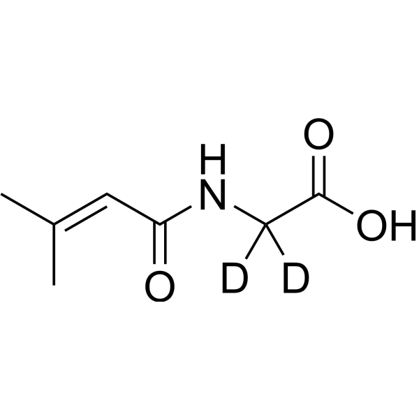
3-Methylcrotonylglycine-d2
3-Methylcrotonylglycine-d2 is the deuterated-labeled 3-Methylcrotonylglycine (HY-113232). 3-Methylcrotonylglycine is an organic acid conjugate associated with leucine catabolism that inhibits respiratory chain complex II-III, mitochondrial creatine kinase, and synaptic membrane Na+, K+-ATPase. 3-Methylcrotonylglycine increases reactive oxygen species levels to mediate oxidative damage, while inhibiting mitochondrial electron transport, reducing tricarboxylic acid cycle flux, and interfering with intracellular energy transport and neuronal ion homeostasis. 3-Methylcrotonylglycine is a neurotoxic metabolite that accumulates in the metabolic pathway of 3-methylcrotonyl-CoA carboxylase deficiency. 3-Methylcrotonylglycine can be used in studies related to 3-methylcrotonyl-CoA carboxylase deficiency.
For research use only. We do not sell to patients.
- CAS No.: 1276197-31-9
- Formula: C7H9D2NO3
- Molecular Weight:159.18
-
Storage:
Please store the product under the recommended conditions in the Certificate of Analysis.

To place orders, for customer services and technical support, please contact: MedChemExpress USA
Tel: 609-228-6898 E-mail: [email protected] [email protected]


-
 Biological Activity
-
Chemical Information
- Protocols
- Purity & Documentation
- References
-
 Help & FAQs


All Endogenous Metabolite Isoforms
More

Biological Activity
Description
Application
1. This compound can be used as a tracer
2. This compound can be used as an internal standard for quantitative analysis by NMR, GC-MS, or LC-MS.


Chemical Information
-
CAS No. 1276197-31-9
-
Unlabeled CAS 33008-07-0
-
Molecular Weight 159.18
-
Formula C7H9D2NO3
-
SMILES
OC(C([2H])([2H])NC(/C=C(C)/C)=O)=O
-
Shipping
Room temperature in continental US; may vary elsewhere.
-
Storage
Please store the product under the recommended conditions in the Certificate of Analysis.

Protocols
-
Neurotoxicity Study
This protocol assesses in vitro neurotoxicity by combining neuronal viability, mitochondrial/metabolic activity, neurite outgrowth, and optional neuronal network function readouts. Calcein-AM or resazurin/PrestoBlue readouts estimate viable or metabolically active cells; βIII-tubulin immunofluorescence detects neuronal morphology and neurite networks; TMRE detects mitochondrial membrane potential; and MEA recordings detect functional changes in neuronal network activity.
-
ROS/oxidative-stress fluorescent staining
ROS/oxidative-stress fluorescent staining uses cell-permeant fluorogenic probes that become fluorescent after oxidation inside cells or tissues; commonly used examples include DCFH-DA/DCFDA for broad cellular oxidant detection, DHE for superoxide-related signal detection, MitoSOX for mitochondrial superoxide-related signal detection, and CellROX probes for oxidative-stress-associated fluorescence readouts. The assay detects probe oxidation rather than a single ROS species unless the probe and analysis method have been chemically validated for that species. DCFH-DA enters cells, is deacetylated by intracellular esterases to DCFH, and produces fluorescent DCF after oxidation, so the readout is used as an operational measure of total cellular oxidative stress rather than a species-specific ROS measurement. DHE and MitoSOX can report superoxide-related oxidation, but red fluorescence alone can include non-specific ethidium-like oxidation products; HPLC or optimized spectral approaches are


Purity & Documentation
References

Calculators
Concentration (start) × Volume (start) = Concentration (final) × Volume (final)


3-Methylcrotonylglycine-d2 Related Classifications
HY-113232S Related Classifications
 Powered by Bioz
Keywords
- 3-Methylcrotonylglycine-d2
- 1276197-31-9
- Isotope-Labeled Compounds
- Endogenous Metabolite
- Na+/K+ ATPase
- Mitochondrial Metabolism
- cerebral cortex
- citric acid cycle
- lipid peroxidation
- young rats
- 3-methylcrotonyl-coa carboxylase deficiency
- synaptic membrane Na+,K+-ATPase
- Wistar rats
- mitochondrial creatine kinase
- glutathione peroxidase
- respiratory chain complex II-III
- Inhibitor
- inhibitor
- inhibit