



5,6-Epoxyeicosatrienoic acid

Based on 1 Customer Validation
5,6-Epoxyeicosatrienoic acid (5,6-EET; (±)5,6-EpETrE) is a fully racemic version of the enantiomeric forms biosynthesized from arachidonic acid by cytochrome P450 enzymes. In solution, 5,6-Epoxyeicosatrienoic acid degrades into 5,6-DiHET and 5,6-δ-lactone, which can be converted to 5,6-DiHET and quantified by GC-MS. In neuroendocrine cells, such as the anterior pituitary and pancreatic islets, 5,6-Epoxyeicosatrienoic acid has been implicated in the mobilization of calcium and hormone secretion. 5,6-Epoxyeicosatrienoic acid is an inhibitor of T-type voltage-gated calcium channels (Cav3) that inhibits isoforms Cav3.1, Cav3.2 (IC50=0.54 μM), and Cav3. and decreases nifedipine-resistant phenylephrine-induced vasoconstriction in isolated mouse mesenteric arteries via Cav3.2 blockade when used at a concentration of 3 μM. In addition, it is a substrate of COX-1 and COX-2.
For research use only. We do not sell to patients.
- Purity : 99.07%
- CAS No.: 87173-80-6
- Formula: C20H32O3
- Molecular Weight:320.47
-
Storage:
Please store the product under the recommended conditions in the Certificate of Analysis.


To place orders, for customer services and technical support, please contact: MedChemExpress USA
Tel: 609-228-6898 E-mail: [email protected] [email protected]


-
 Biological Activity
-
Chemical Information
- Protocols
- Purity & Documentation
- References
-
 Help & FAQs


-
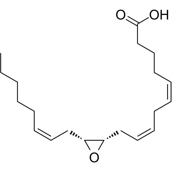
HY-130494
(±)11(12)-EETNLRP3 Inflammasome Inhibitor -
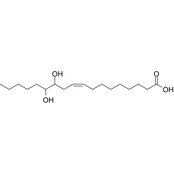
HY-116003
12,13-DiHOMEBAT Activator -
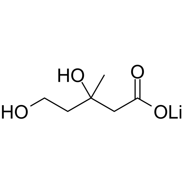
HY-113071A
Mevalonic acid lithium saltEndogenous Metabolite -
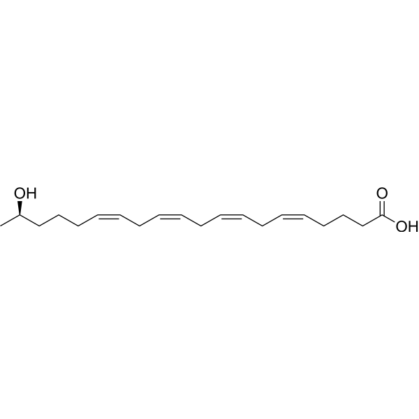
HY-118332
19(R)-HETEVasodilator -

HY-138971
(±)17(18)-EpETE-ethanolamide -

HY-130229
(±)5,6-DHET lactoneLactone -
HY-120954
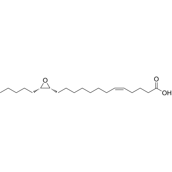
14,15-EE-5(Z)-E14,15-EET structural analogue -
HY-139114
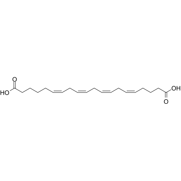
20-Carboxyarachidonic acidPPARalpha/PPARgamma Activator -

HY-W715511
N-Desethyl-N-methyl vardenafilPDE5 Inhibitor -
HY-B0192S
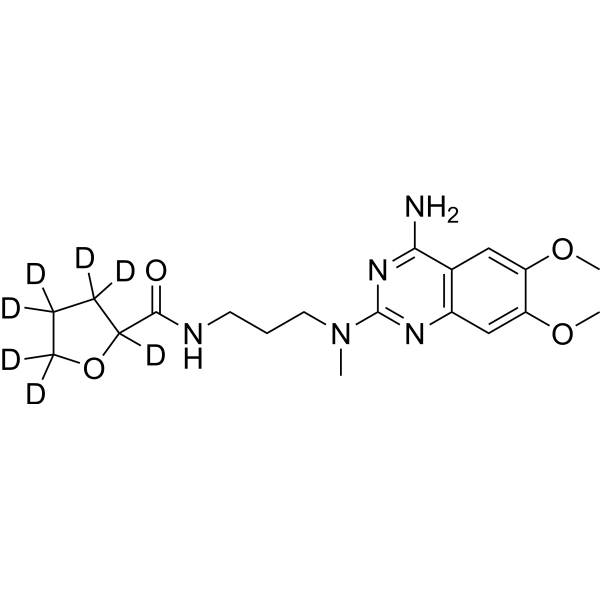
Alfuzosin-d7Stable Isotope -

HY-165067
14,15-EE-8(Z)-EVasodilator Agonist -

HY-165078
(±)11(12)-EpETEComponent of PUFAs


Biological Activity
Description


Chemical Information
-
CAS No. 87173-80-6
-
Appearance Liquid (Density: 0.983±0.06 g/cm3)
-
Molecular Weight 320.47
-
Formula C20H32O3
-
Color Colorless to light yellow
-
SMILES
OC(CCC[C@H]1[C@H](O1)C/C=C\C/C=C\C/C=C\CCCCC)=O
-
Synonyms
5,6-EET; (±)5,6-EpETrE
-
Shipping
Shipping with dry ice.
-
Storage
Please store the product under the recommended conditions in the Certificate of Analysis.

Protocols
-
Cell Cytotoxicity Assay
Cytotoxicity assays are usually based on the assessment of cell membrane damage, which can also be indirectly detected by measuring cell viability. Detection methods include MTT assay, CKK-8 assay, LDH assay and ATP assay, etc.
-
Research Protocol for Endocrine Diseases
Endocrine diseases often arise from disrupted hormone production, hormone signaling, or target-tissue responsiveness; for diabetes-focused endocrine disease models, insulin signaling regulates glucose uptake, hepatic glucose output, lipid metabolism, and β-cell compensation. Type 2 diabetes develops through interacting defects in insulin resistance, β-cell dysfunction, adipose inflammation, hepatic glucose overproduction, altered incretin signaling, and ectopic lipid metabolism. A major unresolved question is whether endocrine dysfunction is driven primarily by target-tissue insulin resistance, intrinsic β-cell failure, immune/inflammatory stress, or combined multi-organ failure that differs by disease stage.
-
Human Islet Cell Culture
The method of preserving islets in vitro, with purified reduced immunogenicity. The steps are islet isolation, islet cell purification, in vitro determination of islet function and islet cell culture.


Purity & Documentation
-
Data Sheet (261 KB)
-
SDS (557 KB)
- English - EN (557 KB)
- Français - FR (557 KB)
- Deutsch - DE (557 KB)
- Norwegian - NO (557 KB)
- Español - ES (557 KB)
- Swedish - SV (557 KB)
- Italian - IT (557 KB)
- Korean - KR (557 KB)
- Portuguese - PT (557 KB)
-
Handling Instructions (2659 KB)
References

Calculators
Concentration (start) × Volume (start) = Concentration (final) × Volume (final)


5,6-Epoxyeicosatrienoic acid Related Classifications
HY-132184 Related Classifications
 Powered by Bioz