



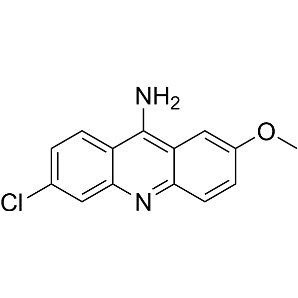
9-Amino-6-chloro-2-methoxyacridine
Based on 3 publication(s) in Google Scholar
9-Amino-6-chloro-2-methoxyacridine (ACMA) is an acridine derivative and Fluorescent probe. 9-Amino-6-chloro-2-methoxyacridine inhibits viral replication, inhibits DNA synthesis and protein metabolism through cell cycle arrest, and induces lipid metabolism regulation, genotoxicity, mutagenicity, and cytotoxicity through DNA strand breaks. 9-Amino-6-chloro-2-methoxyacridine partially intercalates into DNA, forming three distinct complexes, stabilizes the DNA helix, and quenches fluorescence upon intercalation through electron transfer with guanine. 9-Amino-6-chloro-2-methoxyacridine monitors the generation and collapse of the proton motive force, H+ pumping by F1F0-ATP synthase, and the collapse of pH gradients in inverted membrane vesicles. 9-Amino-6-chloro-2-methoxyacridine shows potent anti-BVDV activity. 9-Amino-6-chloro-2-methoxyacridine can be used for research on bovine viral diarrhea virus infection, leishmaniasis, and MRSA infection.
For research use only. We do not sell to patients.
- Purity : 99.88%
- CAS No.: 3548-09-2
- Formula: C14H11ClN2O
- Molecular Weight:258.70
-
Storage:
4°C, protect from light
* In solvent : -80°C, 6 months; -20°C, 1 month (protect from light)

To place orders, for customer services and technical support, please contact: MedChemExpress USA
Tel: 609-228-6898 E-mail: [email protected] [email protected]


-
 Biological Activity
-
Chemical Information
-
Solvent & Solubility
- Protocols
- Purity & Documentation
- References
-
 Help & FAQs



Publications Citing Use of MedChemExpress (MCE) 9-Amino-6-chloro-2-methoxyacridine
MoreAll DNA/RNA Synthesis Isoforms
More

Biological Activity
Description
Cellular Effect
|
Cell Line

|
Type | Value | Description | References |
|---|---|---|---|---|
| MT4 | CC50 |
1.8 μM
|
Cytotoxicity against human MT-4 cells assessed as reduction in cell viability incubated for 96 hrs by MTT method.
Cytotoxicity against human MT-4 cells assessed as reduction in cell viability incubated for 96 hrs by MTT method.
|
21619897 |
| BHK-21 | CC50 |
9 μM
|
Cytotoxicity against Baby Hamster Kidney (BHK-21) cells assessed as reduction in cell viability incubated for 3-4 days by MTT method.
Cytotoxicity against Baby Hamster Kidney (BHK-21) cells assessed as reduction in cell viability incubated for 3-4 days by MTT method.
|
21619897 |
| Vero 76 | CC50 |
10 μM
|
Cytotoxicity against african green monkey Vero-76 cells assessed as reduction in cell viability incubated for 2 to 5 days by crystal violet staining method.
Cytotoxicity against african green monkey Vero-76 cells assessed as reduction in cell viability incubated for 2 to 5 days by crystal violet staining method.
|
21619897 |
| V79 | IC50 |
26.9 μM
|
Cytotoxicity against V79 Chinese hamster cells assessed as reduction in cell viability incubated for 3 hrs by MTT assay.
Cytotoxicity against V79 Chinese hamster cells assessed as reduction in cell viability incubated for 3 hrs by MTT assay.
|
21971327 |
| V79 | IC50 |
5.1 μM
|
Cytotoxicity against V79 Chinese hamster cells assessed as reduction in cell viability incubated for 24 hrs by MTT assay.
Cytotoxicity against V79 Chinese hamster cells assessed as reduction in cell viability incubated for 24 hrs by MTT assay.
|
21971327 |
In Vitro
9-Amino-6-chloro-2-methoxyacridine serves as a fluorescent probe for monitoring the proton motive force in everted membrane vesicles of E. coli[3].
9-Amino-6-chloro-2-methoxyacridine serves as a fluorescent reporter in the E. coli ISO vesicle H+ pumping assay, reporting F-ATPase activity through the inverse relationship between H+ pumping and fluorescence quenching[6].
9-Amino-6-chloro-2-methoxyacridine (3-4 days) exhibits potent anti-BVDV activity in MDBK cells with an EC50 of 0.1 μM, a CC50 of 9.0 μM, and a selectivity index of 90[4].
9-Amino-6-chloro-2-methoxyacridine (96 h) shows cytotoxicity in MT-4 cells with a CC50 of 1.8 μM[4].
ACMA significantly stabilizes the CT-DNA double helix, increasing the melting temperature from 80.0 °C to 86.9 °C at a CD/CP ratio of 0.10[5].
EB displaces ACMA from CT-DNA, confirming that ACMA binds to DNA via intercalation[5].
9-Amino-6-chloro-2-methoxyacridine (1.8-44.5 μM; 3-24 h) induces dose-dependent cytotoxicity in V79 cells, with an IC50 value of 26.9 μM at 3 h exposure and an IC50 value of 5.1 μM at 24 h exposure[5].
MedChemExpress (MCE) has not independently confirmed the accuracy of these methods. They are for reference only. Further protocols information, click here.
-
Cell Line:MDBK cells infected with Bovine Viral Diarrhea Virus (BVDV) strain NADL
-
Concentration:Serial dilutions
-
Incubation Time:3-4 days
-
Result:Exhibited a CC50 of 9.0 μM.
Exhibited an EC50 of 0.1 μM against BVDV.
Yielded a selectivity index (SI) of 90.
-
Cell Line:MT-4 cells
-
Concentration:Serial dilutions
-
Incubation Time:96 h
-
Result:Exhibited a CC50 of 1.8 μM.
-
Cell Line:V79 Chinese hamster cells
-
Concentration:1.8-44.5 μM
-
Incubation Time:3 h and 24 h
-
Result:Decreased the percentage of surviving cells as ACMA concentration was raised.
Dropped cell viability below 50% at concentrations higher than 26.7 μM for 3 h and 8.9 μM for 24 h.
Yielded IC50 values of 26.9 μM (95% CI 13.6-53.3) for 3 h and 5.1 μM (95% CI 3.4-7.5) for 24 h incubation periods.


Chemical Information
-
CAS No. 3548-09-2
-
Appearance Solid
-
Molecular Weight 258.70
-
Formula C14H11ClN2O
-
Color Light yellow to green yellow
-
SMILES
ClC1=CC=C2C(N=C3C=CC(OC)=CC3=C2N)=C1
-
Synonyms
ACMA
-
Shipping
Room temperature in continental US; may vary elsewhere.
-
Storage
4°C, protect from light
* In solvent : -80°C, 6 months; -20°C, 1 month (protect from light)


Publications (3)
-
Journal Impact Factor
-
Most Recent
-
J Cell Biol
2025 Oct 6;224(10):e202503166. PMID: 40923996 -
-


Solvent & Solubility
In Vitro:
DMSO : 25 mg/mL (96.64 mM; ultrasonic and warming and heat to 60°C; Hygroscopic DMSO has a significant impact on the solubility of product, please use newly opened DMSO)
Please refer to the solubility information to select the appropriate solvent. Once prepared, please aliquot and store the solution to prevent product inactivation from repeated freeze-thaw cycles.
Storage method and period of stock solution: -80°C, 6 months; -20°C, 1 month (protect from light). When stored at -80°C, please use it within 6 months. When stored at -20°C, please use it within 1 month.
Please refer to the solubility information to select the appropriate solvent. Once prepared, please aliquot and store the solution to prevent product inactivation from repeated freeze-thaw cycles.
Storage method and period of stock solution: -80°C, 6 months; -20°C, 1 month (protect from light). When stored at -80°C, please use it within 6 months. When stored at -20°C, please use it within 1 month.
Concentration (start) × Volume (start) = Concentration (final) × Volume (final)

Protocols
-
EdU Incorporation Assay (Click Chemistry-Based DNA Synthesis Measurement)
The EdU incorporation assay measures DNA synthesis by adding the thymidine analog 5-ethynyl-2′-deoxyuridine to cells or tissues, where it is incorporated into newly synthesized DNA during S phase. Incorporated EdU is detected by copper-catalyzed azide-alkyne cycloaddition, in which a fluorescent azide covalently reacts with the ethynyl group on EdU, allowing S-phase cells to be detected by fluorescence microscopy, flow cytometry, or high-content imaging. EdU detection does not require DNA denaturation or anti-BrdU antibody access, which preserves sample structure and improves compatibility with immunostaining and multiparameter cytometry compared with BrdU-based detection. EdU can be cytotoxic in a cell-type- and exposure-dependent manner, so pulse duration, concentration, and continuous-labeling designs should be validated for each cell type.
-
Research Protocol for Infectious Diseases
Infectious-disease experiments test how pathogens interact with host barriers, innate immune receptors, inflammatory signaling, pathogen replication, and tissue injury; pattern-recognition receptors such as TLRs, RIG-I-like receptors, NOD-like receptors, and inflammasomes detect microbial molecules and activate NF-κB, interferon, and cytokine responses. The central hypothesis is that infection severity reflects the balance between pathogen burden and host response: protective inflammation restricts pathogen growth, whereas excessive or mislocalized inflammation contributes to tissue damage and disease phenotype. Unresolved questions include which host pathways are protective versus pathogenic, why some infection models fail to translate to human disease, and which combined readouts best predict clinically relevant infection outcomes.
-
Flow cytometric DNA-content cell-cycle staining
Flow cytometric DNA-content cell-cycle staining measures the fluorescence intensity of DNA-bound fluorochromes in single cells or nuclei to estimate DNA content distributions, allowing assignment of populations to G0/G1, S, and G2/M phases by DNA histogram deconvolution. Propidium iodide (PI) intercalates into DNA, and PI fluorescence is proportional to cellular DNA content when staining is performed under conditions that make DNA accessible and minimize non-DNA signal. Cells with G2/M DNA content are expected to show approximately twice the fluorescence intensity of G0/G1 cells, while S-phase cells occupy intermediate fluorescence values. PI-based DNA-content analysis can also detect cells with fractional DNA content, often reported as sub-G1, when DNA fragmentation and extraction during staining reduce retained DNA signal in apoptotic cells. DAPI is an alternative DNA fluorochrome for univariate DNA-content analysis, while bivariate approaches combining DNA content with proliferation
-
Cell Cytotoxicity Assay
Cytotoxicity assays are usually based on the assessment of cell membrane damage, which can also be indirectly detected by measuring cell viability. Detection methods include MTT assay, CKK-8 assay, LDH assay and ATP assay, etc.
-
Mammalian live/dead viability and cytotoxicity staining
Live/dead viability and cytotoxicity staining assays are based on the simultaneous detection of intracellular esterase activity in metabolically active (viable) cells and membrane integrity loss in non-viable cells. In commonly used dual-staining approaches, membrane-permeant fluorogenic substrates are converted by intracellular esterases into fluorescent products in live cells, while impermeant DNA-binding dyes selectively enter cells with compromised plasma membranes and label nucleic acids in dead or dying cells, enabling discrimination between viable and non-viable populations by fluorescence microscopy or flow cytometry.
-
BrdU Incorporation Assay
Bromodeoxyuridine (BrdU) incorporation assay is based on the principle that BrdU, a thymidine analog, is incorporated into newly synthesized DNA during the S phase of the cell cycle, thereby serving as a marker of DNA replication and cellular proliferation. Incorporated BrdU can be detected using anti-BrdU antibodies following DNA denaturation, enabling visualization or quantification of proliferating cells through immunochemical detection methods such as immunofluorescence or immunohistochemistry.
-
Protocol for Cell Cycle
Cell-cycle analysis by flow cytometry measures DNA content in single cells to estimate the fraction of cells in G0/G1, S, and G2/M phases. Propidium iodide intercalates into DNA, and after RNA removal with RNase, fluorescence intensity reflects cellular DNA content: 2N cells are assigned to G0/G1, cells between 2N and 4N to S phase, and 4N cells to G2/M. DNA-content analysis alone cannot reliably separate G0 from G1 or G2 from M. Ki-67 can distinguish quiescent G0 cells from cycling cells, EdU or BrdU incorporation marks active DNA synthesis in S phase, and phospho-histone H3 staining identifies mitotic cells within the 4N population.
-
Genotoxicity/Mutagenicity Study
The bacterial reverse mutation assay detects point mutations that restore amino-acid prototrophy in auxotrophic Salmonella typhimurium or Escherichia coli tester strains; after exposure to a test article, mutagenic activity is read out as an increased number of revertant colonies on minimal agar compared with the vehicle control. The assay uses tester strains with different mutation targets so that base-substitution and frameshift mutagens can be detected, and testing is performed with and without exogenous mammalian metabolic activation because some chemicals require biotransformation to become mutagenic.
-
Cell Viability Determination by MTT Colorimetric Assay
The following protocol uses the MTT colorimetric assay as a classic literature-established method for assessing cell viability/metabolic activity in cultured mammalian cells. MTT[3-(4,5-dimethylthiazol-2-yl)-2,5-diphenyltetrazolium bromide] is reduced by metabolically active cells to a colored formazan product; the amount of formazan is quantified spectrophotometrically and provides an indirect measure of metabolically active viable cells. Importantly, MTT reduction reflects cellular oxidoreductase/metabolic activity rather than an absolute direct count of living cells, so changes in cellular metabolism can alter the signal independently of cell number.


Purity & Documentation
-
Data Sheet (280 KB)
-
SDS (557 KB)
- English - EN (557 KB)
- Français - FR (557 KB)
- Deutsch - DE (557 KB)
- Norwegian - NO (557 KB)
- Español - ES (557 KB)
- Swedish - SV (557 KB)
- Italian - IT (557 KB)
- Korean - KR (557 KB)
- Portuguese - PT (557 KB)
-
Handling Instructions (2659 KB)
References


Complete Stock Solution Preparation Table
Please refer to the solubility information to select the appropriate solvent. Once prepared, please aliquot and store the solution to prevent product inactivation from repeated freeze-thaw cycles.
Storage method and period of stock solution: -80°C, 6 months; -20°C, 1 month (protect from light). When stored at -80°C, please use it within 6 months. When stored at -20°C, please use it within 1 month.
| Optional Solvent | Concentration Solvent Mass | 1 mg | 5 mg | 10 mg | 25 mg |
|---|---|---|---|---|---|
| DMSO | 1 mM | 3.8655 mL | 19.3274 mL | 38.6548 mL | 96.6370 mL |
| 5 mM | 0.7731 mL | 3.8655 mL | 7.7310 mL | 19.3274 mL | |
| 10 mM | 0.3865 mL | 1.9327 mL | 3.8655 mL | 9.6637 mL | |
| 15 mM | 0.2577 mL | 1.2885 mL | 2.5770 mL | 6.4425 mL | |
| 20 mM | 0.1933 mL | 0.9664 mL | 1.9327 mL | 4.8319 mL | |
| 25 mM | 0.1546 mL | 0.7731 mL | 1.5462 mL | 3.8655 mL | |
| 30 mM | 0.1288 mL | 0.6442 mL | 1.2885 mL | 3.2212 mL | |
| 40 mM | 0.0966 mL | 0.4832 mL | 0.9664 mL | 2.4159 mL | |
| 50 mM | 0.0773 mL | 0.3865 mL | 0.7731 mL | 1.9327 mL | |
| 60 mM | 0.0644 mL | 0.3221 mL | 0.6442 mL | 1.6106 mL | |
| 80 mM | 0.0483 mL | 0.2416 mL | 0.4832 mL | 1.2080 mL |


9-Amino-6-chloro-2-methoxyacridine Related Classifications
HY-118155 Related Classifications
 Powered by Bioz
Keywords
- 9-Amino-6-chloro-2-methoxyacridine
- 3548-09-2
- ACMA
- Fluorescent Dye
- DNA/RNA Synthesis
- DNA Alkylator/Crosslinker
- Flavivirus
- BVDV RNA-dependent RNA polymerase (RdRp)
- intracellular amastigotes
- leishmaniasis
- MT-4 cells
- E. coli inverted membrane vesicles
- bovine viral diarrhea virus
- Leishmania infantum promastigotes
- MDBK cells
- MRSA
- CT-DNA
- Inhibitor
- inhibitor
- inhibit