



9-ING-41
Based on 6 publication(s) in Google Scholar
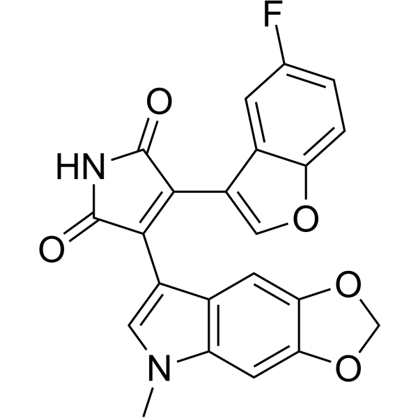
9-ING-41 (Elraglusib) is a maleimide-based ATP-competitive and selective glycogen synthase kinase-3β (GSK-3β) inhibitor with an IC50 of 0.71 μM. 9-ING-41 significantly leads to cell cycle arrest, autophagy and apoptosis in cancer cells. 9-ING-41 has anticancer activity and has the potential for enhancing the antitumor effects of chemotherapeutic agents.
For research use only. We do not sell to patients.
- Purity : 99.13%
- CAS No.: 1034895-42-5
- Formula: C22H13FN2O5
- Molecular Weight:404.35
-
Storage:Powder -20°C, 3 years , 4°C, 2 years ; In solvent -80°C, 2 years , -20°C, 1 year

To place orders, for customer services and technical support, please contact: MedChemExpress USA
Tel: 609-228-6898 E-mail: [email protected] [email protected]


-
 Biological Activity
-
Chemical Information
-
Solvent & Solubility
- Protocols
- Purity & Documentation
- References
-
 Help & FAQs


-

HY-10182
LaduviglusibGSK-3 Inhibitor -
HY-16294
LY2090314GSK-3 Inhibitor -
HY-12012
SB 216763Autophagy Inducer -
HY-14872
TideglusibGSK-3 Inhibitor -
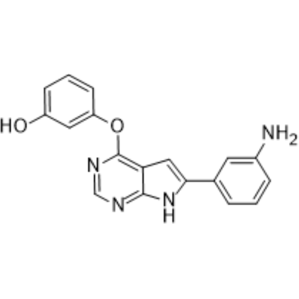
HY-10590
TWS119GSK-3β Inhibitor -
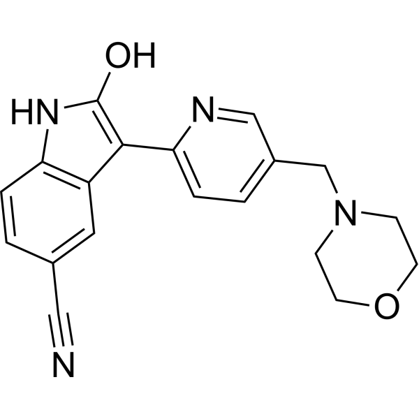
HY-13862
AZD1080GSK-3 Inhibitor -
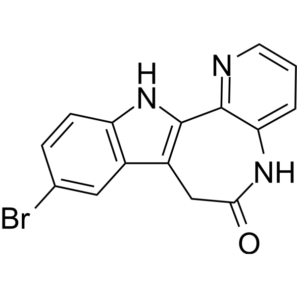
HY-59090
1-AzakenpaulloneGSK-3 Inhibitor -
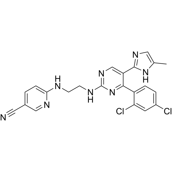
HY-10182G
Laduviglusib (GMP)GSK-3α/β Inhibitor (GMP) -
HY-10580
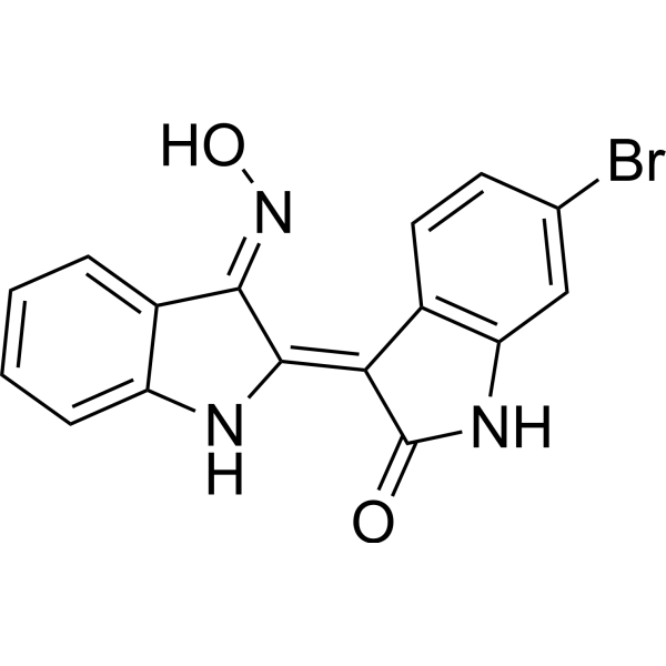
GSK 3 Inhibitor IXATP-Competitive Inhibitor -
HY-13076
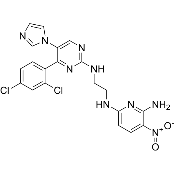
CHIR-98014GSK-3 Inhibitor -

HY-10182R
Laduviglusib (Standard)GSK-3 Inhibitor -
HY-19807
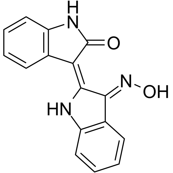
Indirubin-3'-monoximeGSK-3β Inhibitor
Publications Citing Use of MedChemExpress (MCE) 9-ING-41
More Customer Validation & Images

-
Cell Proliferation/Viability Assay

-
Cell Imaging/Staining

-
Cell Proliferation/Viability Assay

-
Histological Imaging/Staining

-
IHC


Biological Activity
Description
IC50 & Target
[1]|
GSK-3β 0.71 μM (IC50) |
Cellular Effect
|
Cell Line

|
Type | Value | Description | References |
|---|---|---|---|---|
| BXPC-3 | IC50 |
1 μM
Compound: 26
|
Growth inhibition of human BxPC3 cells after 72 hrs by MTS assay
Growth inhibition of human BxPC3 cells after 72 hrs by MTS assay
|
[PMID: 19338355] |
| HuP-T3 | IC50 |
0.6 μM
Compound: 26
|
Growth inhibition of human HUPT3 cells after 72 hrs by MTS assay
Growth inhibition of human HUPT3 cells after 72 hrs by MTS assay
|
[PMID: 19338355] |
| MIA PaCa-2 | IC50 |
5 μM
Compound: 26
|
Growth inhibition of human MIAPaCa2 cells after 72 hrs by MTS assay
Growth inhibition of human MIAPaCa2 cells after 72 hrs by MTS assay
|
[PMID: 19338355] |
In Vitro
9-ING-41 (compound 26; 5 μM; for 6, 12, 24, 36 h) results in a pronounced decrease in NFκB-mediated expression of XIAP, the most potent antiapoptotic protein, leading to subsequent apoptosis in BXPC3 pancreatic cancer cells[1].
?9-ING-41 (2, 4 μM; 48 hours) decreases neuroblastoma cell viability induces apoptosis[2].
?
9-ING-41 (1, 2 μM; 24 hours) is a potent cell cycle-blocking agent for lymphoma cells[2].
?
9-ING-41 (10?μM; for 72?hours) increases the expression of LC3, an autophagy marker[3].
?
9-ING-41 (0.5, 1.0, 1.5, 2.0 μM) inhibits the proliferation rate of all TCL and MCL lines with concentrations as low as 1.0 mM[2].
?
9-ING-41 (10?μM; for 72?hours) causes cell cycle blockage at G2/M after 24?hours. 9-ING-41 treatment induces apoptotic cell death in bladder cancer cells[3].
?
9-ING-41 (25?μM; for 96?hours) significantly decreases expression of Cdk1 and Cyclin B1 proteins and leads to a decreased expression of antiapoptotic molecules, Bcl-2 and XIAP[3].
?
9-ING-41 (0.1-1 μM) inhibits GSK-3 leading to a decreased expression of the NF-κB target XIAP and significant apoptosis in neuroblastoma cells as shown by PARP cleavage, an apoptosis marker[4].
MedChemExpress (MCE) has not independently confirmed the accuracy of these methods. They are for reference only. Further protocols information, click here.
-
Cell Line:TCL and MCL lines
-
Concentration:2, 4 μM
-
Incubation Time:48 hours
-
Result:Induced apoptosis.
-
Cell Line:Lymphoma cells (Jeko, Mino, and OCI-Ly cell lines)
-
Concentration:1, 2 μM
-
Incubation Time:24 hours
-
Result:Led to cell cycle arrest in G2/M.
-
Cell Line:T24 cancer cells
-
Concentration:25 μM
-
Incubation Time:24 hours
-
Result:Showed extensive vacuolation and formation of autophagosome like structures in the cytoplasm.
Showed an increased expression of LC3, an autophagy marker.
-
Cell Line:SK-N-DZ and SK-N-BE neuroblastoma cells
-
Concentration:0.1, 1 µM
-
Incubation Time:48 hours
-
Result:Inhibited GSK-3 leading to a decreased expression of the NF-κB target XIAP.
In Vivo
MedChemExpress (MCE) has not independently confirmed the accuracy of these methods. They are for reference only.
-
Animal Model:NSG (NOD.Cg-PrkdcscidIl2rgtm1Wjl/SzJ) mice[2]
-
Dosage:40 mg/kg
-
Administration:Every other day; for 17 days
-
Result:Had single-agent antitumor activity in a mouse model of MCL.
Clinical Trial
| NCT Number | Sponsor | Condition | Start Date |
Phase
|
|---|---|---|---|---|
| NCT01329991 | Plexxikon| | 2011-05 | PHASE1 |


Chemical Information
-
CAS No. 1034895-42-5
-
Appearance Solid
-
Molecular Weight 404.35
-
Formula C22H13FN2O5
-
Color Pink to red
-
SMILES
O=C(C(C1=COC2=CC=C(F)C=C12)=C3C4=CN(C)C5=C4C=C6C(OCO6)=C5)NC3=O
-
Synonyms
Elraglusib
-
Shipping
Room temperature in continental US; may vary elsewhere.
-
Storage
Powder -20°C 3 years 4°C 2 years In solvent -80°C 2 years -20°C 1 year


Publications (6)
-
Journal Impact Factor
-
Most Recent
-
Nature
Perturbing LSD1 and WNT rewires transcription to synergistically induce AML differentiation. [Abstract]2025 Jun;642(8067):508-518. PMID: 40240608
9-ING-41 purchased from MedChemExpress. Usage Cited in: Nature. 2025 Jun;642(8067):508-518. [Abstract]
Analysis of cell viability in THP-1 cells treated with the indicated inhibitors for 5 days by the Cell-Titer-Glo assay.
9-ING-41 purchased from MedChemExpress. Usage Cited in: Nature. 2025 Jun;642(8067):508-518. [Abstract]
Wright-Giemsa-stained cytospins for a DNMT3A-mutant primary AML sample treated with DMSO, IMG-7289 (50 nM), 9-ING-41 (100 nM) and combination of both inhibitors.
-
Adv Sci (Weinh)
Microfluidic Organoid Cultures Derived from Pancreatic Cancer Biopsies for Personalized Testing of Chemotherapy and Immunotherapy. [Abstract]2024 Feb;11(5):e2303088. PMID: 38018486 -
-
Front Immunol
Tumor microenvironment-modulating oncolytic adenovirus combined with GSK-3β inhibitor enhances antitumor immune response against bladder cancer. [Abstract]2024 May 15:15:1360436. PMID: 38812516
9-ING-41 purchased from MedChemExpress. Usage Cited in: Front Immunol. 2024 May 15:15:1360436. [Abstract]
Bladder cancer cells (MB49) and normal cells (NIH3T3) were treated with 0–10 μM of 9-ING-41 for 48 h The cell viability was determined by MTT assay.
9-ING-41 purchased from MedChemExpress. Usage Cited in: Front Immunol. 2024 May 15:15:1360436. [Abstract]
MB49 tumor tissues of mice treated with PBS, 9-ING-41 (10 mg/kg), HY-oAd, and HY-oAd+9-ING-41 were stained with Masson’s trichrome to assess collagen accumulation in the tumor tissues.
9-ING-41 purchased from MedChemExpress. Usage Cited in: Front Immunol. 2024 May 15:15:1360436. [Abstract]
Immunohistochemical staining of CD4+ and CD8+ T cells from subcutaneous MB49 tumor tissue sections obtained after treatment with PBS, 9-ING-41 (10 mg/kg), HY-oAd, and HY-oAd+9-ING-41.
-
Cancer Biol Ther
Potential therapeutic GSK-3β inhibitor 9-ING-41 is active in combination with venetoclax in double-hit lymphoma (DHL). [Abstract]2025 Dec 31;26(1):2581831. PMID: 41220107 -


Solvent & Solubility
In Vitro:
DMSO : 50 mg/mL (123.66 mM; Need ultrasonic; Hygroscopic DMSO has a significant impact on the solubility of product, please use newly opened DMSO)
Please refer to the solubility information to select the appropriate solvent. Once prepared, please aliquot and store the solution to prevent product inactivation from repeated freeze-thaw cycles.
Storage method and period of stock solution: -80°C, 2 years; -20°C, 1 year. When stored at -80°C, please use it within 2 years. When stored at -20°C, please use it within 1 year.
Please refer to the solubility information to select the appropriate solvent. Once prepared, please aliquot and store the solution to prevent product inactivation from repeated freeze-thaw cycles.
Storage method and period of stock solution: -80°C, 2 years; -20°C, 1 year. When stored at -80°C, please use it within 2 years. When stored at -20°C, please use it within 1 year.
Concentration (start) × Volume (start) = Concentration (final) × Volume (final)

Protocols
-
Apoptosis
Apoptosis, also called programmed cell death, is generally characterized by distinct morphological characteristics.
-
TUNEL staining for apoptotic DNA fragmentation
TUNEL staining detects DNA strand breaks by using terminal deoxynucleotidyl transferase to add labeled nucleotides to exposed 3′-OH DNA termini, generating either microscopic staining in fixed cells or tissue sections, or fluorescence/cytometric signal in cell suspensions. TUNEL positivity reflects DNA fragmentation but should not be interpreted alone as definitive apoptosis, because TUNEL can also label necrotic, autolytic, mechanically damaged, or DNA-repair-associated DNA breaks.
-
Cell Cytotoxicity Assay
Cytotoxicity assays are usually based on the assessment of cell membrane damage, which can also be indirectly detected by measuring cell viability. Detection methods include MTT assay, CKK-8 assay, LDH assay and ATP assay, etc.
-
Flow cytometric DNA-content cell-cycle staining
Flow cytometric DNA-content cell-cycle staining measures the fluorescence intensity of DNA-bound fluorochromes in single cells or nuclei to estimate DNA content distributions, allowing assignment of populations to G0/G1, S, and G2/M phases by DNA histogram deconvolution. Propidium iodide (PI) intercalates into DNA, and PI fluorescence is proportional to cellular DNA content when staining is performed under conditions that make DNA accessible and minimize non-DNA signal. Cells with G2/M DNA content are expected to show approximately twice the fluorescence intensity of G0/G1 cells, while S-phase cells occupy intermediate fluorescence values. PI-based DNA-content analysis can also detect cells with fractional DNA content, often reported as sub-G1, when DNA fragmentation and extraction during staining reduce retained DNA signal in apoptotic cells. DAPI is an alternative DNA fluorochrome for univariate DNA-content analysis, while bivariate approaches combining DNA content with proliferation
-
Annexin V plus membrane-impermeant dye apoptosis staining
Annexin V-based apoptosis assays rely on the detection of phosphatidylserine (PS) externalization from the inner leaflet of the plasma membrane to the outer leaflet, an early biochemical hallmark of apoptosis. Fluorescently labeled Annexin V binds PS in a calcium-dependent manner, enabling identification of early apoptotic cells by flow cytometry or fluorescence microscopy. When combined with a membrane-impermeant DNA-binding dye (e. g. , propidium iodide), this approach allows discrimination between viable (Annexin V−/dye−), early apoptotic (Annexin V+/dye−), and late apoptotic or necrotic (Annexin V+/dye+) cell populations by assessing membrane integrity and PS exposure.
-
BrdU Incorporation Assay
Bromodeoxyuridine (BrdU) incorporation assay is based on the principle that BrdU, a thymidine analog, is incorporated into newly synthesized DNA during the S phase of the cell cycle, thereby serving as a marker of DNA replication and cellular proliferation. Incorporated BrdU can be detected using anti-BrdU antibodies following DNA denaturation, enabling visualization or quantification of proliferating cells through immunochemical detection methods such as immunofluorescence or immunohistochemistry.
-
Autophagy
Autophagy is a process in which eukaryotic cells use lysosomes to degrade their own cytoplasmic proteins and damaged organelles under the regulation of autophagy related gene (Atg). Microtubule-associated proteins light chain 3 (LC3) is recognized as autophagy marker, which transfers from cytoplasmic LC3 (LC3-I) to membrane type (LC3-II). LC3-II/I ratio could be detected by Western Blot and fluorescence microscopy.
-
Apoptosis Solutions
Apoptosis is a regulated, generally non-lytic cell-death pathway that removes unwanted, damaged, infected, or abnormal cells through coordinated morphological changes, caspase activation, DNA fragmentation, and membrane remodeling. The intrinsic apoptosis pathway is controlled mainly by mitochondrial outer membrane permeabilization, BCL-2 family proteins, cytochrome c release, apoptosome formation, caspase-9 activation, and downstream executioner caspase-3/7 activation. The extrinsic apoptosis pathway is initiated by death receptors such as Fas, TNFR, and TRAIL receptors, which recruit adaptor proteins and activate caspase-8 before engaging executioner caspases or mitochondrial amplification through BID cleavage. Apoptosis is linked to many phenotypes, including cancer cell killing, tissue homeostasis, immune regulation, neurodegeneration, infection response, and treatment-induced cytotoxicity; unresolved questions include how apoptosis interacts with necroptosis, pyroptosis, ferroptos
-
Protocol for Cell Cycle
Cell-cycle analysis by flow cytometry measures DNA content in single cells to estimate the fraction of cells in G0/G1, S, and G2/M phases. Propidium iodide intercalates into DNA, and after RNA removal with RNase, fluorescence intensity reflects cellular DNA content: 2N cells are assigned to G0/G1, cells between 2N and 4N to S phase, and 4N cells to G2/M. DNA-content analysis alone cannot reliably separate G0 from G1 or G2 from M. Ki-67 can distinguish quiescent G0 cells from cycling cells, EdU or BrdU incorporation marks active DNA synthesis in S phase, and phospho-histone H3 staining identifies mitotic cells within the 4N population.
-
Lysosome and acidic-vesicle live-cell staining
Lysosome and acidic-vesicle live-cell staining detects acidic intracellular compartments by using membrane-permeant acidotropic probes that accumulate in low-pH vesicles, including lysosomes, late endosomes, autolysosomes, and acidic phagosomes. LysoTracker staining is commonly used as an intensity-based readout of acidic lysosomal compartment abundance or enlargement, while acridine orange produces green fluorescence in less concentrated compartments and red fluorescence after concentration-dependent accumulation in acidic vesicular organelles. Loss or reduction of acridine-orange red signal can be used as a readout of lysosomal membrane permeabilization or reduced acidic-vesicle integrity. This protocol is designed for live cultured cells and can be adapted for fluorescence microscopy, high-content imaging, plate-reader readout, or flow cytometry when the selected literature supports the readout. Because these dyes report acidotropic accumulation rather than lysosome identity alone,
-
Macroautophagy Solutions
Macroautophagy is a conserved lysosome-dependent degradation pathway in which cytoplasmic material is sequestered into double-membrane autophagosomes and delivered to lysosomes for degradation and recycling. The pathway supports cellular homeostasis during nutrient limitation, organelle stress, protein-aggregate accumulation, infection, differentiation, and tissue remodeling by coupling cargo sequestration, autophagosome maturation, lysosomal fusion, and degradation of cargo-derived macromolecules. The core molecular sequence includes initiation by nutrient- and stress-regulated autophagy machinery, autophagosome nucleation, LC3/ATG8-family conjugation to autophagosomal membranes, cargo selection through receptors such as SQSTM1/p62, autophagosome-lysosome fusion, and lysosomal degradation. LC3 was identified as a mammalian homolog of yeast Atg8 that localizes to autophagosomal membranes after processing, and p62/SQSTM1 was shown to connect ubiquitinated cargo with autophagic degradati


Purity & Documentation
-
Data Sheet (289 KB)
-
SDS (644 KB)
- English - EN (644 KB)
- Français - FR (644 KB)
- Deutsch - DE (644 KB)
- Norwegian - NO (644 KB)
- Español - ES (644 KB)
- Swedish - SV (644 KB)
- Italian - IT (644 KB)
- Korean - KR (644 KB)
- Portuguese - PT (644 KB)
-
Handling Instructions (2659 KB)
References
[1]. Irina N Gaisina, et al. From a natural product lead to the identification of potent and selective benzofuran-3-yl-(indol-3-yl)maleimides as glycogen synthase kinase 3beta inhibitors that suppress proliferation and survival of pancreatic cancer cells. J Med Chem. 2009 Apr 9;52(7):1853-63. [Content Brief]
[2]. Wu X, et al. Targeting glycogen synthase kinase 3 for therapeutic benefit in lymphoma. Blood. 2019 Jul 25;134(4):363-373. [Content Brief]
[3]. Hiroo Kuroki, et al. 9-ING-41, a small molecule inhibitor of GSK-3beta, potentiates the effects of anticancer therapeutics in bladder cancer. Sci Rep. 2019 Dec 27;9(1):19977. [Content Brief]
[4]. Ugolkov AV, et al. 9-ING-41, a small-molecule glycogen synthase kinase-3 inhibitor, is active in neuroblastoma. Anticancer Drugs. 2018 Sep;29(8):717-724. [Content Brief]


Complete Stock Solution Preparation Table
Please refer to the solubility information to select the appropriate solvent. Once prepared, please aliquot and store the solution to prevent product inactivation from repeated freeze-thaw cycles.
Storage method and period of stock solution: -80°C, 2 years; -20°C, 1 year. When stored at -80°C, please use it within 2 years. When stored at -20°C, please use it within 1 year.
| Optional Solvent | Concentration Solvent Mass | 1 mg | 5 mg | 10 mg | 25 mg |
|---|---|---|---|---|---|
| DMSO | 1 mM | 2.4731 mL | 12.3655 mL | 24.7310 mL | 61.8276 mL |
| 5 mM | 0.4946 mL | 2.4731 mL | 4.9462 mL | 12.3655 mL | |
| 10 mM | 0.2473 mL | 1.2366 mL | 2.4731 mL | 6.1828 mL | |
| 15 mM | 0.1649 mL | 0.8244 mL | 1.6487 mL | 4.1218 mL | |
| 20 mM | 0.1237 mL | 0.6183 mL | 1.2366 mL | 3.0914 mL | |
| 25 mM | 0.0989 mL | 0.4946 mL | 0.9892 mL | 2.4731 mL | |
| 30 mM | 0.0824 mL | 0.4122 mL | 0.8244 mL | 2.0609 mL | |
| 40 mM | 0.0618 mL | 0.3091 mL | 0.6183 mL | 1.5457 mL | |
| 50 mM | 0.0495 mL | 0.2473 mL | 0.4946 mL | 1.2366 mL | |
| 60 mM | 0.0412 mL | 0.2061 mL | 0.4122 mL | 1.0305 mL | |
| 80 mM | 0.0309 mL | 0.1546 mL | 0.3091 mL | 0.7728 mL | |
| 100 mM | 0.0247 mL | 0.1237 mL | 0.2473 mL | 0.6183 mL |


9-ING-41 Related Classifications
HY-113914 Related Classifications
 Powered by Bioz