



Candesartan Cilexetil
Based on 6 publication(s) in Google Scholar
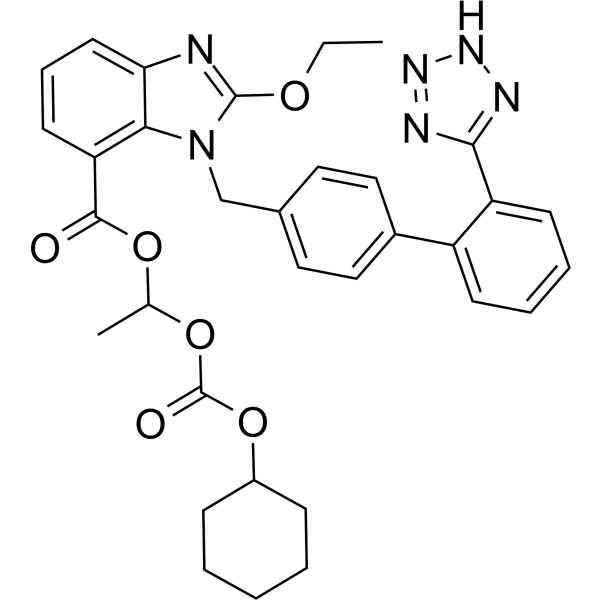
Candesartan Cilexetil (TCV-116) is an angiotensin II receptor inhibitor. Candesartan Cilexetil ameliorates the pulmonary fibrosis and has antiviral and skin wound healing effect. Candesartan Cilexetil can be used for the research of high blood pressure.
For research use only. We do not sell to patients.
- Purity : 99.66%
- CAS No.: 145040-37-5
- Formula: C33H34N6O6
- Molecular Weight:610.66
-
Storage:Powder -20°C, 3 years , 4°C, 2 years ; In solvent -80°C, 2 years , -20°C, 1 year

To place orders, for customer services and technical support, please contact: MedChemExpress USA
Tel: 609-228-6898 E-mail: [email protected] [email protected]


-
 Biological Activity
-
Chemical Information
-
Solvent & Solubility
- Protocols
- Purity & Documentation
- References
-
 Help & FAQs


-
HY-18204
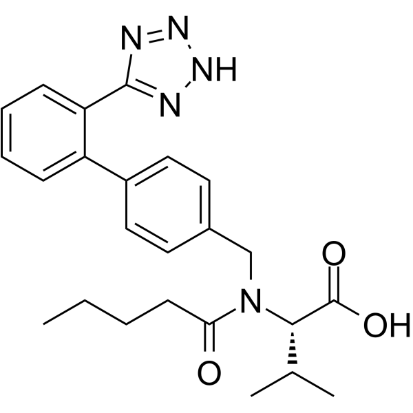
ValsartanAngiotensin II Receptor Antagonist -
HY-B0205
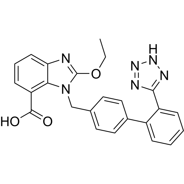
CandesartanAngiotensin II AT1-Receptor Blocker -
HY-14519
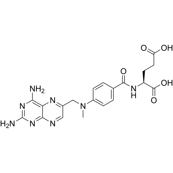
MethotrexateDihydrofolate Reductase Inhibitor -
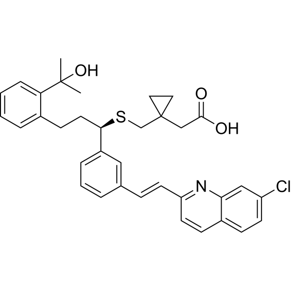
HY-13315A
MontelukastCysLT1 Antagonist -
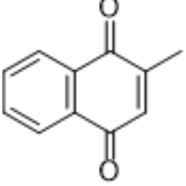
HY-B0332
MenadioneEndogenous Metabolite -
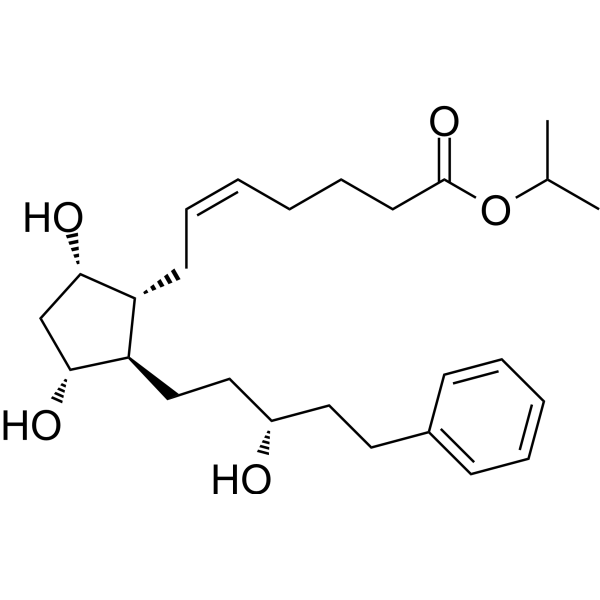
HY-B0577
LatanoprostProstaglandin Receptor Agonist -
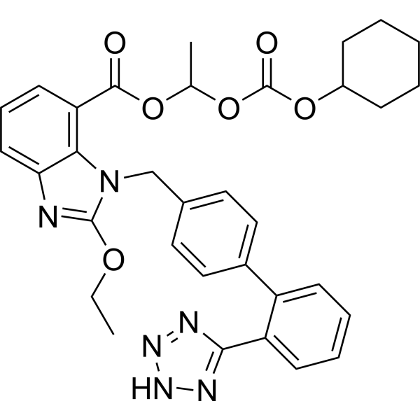
HY-17505R
Candesartan Cilexetil (Standard)Angiotensin II receptor Inhibitor -
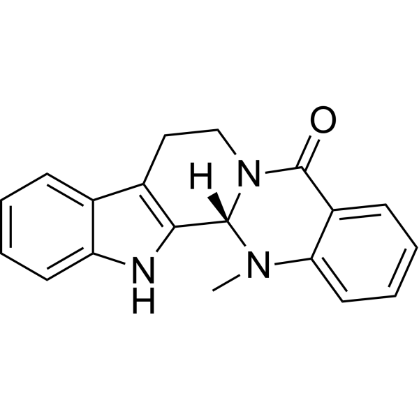
HY-N0114
EvodiamineAlkaloid -
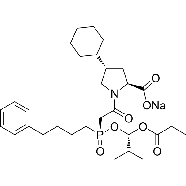
HY-B0382
Fosinopril sodiumACE Inhibitor -
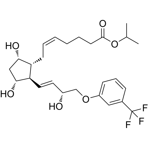
HY-B0584
TravoprostFP Prostaglandin Receptor Agonist -
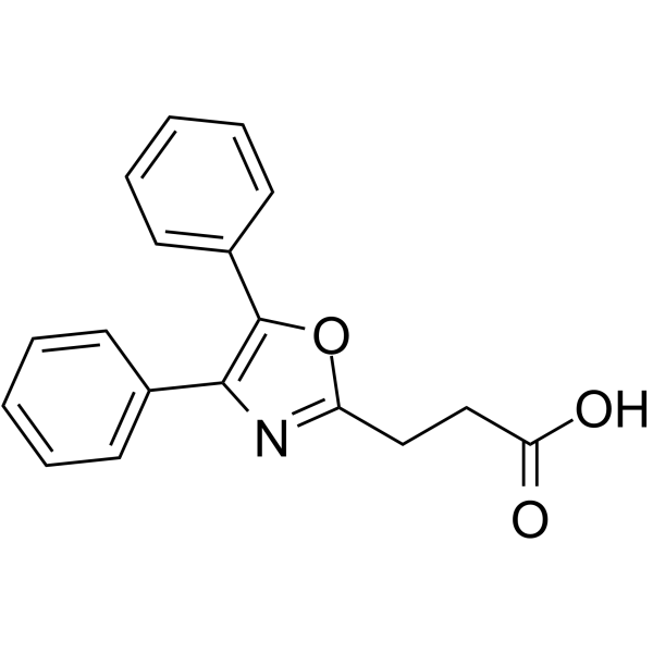
HY-B0808
OxaprozinCOX Inhibitor -
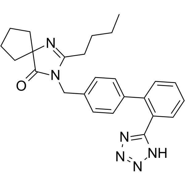
HY-B0202
IrbesartanAT1 Receptor Blocker
Publications Citing Use of MedChemExpress (MCE) Candesartan Cilexetil
MoreAll Angiotensin Receptor Isoforms
More

Biological Activity
Description
Cellular Effect
|
Cell Line

|
Type | Value | Description | References |
|---|---|---|---|---|
| 16HBE14o- | IC50 |
>100 μM
Compound: 1; CDC, MT04D0301
|
Cytotoxicity against human 16HBE cells after 48 hrs by CCK-8 assay
Cytotoxicity against human 16HBE cells after 48 hrs by CCK-8 assay
|
[PMID: 31732254] |
| A-431 | IC50 |
2.6 μM
Compound: Candesartan cilexetil
|
Inhibition of IDO in human A431 cells assessed as kynurenine production after 24 hrs
Inhibition of IDO in human A431 cells assessed as kynurenine production after 24 hrs
|
10.1039/C2MD00278G |
| A549 | IC50 |
63.93 μM
Compound: 1; CDC, MT04D0301
|
Antiproliferative activity against human A549 cells measured after 48 hrs by CCK8 assay
Antiproliferative activity against human A549 cells measured after 48 hrs by CCK8 assay
|
[PMID: 31732254] |
| A549 | IC50 |
65.92 μM
Compound: 1; CDC
|
Antiproliferative activity against human A549 cells incubated for 48 hrs by CCK-8 assay
Antiproliferative activity against human A549 cells incubated for 48 hrs by CCK-8 assay
|
[PMID: 33129593] |
| BEAS-2B | IC50 |
79.76 μM
Compound: 1; CDC
|
Cytotoxicity against human BEAS-2B cells incubated for 48 hrs by CCK-8 assay
Cytotoxicity against human BEAS-2B cells incubated for 48 hrs by CCK-8 assay
|
[PMID: 33129593] |
| BEAS-2B | IC50 |
89.94 μM
Compound: 1; CDC, MT04D0301
|
Cytotoxicity against human BEAS2B cells after 48 hrs by CCK-8 assay
Cytotoxicity against human BEAS2B cells after 48 hrs by CCK-8 assay
|
[PMID: 31732254] |
| EKVX | IC50 |
>100 μM
Compound: 1; CDC, MT04D0301
|
Antiproliferative activity against human EKVX cells measured after 48 hrs by CCK8 assay
Antiproliferative activity against human EKVX cells measured after 48 hrs by CCK8 assay
|
[PMID: 31732254] |
| HepG2 | IC50 |
>100 μM
Compound: 1; CDC, MT04D0301
|
Antiproliferative activity against human HepG2 cells measured after 48 hrs by CCK8 assay
Antiproliferative activity against human HepG2 cells measured after 48 hrs by CCK8 assay
|
[PMID: 31732254] |
| Huh-7 | IC50 |
>100 μM
Compound: 1; CDC, MT04D0301
|
Antiproliferative activity against human HuH7 cells measured after 48 hrs by CCK8 assay
Antiproliferative activity against human HuH7 cells measured after 48 hrs by CCK8 assay
|
[PMID: 31732254] |
| MCF7 | IC50 |
>100 μM
Compound: 1; CDC, MT04D0301
|
Antiproliferative activity against human MCF7 cells measured after 48 hrs by CCK8 assay
Antiproliferative activity against human MCF7 cells measured after 48 hrs by CCK8 assay
|
[PMID: 31732254] |
| MGC-803 | IC50 |
57.14 μM
Compound: 1; CDC, MT04D0301
|
Antiproliferative activity against human MGC803 cells measured after 48 hrs by CCK8 assay
Antiproliferative activity against human MGC803 cells measured after 48 hrs by CCK8 assay
|
[PMID: 31732254] |
| MKN-45 | IC50 |
68.87 μM
Compound: 1; CDC, MT04D0301
|
Antiproliferative activity against human MKN45 cells measured after 48 hrs by CCK8 assay
Antiproliferative activity against human MKN45 cells measured after 48 hrs by CCK8 assay
|
[PMID: 31732254] |
| NCI-H1299 | IC50 |
45.02 μM
Compound: 1; CDC
|
Antiproliferative activity against human H1299 cells incubated for 48 hrs by CCK-8 assay
Antiproliferative activity against human H1299 cells incubated for 48 hrs by CCK-8 assay
|
[PMID: 33129593] |
| NCI-H1299 | IC50 |
45.11 μM
Compound: 1; CDC, MT04D0301
|
Antiproliferative activity against human H1299 cells measured after 48 hrs by CCK8 assay
Antiproliferative activity against human H1299 cells measured after 48 hrs by CCK8 assay
|
[PMID: 31732254] |
| PLC | IC50 |
60.21 μM
Compound: 1; CDC, MT04D0301
|
Antiproliferative activity against human PLC cells measured after 48 hrs by CCK8 assay
Antiproliferative activity against human PLC cells measured after 48 hrs by CCK8 assay
|
[PMID: 31732254] |
| T47D | IC50 |
64.35 μM
Compound: 1; CDC, MT04D0301
|
Antiproliferative activity against human T47D cells measured after 48 hrs by CCK8 assay
Antiproliferative activity against human T47D cells measured after 48 hrs by CCK8 assay
|
[PMID: 31732254] |
In Vitro
Candesartan Cilexetil (0.1-10 μM, 24 h) exhibits antiviral effects against ZIKV in HUVEC cells, HEK293T cells[1].
MedChemExpress (MCE) has not independently confirmed the accuracy of these methods. They are for reference only. Further protocols information, click here.
-
Cell Line:HUVEC cells, HEK293T cells
-
Concentration:0.1 μM, 1 μM, 5 μM, 10 μM
-
Incubation Time:24 h
-
Result:Showed a dose-dependent antiviral effect in ZIKV-infected HUVEC and HEK293T cells.
In Vivo
Candesartan Cilexetil (1-10 mg/kg, Oral, once a day for 15 days) has an effect of skin wound healing in rats[3].
Candesartan Cilexetil (1-10 mg/kg, Administered through a stomach tube, once a day for 4-12 weeks) inhibits the increase in blood pressure in spontaneously hypertensive rats[5].
Candesartan Cilexetil (10 mg/kg, Oral, supplemented in drinking water, for 2-23 days) inhibits the synthesis of TGF-b1 and ameliorates the pulmonary fibrosis in bleomycin (HY-108345) induced pulmonary fibrosis model rats[6].
MedChemExpress (MCE) has not independently confirmed the accuracy of these methods. They are for reference only.
-
Animal Model:Dahl salt-sensitive hypertensive rats[2]
-
Dosage:5 mg/kg
-
Administration:Oral gavage (p.o.)
-
Result:Increased body weight and decreased left ventricular end-diastolic diameter.
Decreased the level of iNOS mRNA and ADM mRNA in the left ventricle (LV).
Increased the eNOS protein mass in the LV and decreased the immunoreactive ADM contents of LV.
Enhanced immunoreactivity for eNOS.
Significantly decreased the degree of perivascular fibrosis.
-
Animal Model:Skin wound model rat [3]
-
Dosage:1 mg/kg, 10 mg/kg
-
Administration:Oral
-
Result:Showed 100% re-epithelization of the wound at 1 mg/kg.
Significantly suppressed angiogenesis at 10 mg/kg.
Clinical Trial
| NCT Number | Sponsor | Condition | Start Date |
Phase
|
|---|---|---|---|---|
| NCT01329991 | Plexxikon| | 2011-05 | PHASE1 |


Chemical Information
-
CAS No. 145040-37-5
-
Appearance Solid
-
Molecular Weight 610.66
-
Formula C33H34N6O6
-
Color White to off-white
-
SMILES
O=C(C1=C2C(N=C(OCC)N2CC3=CC=C(C4=CC=CC=C4C5=NNN=N5)C=C3)=CC=C1)OC(OC(OC6CCCCC6)=O)C
-
Synonyms
TCV-116
-
Shipping
Room temperature in continental US; may vary elsewhere.
-
Storage
Powder -20°C 3 years 4°C 2 years In solvent -80°C 2 years -20°C 1 year


Publications (6)
-
Journal Impact Factor
-
Most Recent
-
Cell Death Discov
Single cell RNA sequencing reveals the role of local renin-angiotensin system in regulating ovarian physiological cycle and promoting PCOS. [Abstract]2025 May 27;11(1):255. PMID: 40425574 -
EXCLI J
Candesartan cilexetil as a repurposed therapeutic candidate for nasopharyngeal carcinoma: Integrated in vitro and in silico analyses. [Abstract]2026 Jan 16:25:204-221. PMID: 41768859 -
Int J Mol Sci
2026 Mar 11;27(6):2587. PMID: 41898448 -
-
Biomed Pharmacother
A low-salt diet with candesartan administration is associated with acute kidney injury in nephritis by increasing nitric oxide. [Abstract]2023 May:161:114484. PMID: 36921530 -


Solvent & Solubility
In Vitro:
DMSO : ≥ 50 mg/mL (81.88 mM; Hygroscopic DMSO has a significant impact on the solubility of product, please use newly opened DMSO)
* "≥" means soluble, but saturation unknown.
Please refer to the solubility information to select the appropriate solvent. Once prepared, please aliquot and store the solution to prevent product inactivation from repeated freeze-thaw cycles.
Storage method and period of stock solution: -80°C, 2 years; -20°C, 1 year. When stored at -80°C, please use it within 2 years. When stored at -20°C, please use it within 1 year.
Please refer to the solubility information to select the appropriate solvent. Once prepared, please aliquot and store the solution to prevent product inactivation from repeated freeze-thaw cycles.
Storage method and period of stock solution: -80°C, 2 years; -20°C, 1 year. When stored at -80°C, please use it within 2 years. When stored at -20°C, please use it within 1 year.
Concentration (start) × Volume (start) = Concentration (final) × Volume (final)
In Vivo:
Select the appropriate dissolution method based on your experimental animal and administration route.
- For the following dissolution methods, please ensure to first prepare a clear stock solution using an In Vitro approach and then sequentially add co-solvents:
- To ensure reliable experimental results, the clarified stock solution can be appropriately stored based on storage conditions. As for the working solution for In Vivo experiments, it is recommended to prepare freshly and use it on the same day.
- The percentages shown for the solvents indicate their volumetric ratio in the final prepared solution. If precipitation or phase separation occurs during preparation, heat and/or sonication can be used to aid dissolution.
Add each solvent one by one: 10% DMSO 40% PEG300 5% Tween-80 45% Saline
Solubility: ≥ 2.5 mg/mL (4.09 mM); Clear solution
This protocol yields a clear solution of ≥ 2.5 mg/mL (saturation unknown).
Taking 1 mL working solution as an example, add 100 μL DMSO stock solution (25.0 mg/mL) to 400 μL PEG300, and mix evenly; then add 50 μL Tween-80 and mix evenly; then add 450 μL Saline to adjust the volume to 1 mL.
Preparation of Saline: Dissolve 0.9 g sodium chloride in ddH₂O and dilute to 100 mL to obtain a clear Saline solution.
In Vivo Dissolution Calculator
Please enter the basic information of animal experiments:
-
-
-
-
Recommended: Prepare an additional quantity of animals to account for potential losses during experiments.
Please enter your animal formula composition:
-
%DMSO +
Recommended: Keep the proportion of DMSO in working solution below 2% if your animal is weak.
-
%+
-
+%Tween-80 + +
-
%Saline +
The co-solvents required include: DMSO, . All of co-solvents are available by MedChemExpress (MCE). , Tween 80. All of co-solvents are available by MedChemExpress (MCE).
Working solution concentration: 0.22 mg/mL
Method for preparing stock solution: mg drug dissolved in μL DMSO. Stock solution concentration: mg/mL.
1. Take μL DMSO stock solution;
2. Add μL .
μL , mix evenly;
3. Then add μL Tween 80, mix evenly;
4. Then add μL
Please ensure that the stock solution in the first step is dissolved to a clear state, and add co-solvents in sequence. You can use ultrasonic heating (ultrasonic cleaner, recommended frequency 20-40 kHz), vortexing, etc. to assist dissolution.

Protocols
-
Fibrosis/Collagen Morphometry
Fibrosis and collagen morphometry is based on the quantitative visualization of fibrillar collagen deposition in tissue sections using histochemical stains such as Sirius Red (Picrosirius Red) or Masson's trichrome, followed by image-based or polarization-enhanced analysis to estimate collagen proportional area as a surrogate of extracellular matrix accumulation during fibrotic remodeling. Sirius Red combined with polarized light microscopy enhances detection of collagen fibers due to birefringence properties, enabling more specific visualization of collagen type I and III fibrils compared to conventional bright-field histology, while whole-section or region-restricted digital morphometry reduces field-selection bias in fibrosis assessment. Alternative quantitative approaches include second harmonic generation (SHG) and two-photon excited fluorescence microscopy, which enable label-free detection of fibrillar collagen and have been validated against histological staining and biochemica
-
Collagen: Sirius Red Staining
Sirius Red or picrosirius red staining is a histochemical method for visualizing collagen-rich extracellular matrix in tissue sections, and collagen fibers are detected as red-stained structures under bright-field microscopy with enhanced birefringence under polarized light. Picrosirius red is useful for assessing total collagen organization, distribution, and fibrosis burden, but polarized color should not be interpreted as a definitive collagen type I versus type III readout because color is affected by fiber orientation, thickness, and packing.
-
Research Protocol for Infectious Diseases
Infectious-disease experiments test how pathogens interact with host barriers, innate immune receptors, inflammatory signaling, pathogen replication, and tissue injury; pattern-recognition receptors such as TLRs, RIG-I-like receptors, NOD-like receptors, and inflammasomes detect microbial molecules and activate NF-κB, interferon, and cytokine responses. The central hypothesis is that infection severity reflects the balance between pathogen burden and host response: protective inflammation restricts pathogen growth, whereas excessive or mislocalized inflammation contributes to tissue damage and disease phenotype. Unresolved questions include which host pathways are protective versus pathogenic, why some infection models fail to translate to human disease, and which combined readouts best predict clinically relevant infection outcomes.
-
Cell migration
Cell migration is a method that plays an important role in wound healing, cell differentiation, embryonic development, etc.
-
Connective Tissue: Masson's Trichrome/Collagen Trichrome Staining
Masson’s Trichrome (collagen/trichrome staining) is a histological technique that differentially stains tissue compartments using sequential acidic dyes to distinguish collagen from muscle and cytoplasmic components based on dye affinity and tissue permeability differences, enabling visualization of fibrosis and connective tissue architecture in histological sections. The classical formulation typically uses Weigert's iron hematoxylin for nuclear staining, Biebrich scarlet-acid fuchsin for cytoplasm and muscle, and aniline blue (or light green variants) for collagen, producing a characteristic blue/green collagen signal contrasted against red cytoplasm and dark nuclei. The staining principle relies on selective displacement of smaller dye molecules by larger anionic dyes in collagen-rich regions under controlled acidified conditions, which enhances collagen-specific dye retention. This property makes the method widely used for fibrosis assessment in organs such as heart, liver, lung, a
-
Cell-Exclusion Zone Migration Assay
The Cell-Exclusion Zone (CEZ) migration assay is an in vitro 2D cell migration method in which a defined cell-free area is created using removable physical barriers such as silicone stoppers, allowing cells to be seeded around the barrier and subsequently migrate into the cleared zone after barrier removal. This approach enables quantification of collective cell migration by monitoring repopulation of the initially cell-free region over time using microscopy-based imaging. Compared with scratch-based wound healing assays, barrier-based exclusion methods are designed to avoid mechanical damage to the extracellular matrix and reduce injury-induced effects on boundary cells, thereby improving interpretability of migration behavior in vitro. The assay readout is typically the progressive reduction in the cell-free area or the number of cells invading the exclusion zone, reflecting coordinated cell motility relevant to physiological processes such as wound healing, epithelial repair, and ca


Purity & Documentation
-
Data Sheet (288 KB)
-
SDS (479 KB)
- English - EN (479 KB)
- Français - FR (479 KB)
- Deutsch - DE (479 KB)
- Norwegian - NO (479 KB)
- Español - ES (479 KB)
- Swedish - SV (479 KB)
- Italian - IT (479 KB)
- Korean - KR (479 KB)
- Portuguese - PT (479 KB)
-
Handling Instructions (2659 KB)
References
[1]. Loe M W C, et al. Antiviral activity of the FDA-approved drug candesartan cilexetil against Zika virus infection [J]. Antiviral Research, 2019, 172: 104637. [Content Brief]
[2]. Kobayashi N, et al. Effects of TCV-116 on expression of NOS and adrenomedullin in failing heart of Dahl salt-sensitive rats [J]. Atherosclerosis, 2001, 156(2): 255-265. [Content Brief]
[3]. Takeda H, et al. Effects of angiotensin II receptor signaling during skin wound healing [J]. The American journal of pathology, 2004, 165(5): 1653-1662. [Content Brief]
[4]. McClellan K J, et al. Candesartan cilexetil: a review of its use in essential hypertension [J]. Drugs, 1998, 56: 847-869. [Content Brief]
[5]. Kojima M, et al. Angiotensin II receptor antagonist TCV-116 induces regression of hypertensive left ventricular hypertrophy in vivo and inhibits the intracellular signaling pathway of stretch-mediated cardiomyocyte hypertrophy in vitro [J]. Circulation, 1994, 89(5): 2204-2211. [Content Brief]
[6]. Otsuka M, et al. Reduction of bleomycin induced lung fibrosis by candesartan cilexetil, an angiotensin II type 1 receptor antagonist [J]. Thorax, 2004, 59(1): 31-38. [Content Brief]


Complete Stock Solution Preparation Table
Please refer to the solubility information to select the appropriate solvent. Once prepared, please aliquot and store the solution to prevent product inactivation from repeated freeze-thaw cycles.
Storage method and period of stock solution: -80°C, 2 years; -20°C, 1 year. When stored at -80°C, please use it within 2 years. When stored at -20°C, please use it within 1 year.
| Optional Solvent | Concentration Solvent Mass | 1 mg | 5 mg | 10 mg | 25 mg |
|---|---|---|---|---|---|
| DMSO | 1 mM | 1.6376 mL | 8.1879 mL | 16.3757 mL | 40.9393 mL |
| 5 mM | 0.3275 mL | 1.6376 mL | 3.2751 mL | 8.1879 mL | |
| 10 mM | 0.1638 mL | 0.8188 mL | 1.6376 mL | 4.0939 mL | |
| 15 mM | 0.1092 mL | 0.5459 mL | 1.0917 mL | 2.7293 mL | |
| 20 mM | 0.0819 mL | 0.4094 mL | 0.8188 mL | 2.0470 mL | |
| 25 mM | 0.0655 mL | 0.3275 mL | 0.6550 mL | 1.6376 mL | |
| 30 mM | 0.0546 mL | 0.2729 mL | 0.5459 mL | 1.3646 mL | |
| 40 mM | 0.0409 mL | 0.2047 mL | 0.4094 mL | 1.0235 mL | |
| 50 mM | 0.0328 mL | 0.1638 mL | 0.3275 mL | 0.8188 mL | |
| 60 mM | 0.0273 mL | 0.1365 mL | 0.2729 mL | 0.6823 mL | |
| 80 mM | 0.0205 mL | 0.1023 mL | 0.2047 mL | 0.5117 mL |


Candesartan Cilexetil Related Classifications
HY-17505 Related Classifications
 Powered by Bioz