



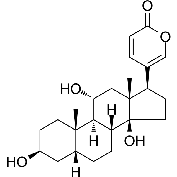
Gamabufotalin
Based on 4 publication(s) in Google Scholar
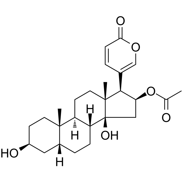
Gamabufotalin (Gamabufagin), a main active compound isolated from Chinese medicine Chansu, has been shown to strongly inhibit cancer cell growth and inflammatory response. Gamabufotalin could inhibite angiogenesis by inhibiting the activation of VEGFR-2 signaling pathways.
For research use only. We do not sell to patients.
- Purity : 99.30%
- CAS No.: 465-11-2
- Formula: C24H34O5
- Molecular Weight:402.52
-
Storage:Powder -20°C, 3 years , 4°C, 2 years ; In solvent -80°C, 2 years , -20°C, 1 year

To place orders, for customer services and technical support, please contact: MedChemExpress USA
Tel: 609-228-6898 E-mail: [email protected] [email protected]


-
 Biological Activity
-
Chemical Information
-
Solvent & Solubility
- Protocols
- Purity & Documentation
- References
-
 Help & FAQs


Publications Citing Use of MedChemExpress (MCE) Gamabufotalin
More Customer Validation & Images

-
Cell Proliferation/Viability Assay
-
Cell Proliferation/Viability Assay
-
WB
-
IF
All VEGFR Isoforms
More

Biological Activity
Description
Cellular Effect
|
Cell Line

|
Type | Value | Description | References |
|---|---|---|---|---|
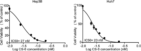
| Hep 3B2 | IC50 |
27 nM
Compound: CS-6
|
Antiproliferative activity against human Hep3B cells assessed as cell viability incubated for 24 hrs by CCK-8
Antiproliferative activity against human Hep3B cells assessed as cell viability incubated for 24 hrs by CCK-8
|
[PMID: 37043698] |
| HL-60 | IC50 |
0.014 μg/mL
Compound: 20
|
Cytotoxicity against human HL60 cells after 72 hrs by MTT assay
Cytotoxicity against human HL60 cells after 72 hrs by MTT assay
|
[PMID: 11575946] |
| Huh-7 | IC50 |
23 nM
Compound: CS-6
|
Antiproliferative activity against human Huh-7 cells assessed as cell viability incubated for 24 hrs by CCK-8
Antiproliferative activity against human Huh-7 cells assessed as cell viability incubated for 24 hrs by CCK-8
|
[PMID: 37043698] |
| KB | IC50 |
0.75 μg/mL
Compound: 20
|
Cytotoxicity against human KB cells after 72 hrs by MTT assay
Cytotoxicity against human KB cells after 72 hrs by MTT assay
|
[PMID: 11575946] |
| MH60 | IC50 |
>25 μg/mL
Compound: 20
|
Cytotoxicity against mouse MH60 cells after 72 hrs by MTT assay
Cytotoxicity against mouse MH60 cells after 72 hrs by MTT assay
|
[PMID: 11575946] |
In Vitro
Gamabufotalin (0-500 nM, 48 h) inhibits cell viability of human lung cancer A549, H1299, H322 cells, and inhibits colony formation and migration (0-100 nM, 48 h), with no cytotoxicity in human normal lung cell line (HLF cells)[1].
Gamabufotalin (0-100 nM, 12-48 h) inhibits COX-2 expression and inhibits NF-κB and p300 translocation in A549 cells[1].
Gamabufotalin (0-100 nM, 12-48 h) induces apoptosis by activating the cytochrome c release and caspase-dependent apoptotic pathway in A549 cells[1].
Gamabufotalin (0-100 nM, 48 h) induces hyperphosphorylation of p38, increases the expression of ATP1A3 and decreases AQP4 expression in U87 cells[3].
Gamabufotalin (0-50 nM, 24 h) inhibits VEGF (50 ng/mL)-induced angiogenesis in an HUVECs in vitro angiogenesis tube formation assay[4].
MedChemExpress (MCE) has not independently confirmed the accuracy of these methods. They are for reference only. Further protocols information, click here.
-
Cell Line:A549 cells
-
Concentration:0-100 nM
-
Incubation Time:12-48 h
-
Result:Increase the expression levels of the cleaved caspase-3, caspase-9 and PARP.
Induced the release of cyt c from mitochondria to cytosol.
-
Cell Line:A549 cells
-
Concentration:0-100 nM
-
Incubation Time:12-48 h
-
Result:Inhibited translocation of the NF-κB p65/p50 proteins from cell cytoplasm to nucleus, and induced p300 into the cytoplasm.
In Vivo
Gamabufotalin (1 mg/kg, i.p., three times per week) together with Temozolomide (HY-17364) (20 mg/kg. i.p., three times per week) shows a synergistic antitumor effect, and inhibits tumor growth and prolongs mice survival in mice U87 xenografts[3].
MedChemExpress (MCE) has not independently confirmed the accuracy of these methods. They are for reference only.
-
Animal Model:A549-xenografts mice[1]
-
Dosage:5 and 20 mg/kg/day
-
Administration:i.p., for 17 days
-
Result:Reduced tumor volume and the tumor weights.
Decreased COX-2 and p-p65 level in tumors.


Chemical Information
-
CAS No. 465-11-2
-
Appearance Solid
-
Molecular Weight 402.52
-
Formula C24H34O5
-
Color White to off-white
-
SMILES
C[C@]([C@@H](C(C=C1)=COC1=O)CC2)(C[C@@H](O)[C@@]3([H])[C@@]4([H])CC[C@@]5([H])[C@@]3(CC[C@H](O)C5)C)[C@]24O
-
Synonyms
Gamabufagin
-
Structure Classification
-
Initial Source
toad
-
Shipping
Room temperature in continental US; may vary elsewhere.
-
Storage
Powder -20°C 3 years 4°C 2 years In solvent -80°C 2 years -20°C 1 year


Publications (4)
-
Journal Impact Factor
-
Most Recent
-
Nat Microbiol
2023 Jan;8(1):121-134. PMID: 36604514 -
Phytomedicine
Compound-composed Chinese medicine of Huachansu triggers apoptosis of gastric cancer cells through increase of reactive oxygen species levels and suppression of proteasome activities. [Abstract]2024 Jan:123:155169. PMID: 37992493 -
Front Pharmacol
Gamabufotalin Inhibits Osteoclastgenesis and Counteracts Estrogen-Deficient Bone Loss in Mice by Suppressing RANKL-Induced NF-κB and ERK/MAPK Pathways. [Abstract]2021 Apr 23:12:629968. PMID: 33967763 -
J Nat Prod
Gamabufotalin Induces Apoptosis and Cytoprotective Autophagy through the mTOR Signaling Pathway in Hepatocellular Carcinoma. [Abstract]2023 Apr 28;86(4):966-978. PMID: 37043698
Gamabufotalin purchased from MedChemExpress. Usage Cited in: J Nat Prod. 2023 Apr 28;86(4):966-978. [Abstract]
Gamabufotalin (CS-6; 10-1000 nM; 48 h) reduces the viability of Hep3B and Huh7 cells in a dose-dependent manner.
Gamabufotalin purchased from MedChemExpress. Usage Cited in: J Nat Prod. 2023 Apr 28;86(4):966-978. [Abstract]
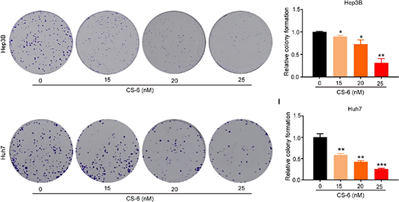
Gamabufotalin (CS-6; 15, 20, 25 nM; 48 h) inhibits the proliferation of Hep3B and Huh7 cells in a dose-dependent manner.
Gamabufotalin purchased from MedChemExpress. Usage Cited in: J Nat Prod. 2023 Apr 28;86(4):966-978. [Abstract]
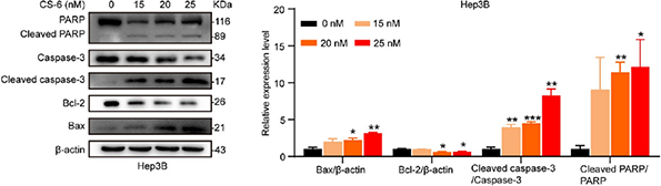
Gamabufotalin (CS-6; 15, 20, 25 nM; 48 h) significantly increases the expression of cleaved PARP/PARP, caspase-3/caspase-3, and Bax and decreases the expression of Bcl-2 in Hep3B cells.
Gamabufotalin purchased from MedChemExpress. Usage Cited in: J Nat Prod. 2023 Apr 28;86(4):966-978. [Abstract]
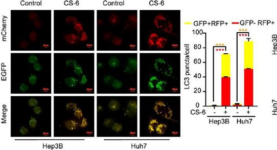
Gamabufotalin (CS-6; 25 nM; 15 min) significantly increases the formation of yellow-spotted autophagosomes (GFP+RFP+) and red-spotted autolysosomes (GFP-RFP+) in Hep3B and Huh7 cells.


Solvent & Solubility
In Vitro:
DMSO : 50 mg/mL (124.22 mM; Need ultrasonic; Hygroscopic DMSO has a significant impact on the solubility of product, please use newly opened DMSO)
Please refer to the solubility information to select the appropriate solvent. Once prepared, please aliquot and store the solution to prevent product inactivation from repeated freeze-thaw cycles.
Storage method and period of stock solution: -80°C, 2 years; -20°C, 1 year. When stored at -80°C, please use it within 2 years. When stored at -20°C, please use it within 1 year.
Please refer to the solubility information to select the appropriate solvent. Once prepared, please aliquot and store the solution to prevent product inactivation from repeated freeze-thaw cycles.
Storage method and period of stock solution: -80°C, 2 years; -20°C, 1 year. When stored at -80°C, please use it within 2 years. When stored at -20°C, please use it within 1 year.
Concentration (start) × Volume (start) = Concentration (final) × Volume (final)
In Vivo:
Select the appropriate dissolution method based on your experimental animal and administration route.
- For the following dissolution methods, please ensure to first prepare a clear stock solution using an In Vitro approach and then sequentially add co-solvents:
- To ensure reliable experimental results, the clarified stock solution can be appropriately stored based on storage conditions. As for the working solution for In Vivo experiments, it is recommended to prepare freshly and use it on the same day.
- The percentages shown for the solvents indicate their volumetric ratio in the final prepared solution. If precipitation or phase separation occurs during preparation, heat and/or sonication can be used to aid dissolution.
Add each solvent one by one: 10% DMSO 40% PEG300 5% Tween-80 45% Saline
Solubility: ≥ 2.5 mg/mL (6.21 mM); Clear solution
This protocol yields a clear solution of ≥ 2.5 mg/mL (saturation unknown).
Taking 1 mL working solution as an example, add 100 μL DMSO stock solution (25.0 mg/mL) to 400 μL PEG300, and mix evenly; then add 50 μL Tween-80 and mix evenly; then add 450 μL Saline to adjust the volume to 1 mL.
Preparation of Saline: Dissolve 0.9 g sodium chloride in ddH₂O and dilute to 100 mL to obtain a clear Saline solution.
Add each solvent one by one: 10% DMSO 90% (20% SBE-β-CD in Saline)
Solubility: ≥ 2.5 mg/mL (6.21 mM); Clear solution
This protocol yields a clear solution of ≥ 2.5 mg/mL (saturation unknown).
Taking 1 mL working solution as an example, add 100 μL DMSO stock solution (25.0 mg/mL) to 900 μL 20% SBE-β-CD in Saline, and mix evenly.
Preparation of 20% SBE-β-CD in Saline (4°C, storage for one week): 2 g SBE-β-CD powder is dissolved in 10 mL Saline, completely dissolve until clear.
In Vivo Dissolution Calculator
Please enter the basic information of animal experiments:
-
-
-
-
Recommended: Prepare an additional quantity of animals to account for potential losses during experiments.
Please enter your animal formula composition:
-
%DMSO +
Recommended: Keep the proportion of DMSO in working solution below 2% if your animal is weak.
-
%+
-
+%Tween-80 + +
-
%Saline +
The co-solvents required include: DMSO, . All of co-solvents are available by MedChemExpress (MCE). , Tween 80. All of co-solvents are available by MedChemExpress (MCE).
Working solution concentration: 0.22 mg/mL
Method for preparing stock solution: mg drug dissolved in μL DMSO. Stock solution concentration: mg/mL.
1. Take μL DMSO stock solution;
2. Add μL .
μL , mix evenly;
3. Then add μL Tween 80, mix evenly;
4. Then add μL
Please ensure that the stock solution in the first step is dissolved to a clear state, and add co-solvents in sequence. You can use ultrasonic heating (ultrasonic cleaner, recommended frequency 20-40 kHz), vortexing, etc. to assist dissolution.

Protocols
-
Human pluripotent stem cell endothelial-cell differentiation
Human pluripotent stem cell endothelial differentiation is based on stepwise developmental patterning: early activation of WNT/GSK3β inhibition promotes mesodermal or vascular progenitor entry, followed by endothelial specification using VEGF-related signaling, BMP4, FGF2, Notch modulation, or cAMP depending on the published protocol. Endothelial differentiation is read out by acquisition of CD31, CD34, VE-cadherin/CD144, KDR/VEGFR2, vWF, Tie2, NOS3, acetylated LDL uptake, tube/network formation, barrier function, and in vivo vessel-forming capacity where tested.
-
Vascular/Branching Fractal Analysis
Vascular/branching fractal analysis quantifies the geometric complexity of vessel trees or vascular networks from segmented 2D images, commonly by converting vessels into binary and/or skeletonized maps and estimating fractal dimension using box-counting or related approaches. Fractal dimension is interpreted as an image-derived readout of vascular branching complexity, space filling, or density, and has been applied to retinal photographs, fluorescein angiography, OCT angiography, capillary perfusion maps, and in vitro Matrigel angiogenesis networks. The assay readout is generated from vessel-positive pixels after image preprocessing, vessel segmentation, binarization, and optional skeletonization; reported outputs include fractal dimension, vessel density, branchpoint density, endpoint density, vessel length density, tortuosity, and generation-based branching metrics when VESGEN-style analysis is used. The biological interpretation is limited to quantitative vascular patterning and s
-
Protocol for Cell Counting and Cell Density Analysis
Cell counting and cell-density analysis estimate the number of cells in a known volume or field area. Manual hemocytometer counting uses a chamber of defined geometry to convert counted cells into cells/mL, while automated counters and image-analysis workflows detect cell objects from optical, brightfield, fluorescence, impedance, or digital-image features. Trypan blue viability counting is based on dye exclusion: viable cells with intact membranes exclude dye, while non-viable cells with compromised membranes stain blue. The readout is total cell density, viable-cell density, dead-cell density, and percent viability. Cell density can also be estimated from microscopy images by counting objects per image area, from flow cytometry using calibrated volume or reference particles, or from in situ microscopy in bioreactors after calibration against reference methods such as hemocytometer or flow cytometry.
-
Somatic Cell Culture
A method of simulating the in vivo environment in vitro to maintain the cell growth, differentation and main functions.
-
Endothelial Tube Formation Assay
Endothelial tube formation assay evaluates the ability of endothelial cells to attach, migrate, align, and organize into capillary-like networks when cultured on gelled basement membrane extract or Matrigel; the readout is the morphology and quantity of tube-like networks, which reflects an in vitro endothelial morphogenesis step related to angiogenesis. Basement membrane extract/Matrigel provides laminin-rich extracellular matrix cues that support endothelial differentiation into capillary-like structures, but it can contain biologically active growth factors, so growth-factor-reduced matrix is preferred when testing defined angiogenic stimulators or inhibitors.
-
CCK-8/WST-8 Cell Proliferation Assay
The CCK-8/WST-8 assay is based on the reduction of the water-soluble tetrazolium salt WST-8 to a water-soluble formazan product by cellular dehydrogenases in metabolically active cells, where the generated formazan amount is proportional to the number of living cells and is quantified by measuring absorbance in the visible range, providing a colorimetric readout for cell viability and proliferation assessment. This class of tetrazolium-based assays improves upon earlier MTT-based systems by producing a water-soluble formazan, eliminating the need for organic solubilization steps and enabling direct spectrophotometric measurement in culture medium.
-
Cell Counting-Based Growth Curve Assay
Cell counting-based growth curve assays quantify cell proliferation by directly measuring changes in viable cell number over time using manual or automated counting methods such as hemocytometer-based counting or instrument-assisted cell enumeration, enabling construction of growth curves that reflect population expansion dynamics in response to culture conditions. A widely used approach is trypan blue exclusion with hemocytometer counting, where membrane-compromised (non-viable) cells take up the dye, allowing discrimination between viable and non-viable cells while simultaneously enabling total cell number quantification. Repeated sampling across time points allows estimation of proliferation rate, growth phases, and comparative growth kinetics between experimental conditions.
-
Research Protocol for Inflammation-related Diseases
The NLRP3 inflammasome is a cytosolic innate immune signaling platform that integrates priming signals and danger-signal activation to promote caspase-1 activation, maturation of IL-1β and IL-18, and gasdermin D-mediated pyroptotic cell death. The core experimental logic is to determine whether inflammatory disease phenotypes are driven by increased NLRP3 expression, ASC-containing inflammasome assembly, caspase-1 cleavage, GSDMD cleavage, and extracellular release of IL-1β/IL-18 rather than by nonspecific cell injury alone. The pathway is strongly linked to inflammation-related disease phenotypes because monosodium urate crystals activate NALP3/NLRP3 inflammasome signaling in gout-like crystal inflammation, cholesterol crystals activate NLRP3 inflammasomes in atherogenesis models, and DSS-induced intestinal inflammation has been reported to involve NLRP3 inflammasome activity. However, experimental colitis studies also show context-dependent protective effects of NLRP3 inflammasome co
-
MTT Cell Proliferation Assay
The MTT assay is a colorimetric endpoint assay for estimating viable cell number, cell growth, cytotoxicity, or cell activation in cultured mammalian cells. Living cells reduce the yellow tetrazolium salt MTT into purple/blue formazan, while dead cells do not generate the same signal; the resulting color can be quantified with a multiwell spectrophotometer. MTT reduction is commonly interpreted as a readout of metabolic activity that often correlates with viable cell number, but it should not be treated as a direct cell-counting method unless the assay is optimized for the cell type and experimental condition. Studies show that MTT reduction can involve mitochondrial and non-mitochondrial reducing systems, and formazan may accumulate in intracellular lipid droplets rather than simply marking mitochondria.
-
Endothelial Cell Migration/Angiogenic Sprouting Assay
Endothelial cell migration and angiogenic sprouting assays are in vitro (and partially ex vivo-adapted) functional models that quantify the ability of endothelial cells to undergo coordinated migration, extracellular matrix invasion, and multicellular organization into capillary-like sprouts in response to pro-angiogenic stimuli such as VEGF, bFGF, or conditioned microenvironments. These assays are used to model early angiogenic events including tip-cell formation, directional migration, and lumen-like sprout extension, which collectively reflect angiogenic activation and vascular morphogenesis processes observed in vivo.
-
Cell Viability Determination by MTT Colorimetric Assay
The following protocol uses the MTT colorimetric assay as a classic literature-established method for assessing cell viability/metabolic activity in cultured mammalian cells. MTT[3-(4,5-dimethylthiazol-2-yl)-2,5-diphenyltetrazolium bromide] is reduced by metabolically active cells to a colored formazan product; the amount of formazan is quantified spectrophotometrically and provides an indirect measure of metabolically active viable cells. Importantly, MTT reduction reflects cellular oxidoreductase/metabolic activity rather than an absolute direct count of living cells, so changes in cellular metabolism can alter the signal independently of cell number.


Purity & Documentation
-
Data Sheet (284 KB)
-
SDS (251 KB)
- English - EN (251 KB)
- Français - FR (251 KB)
- Deutsch - DE (251 KB)
- Norwegian - NO (251 KB)
- Español - ES (251 KB)
- Swedish - SV (251 KB)
- Italian - IT (251 KB)
- Korean - KR (251 KB)
- Portuguese - PT (251 KB)
-
Handling Instructions (2659 KB)
References
[1]. Yu Z, et al. Gamabufotalin, a bufadienolide compound from toad venom, suppresses COX-2 expression through targeting IKKβ/NF-κB signaling pathway in lung cancer cells. Mol Cancer. 2014 Aug 31;13:203. [Content Brief]
[2]. Dong Y, et al. Bufadienolide compounds sensitize human breast cancer cells to TRAIL-induced apoptosis via inhibition of STAT3/Mcl-1 pathway. Apoptosis. 2011 Apr;16(4):394-403. [Content Brief]
[3]. Lan YL, et al. Gamabufotalin induces a negative feedback loop connecting ATP1A3 expression and the AQP4 pathway to promote temozolomide sensitivity in glioblastoma cells by targeting the amino acid Thr794. Cell Prolif. 2020;53(1):e12732. [Content Brief]
[4]. Tang N, et al. Gamabufotalin, a major derivative of bufadienolide, inhibits VEGF-induced angiogenesis by suppressing VEGFR-2 signaling pathway. Oncotarget. 2016;7(3):3533-3547. [Content Brief]


Complete Stock Solution Preparation Table
Please refer to the solubility information to select the appropriate solvent. Once prepared, please aliquot and store the solution to prevent product inactivation from repeated freeze-thaw cycles.
Storage method and period of stock solution: -80°C, 2 years; -20°C, 1 year. When stored at -80°C, please use it within 2 years. When stored at -20°C, please use it within 1 year.
| Optional Solvent | Concentration Solvent Mass | 1 mg | 5 mg | 10 mg | 25 mg |
|---|---|---|---|---|---|
| DMSO | 1 mM | 2.4843 mL | 12.4217 mL | 24.8435 mL | 62.1087 mL |
| 5 mM | 0.4969 mL | 2.4843 mL | 4.9687 mL | 12.4217 mL | |
| 10 mM | 0.2484 mL | 1.2422 mL | 2.4843 mL | 6.2109 mL | |
| 15 mM | 0.1656 mL | 0.8281 mL | 1.6562 mL | 4.1406 mL | |
| 20 mM | 0.1242 mL | 0.6211 mL | 1.2422 mL | 3.1054 mL | |
| 25 mM | 0.0994 mL | 0.4969 mL | 0.9937 mL | 2.4843 mL | |
| 30 mM | 0.0828 mL | 0.4141 mL | 0.8281 mL | 2.0703 mL | |
| 40 mM | 0.0621 mL | 0.3105 mL | 0.6211 mL | 1.5527 mL | |
| 50 mM | 0.0497 mL | 0.2484 mL | 0.4969 mL | 1.2422 mL | |
| 60 mM | 0.0414 mL | 0.2070 mL | 0.4141 mL | 1.0351 mL | |
| 80 mM | 0.0311 mL | 0.1553 mL | 0.3105 mL | 0.7764 mL | |
| 100 mM | 0.0248 mL | 0.1242 mL | 0.2484 mL | 0.6211 mL |


Gamabufotalin Related Classifications
HY-N0883 Related Classifications
 Powered by Bioz