



Based on 1 Customer Validation
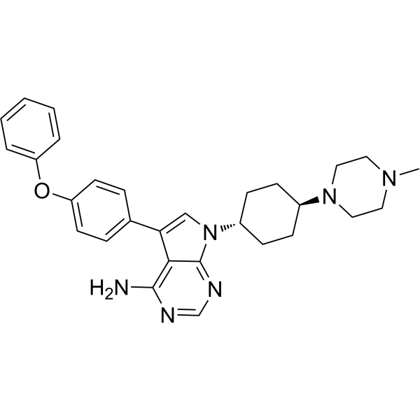
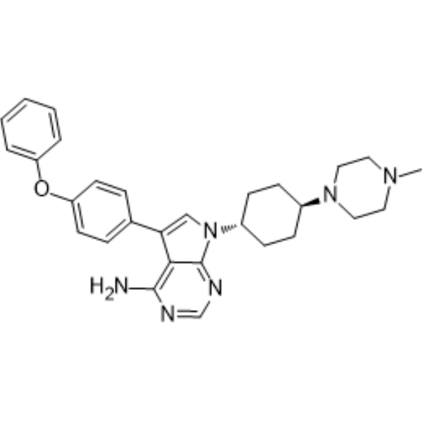
A 419259 GMP is the GMP grade A 419259 (HY-15764), inducing cell apoptosis. GMP-grade small molecules can be used as auxiliary reagents in cell therapy. A 419259 (RK-20449) is a broad-spectrum pyrrole-pyrimidine inhibitor targeting Src, Lck, and Lyn with IC50s of 9 nM, <3 nM, and <3 nM, respectively.
For research use only. We do not sell to patients.
- CAS No.: 364042-47-7
- Formula: C29H34N6O
- Molecular Weight:482.63
-
Storage:
Please store the product under the recommended conditions in the Certificate of Analysis.

To place orders, for customer services and technical support, please contact: MedChemExpress USA
Tel: 609-228-6898 E-mail: [email protected] [email protected]


-
 Biological Activity
-
Chemical Information
-
Solvent & Solubility
- Protocols
- Purity & Documentation
- References
-
 Help & FAQs


-
HY-15764
A 419259Src Inhibitor -
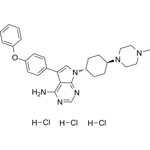
HY-15764A
A 419259 trihydrochlorideSrc Inhibitor -
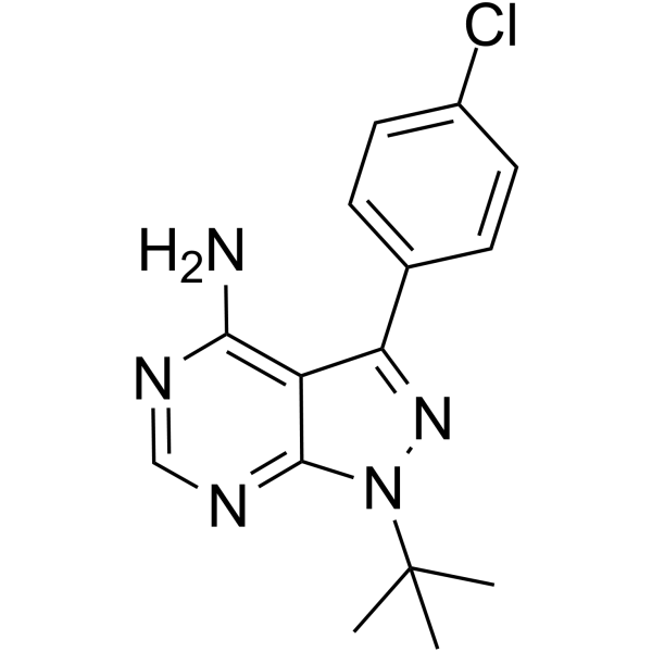
HY-13805
PP2Src Inhibitor -
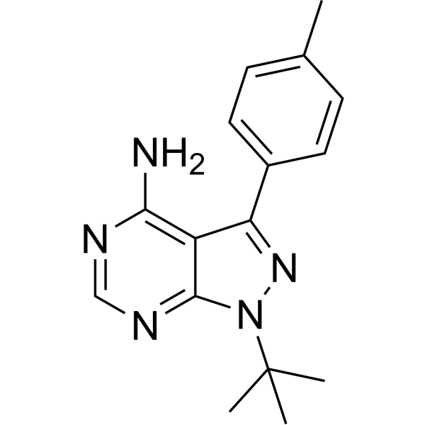
HY-13804
PP1Src Inhibitor -
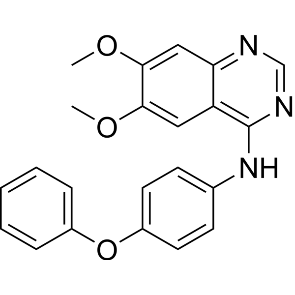
HY-101053
Src Inhibitor 1Src Inhibitor -
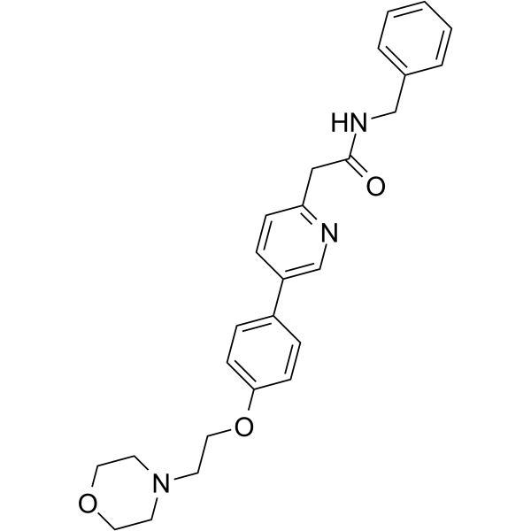
HY-10340
TirbanibulinSrc Inhibitor -
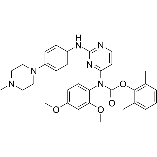
HY-12299
WH-4-023Src Inhibitor -
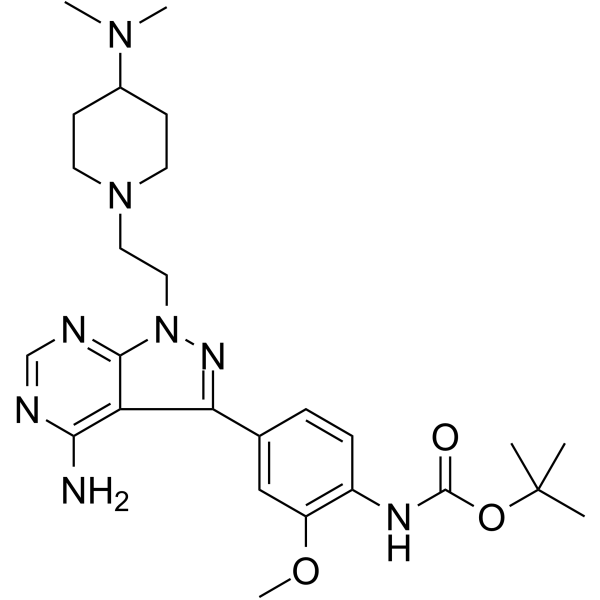
HY-112096
eCF506YES1/SRC inhibitor -
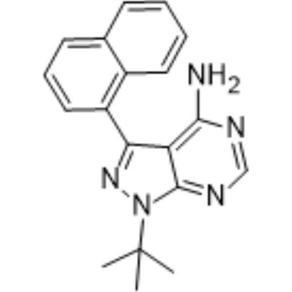
HY-13941
1-Naphthyl PP1Src Inhibitor -
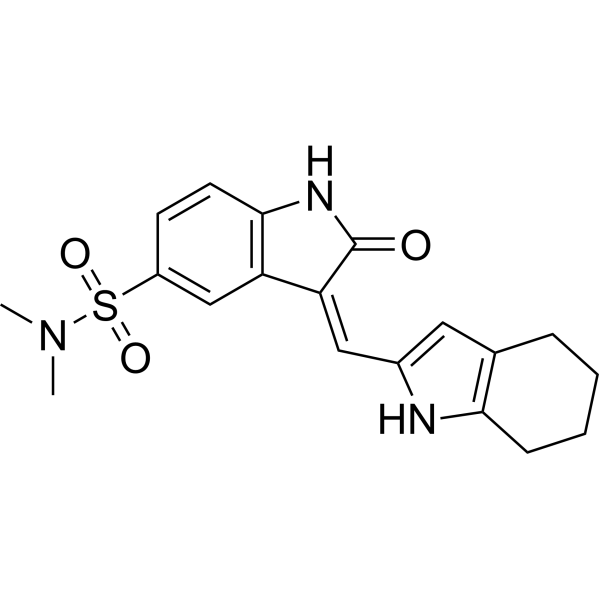
HY-B0789
SU6656Src Inhibitor -
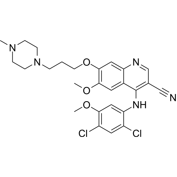
HY-10158
BosutinibSrc/Abl Inhibitor -
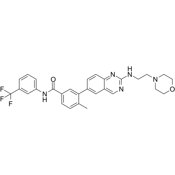
HY-18303
AMG-47aLck Inhibitor


Biological Activity
Description
IC50 & Target
IC50: 9 nM (Src), <3 nM (Lck), <3 nM (Lyn), 3 μM (Abl)[1]
Cellular Effect
|
Cell Line

|
Type | Value | Description | References |
|---|---|---|---|---|
| MV4-11 | GI50 |
40 nM
Compound: RK-20449
|
Growth inhibition of human MV4-11 cells after 3 days by Alamar blue assay
Growth inhibition of human MV4-11 cells after 3 days by Alamar blue assay
|
[PMID: 29037944] |
In Vitro
A419259 is a second-generation pyrrolopyrimidine that blocks proliferation and induces apoptosis in CML cell lines. It induces apoptosis in K-562 cells and also inhibits Meg-01 proliferation (IC50=0.1 μM)[1].
In the absence of IL-3, A-419259 strongly inhibits DAGM/Bcr-Abl cell proliferation (IC50=0.1-0.3 μM)[1].
A-419259 also inhibits overall SFK activity in CML cell lines and blocks Src kinase activation (IC50=0.1-0.3 μM)[1].
A 419259 GMP (1 μM; 16 h) inhibits endogenous SFK (c-Src and Lck) activity, thereby inhibiting Src-driven differentiation of mES cells toward primitive ectoderm-like cells[2].
A 419259 GMP (0.3, 1 μM; 5 days) has no effect on undifferentiated colony morphology of hES cells grown in mTeSR medium[2].
MedChemExpress (MCE) has not independently confirmed the accuracy of these methods. They are for reference only.


Chemical Information
-
CAS No. 364042-47-7
-
Appearance Solid
-
Molecular Weight 482.63
-
Formula C29H34N6O
-
Color White to light yellow
-
SMILES
NC1=C2C(N([C@H]3CC[C@H](N4CCN(C)CC4)CC3)C=C2C5=CC=C(OC6=CC=CC=C6)C=C5)=NC=N1
-
Synonyms
RK-20449 (GMP)
-
Shipping
Room temperature in continental US; may vary elsewhere.
-
Storage
Please store the product under the recommended conditions in the Certificate of Analysis.


Solvent & Solubility
In Vitro:
DMSO : 100 mg/mL (207.20 mM; Need ultrasonic and warming; Hygroscopic DMSO has a significant impact on the solubility of product, please use newly opened DMSO)
Concentration (start) × Volume (start) = Concentration (final) × Volume (final)


Purity & Documentation
-
Data Sheet (275 KB)
-
SDS (251 KB)
- English - EN (251 KB)
- Français - FR (251 KB)
- Deutsch - DE (251 KB)
- Norwegian - NO (251 KB)
- Español - ES (251 KB)
- Swedish - SV (251 KB)
- Italian - IT (251 KB)
- Korean - KR (251 KB)
- Portuguese - PT (251 KB)
-
Handling Instructions (2659 KB)
References
[1]. Wilson MB, et al. Selective pyrrolo-pyrimidine inhibitors reveal a necessary role for Src family kinases in Bcr-Abl signal transduction and oncogenesis. Oncogene. 2002 Nov 21;21(53):8075-88. [Content Brief]
[2]. Zhang X, et al. Src-family tyrosine kinase activities are essential for differentiation of human embryonic stem cells. Stem Cell Res. 2014 Nov;13(3 Pt A):379-89. [Content Brief]


Complete Stock Solution Preparation Table
| Optional Solvent | Concentration Solvent Mass | 1 mg | 5 mg | 10 mg | 25 mg |
|---|---|---|---|---|---|
| DMSO | 1 mM | 2.0720 mL | 10.3599 mL | 20.7198 mL | 51.7995 mL |
| 5 mM | 0.4144 mL | 2.0720 mL | 4.1440 mL | 10.3599 mL | |
| 10 mM | 0.2072 mL | 1.0360 mL | 2.0720 mL | 5.1800 mL | |
| 15 mM | 0.1381 mL | 0.6907 mL | 1.3813 mL | 3.4533 mL | |
| 20 mM | 0.1036 mL | 0.5180 mL | 1.0360 mL | 2.5900 mL | |
| 25 mM | 0.0829 mL | 0.4144 mL | 0.8288 mL | 2.0720 mL | |
| 30 mM | 0.0691 mL | 0.3453 mL | 0.6907 mL | 1.7267 mL | |
| 40 mM | 0.0518 mL | 0.2590 mL | 0.5180 mL | 1.2950 mL | |
| 50 mM | 0.0414 mL | 0.2072 mL | 0.4144 mL | 1.0360 mL | |
| 60 mM | 0.0345 mL | 0.1727 mL | 0.3453 mL | 0.8633 mL | |
| 80 mM | 0.0259 mL | 0.1295 mL | 0.2590 mL | 0.6475 mL | |
| 100 mM | 0.0207 mL | 0.1036 mL | 0.2072 mL | 0.5180 mL |


A 419259 (GMP) Related Classifications
HY-15764G Related Classifications
 Powered by Bioz