



Alemtuzumab

Based on 1 Customer Validation
Alemtuzumab (Campath-IH) is a humanized monoclonal antibody against CD52. Alemtuzumab does not cross-react with murine CD52. Alemtuzumab selectively targets the CD52 antigen to induce profound lymphocyte depletion, followed by recovery of T and B cells with regulatory phenotypes. Alemtuzumab is capable of complement-dependent cytotoxicity and antibody-dependent cell-mediated cytotoxicity (ADCC), as well as induction of apoptosis. Alemtuzumab has the potential for B-cell chronic lymphocytic leukaemia research.
연구목적의 판매만을 진행합니다. 환자를 대상으로 한 판매는 하지 않습니다.
- Purity : 99.719%
- CAS No.: 216503-57-0
- 분자량:146.06 kDa
-
보관:
Please store the product under the recommended conditions in the Certificate of Analysis.


To place orders, for customer services and technical support, please contact: MedChemExpress USA
Tel: 609-228-6898 E-mail: [email protected] [email protected]


-
 Biological Activity
-
Chemical Information
- Protocol
- 순도&문서
- References
-
 Help & FAQs


-

HY-P9907
TrastuzumabHER2 Blocking Antibody -

HY-P9906
BevacizumabVEGF-A Inhibitor -

HY-P9915
DaratumumabAnti-CD38 Monoclonal Antibody -

HY-P9970
InfliximabTNF-α Inhibitor -

HY-P9905
CetuximabEGFR Inhibitor -

HY-P9913
RituximabCD20 Inhibitor -

HY-P9908
AdalimumabTNF-α Inhibitor -

HY-P99001
Human IgG1 kappa, Isotype ControlIgG1κ Isotype Control -

HY-108847
EtanerceptTNF Inhibitor -

HY-P9975
TheralizumabCD28 Superagonist -

HY-P9976
IsatuximabAnti-CD38 Monoclonal Antibody -

HY-P99187
BococizumabAnti-human PCSK9 Antibody


Biological Activity
제품 설명
Isotype
Human IgG1 kappa
Recommend Isotype Controls
Species Reactivity
Human
IC50 & Target
CD52
In Vivo
MedChemExpress (MCE) has not independently confirmed the accuracy of these methods. They are for reference only.
-
Animal Model:hCD52 transgenic mice[2]
-
Dosage:0.5-5 mg/kg
-
Administration:IP; a single dose
-
Result:Resulted in the dose-dependent depletion of B and T lymphocytes in the peripheral blood and lymphoid organs.
Clinical Trial
| NCT Number | Sponsor | Condition | Start Date |
Phase

|
|---|---|---|---|---|
| NCT01329991 | 2011-05 | PHASE1 |
Gene ID
Accession
Conjugated
Unconjugated
Reconsititution
The product can be reconstituted/diluted with sterile PBS or saline.
Format
-
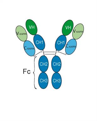
Human IgG1 kappa
신청
ELISA, FACS, Functional assay
Verified Bioactivity
-
-
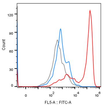
 Immobilized CD52 Protein, Human (His, HY-P72123) can bind Alemtuzumab. The EC50 for this effect is 1.1 ng/mL.


Chemical Information
-
CAS No. 216503-57-0
-
Appearance Liquid
-
분자량 146.06 kDa
-
Color Colorless to light yellow
-
SMILES
[Alemtuzumab]
-
Synonyms
Campath-IH
-
선적
Shipping with dry ice.
-
Formulation
Please refer to the lot-specific COA for specific buffer information.
-
보관
Please store the product under the recommended conditions in the Certificate of Analysis.


순도&문서
-
Data Sheet (259 KB)
-
SDS (251 KB)
- English - EN (251 KB)
- Français - FR (251 KB)
- Deutsch - DE (251 KB)
- Norwegian - NO (251 KB)
- Español - ES (251 KB)
- Swedish - SV (251 KB)
- Italian - IT (251 KB)
- Korean - KR (251 KB)
- Portuguese - PT (251 KB)
-
Inhibitory Antibodies User Guide (603 KB)
References
[1]. Kashani N, et al. Immune Regulatory Cell Bias Following Alemtuzumab Treatment in Relapsing-Remitting Multiple Sclerosis. Front Immunol. 2021;12:706278. Published 2021 Oct 28. [Content Brief]
[2]. Yanping Hu, et al. Investigation of the mechanism of action of alemtuzumab in a human CD52 transgenic mouse model. Immunology. 2009 Oct;128(2):260-70. [Content Brief]

Calculators
Concentration (start) × Volume (start) = Concentration (final) × Volume (final)


Alemtuzumab Related Classifications
HY-P9948 Related Classifications
 Powered by Bioz