



Anti-Mouse VEGFR-2 Antibody (DC101)

Based on 1 Customer Validation
Anti-Mouse VEGFR-2 Antibody (DC101) is a rat anti-mouse VEGFR2 monoclonal antibody. Anti-Mouse VEGFR-2 Antibody (DC101) inhibits tumor angiogenesis by blocking the binding of VEGF and VEGFR2. Anti-Mouse VEGFR-2 Antibody (DC101) promotes immune cell infiltration and induces tumor cell apoptosis. Anti-Mouse VEGFR-2 Antibody (DC101) can be used for researches on various types of cancer such as melanoma, lung cancer and breast cancer .
For research use only. We do not sell to patients.
- Purity : 99%
- Molecular Weight:150 kDa
-
Storage:
Please store the product under the recommended conditions in the Certificate of Analysis.

To place orders, for customer services and technical support, please contact: MedChemExpress USA
Tel: 609-228-6898 E-mail: [email protected] [email protected]


-
 Biological Activity
-
Chemical Information
- Protocols
- Purity & Documentation
- References
-
 Help & FAQs




All VEGFR Isoforms
More

Biological Activity
Description
Isotype
Rat IgG1 kappa
Recommend Isotype Controls
Species Reactivity
Mouse
|
VEGFR2 |
In Vivo
Anti-Mouse VEGFR-2 Antibody (DC101) (10 mg/kg, i.p., on day 2, 5 and 8) significantly inhibits tumor growth without significant toxicity in female BALB/c mice bearing H22 tumors[2].
Anti-Mouse VEGFR-2 Antibody (DC101) (40 mg/kg, i.g., once every three days, for 4 weeks) significantly inhibits tumor growth and increases immune cell infiltration in female C57BL/6 mice bearing B16-OVA tumors[3].
Anti-Mouse VEGFR-2 Antibody (DC101) (40 mg/kg, i.p., three times every week, for 21 days) significantly inhibits wound induced tumors in transgenic mice expressing CER of HPV8[4].
Anti-Mouse VEGFR-2 Antibody (DC101) (0.8 mg/mouse, i.p., twice weekly, for 2-3 weeks) inhibits tumor growth and increases T cell infiltration in FVB mice and Neu-N mice bearing NT2.5 tumors[5].
Anti-Mouse VEGFR-2 Antibody (DC101) (100 or 400 μg, i.p., once every three days, for 21 days) significantly reduces angiogenesis and endothelial cell count in female C57BL/6 mice injected with bFGF and VEGF[6].
Anti-Mouse VEGFR-2 Antibody (DC101) (800 μg, i.p., once every three days) significantly inhibits the growth of various tumors[6].
MedChemExpress (MCE) has not independently confirmed the accuracy of these methods. They are for reference only.
-
Animal Model:1×10⁶ 253J B-V cells injected male athymic BALB/c nude mice (8-12 weeks)[1]
-
Dosage:1 mg/dose, combined with Paclitaxel (HY-B0015) 10 mg/kg
-
Administration:Intraperitoneal injection (i.p.), twice weekly for 4 weeks
-
Result:Significantly prolonged the survival period of mice.
Significantly reduced tumor microvascular density.
Enhanced apoptosis of tumor cells and endothelial cells.
-
Animal Model:2×10⁶ H22 cells injected female BALB/c mice (6-8 weeks, 20 g)[2]
-
Dosage:10 mg/kg, combined with CA4 NPs (45 mg/kg) and anti-PD-1 (100 μg/mouse)
-
Administration:Intraperitoneal injection (i.p.) on day 2, 5 and 8
-
Result:Increased the number of CD8+ T cells within the tumor.
Significantly improved vascular perfusion.
Reduced the level of IFN-γ and TNF-α.
Had good safety in vivo.
-
Animal Model:5×10⁵ B16-OVA cells injected female C57BL/6 mice (6-8 weeks)[3]
-
Dosage:40 mg/kg
-
Administration:Oral gavage (i.g.), once every three days for 4 weeks
-
Result:Significantly inhibited tumor volume.
Increased infiltration of CD3+ and CD8+ T cells.
Increased infiltration of B cells (CD19+) and dendritic cells (CD11c+).
Enhanced the expression of IFN-γ, perforin, and granzyme B in CD8+ T cells.
Reduced vascular density and increased perivascular cell coverage.
Significantly increased the density of PNAd+HEV and promoted the formation of tertiary lymphoid structures (TLS).
Upregulated PD-L1 expression in tumor cells and CD45+ immune cells, as well as PD-1 expression in CD3+ T cells.
-
Animal Model:Transgenic mice expressing CER of HPV8[4]
-
Dosage:40 mg/kg
-
Administration:Intraperitoneal injection (i.p.), three times every week for 21 days
-
Result:Reduced Ki-67+ proliferating cells and CD31+ vascular density.
-
Animal Model:5×10⁶ NT2.5 cells injected FVB mice and Neu-N mice[5]
-
Dosage:0.8 mg/mouse
-
Administration:Intraperitoneal injection (i.p.), twice weekly for 2-3 weeks
-
Result:Increased CD4+ and CD8+ T cell infiltration.
Significantly inhibited tumor volume.
Decreased angiogenesis and increased tumor cell apoptosis.
-
Animal Model:Female C57BL/6 injected with bFGF (500 ng) and VEGF (10 μg)[6]
-
Dosage:100 or 400 μg
-
Administration:Intraperitoneal injection (i.p.), once every three days for 21 days
-
Result:Significantly reduced angiogenesis and endothelial cell count in matrigel plug model.
-
Animal Model:2×10⁶ Lewis lung cells or 1×10⁵ B16 cells injected female C57BL/6 mice (5-6 weeks), 1×10⁵ 4T1 cells injected female BALB/c mice (5-6 weeks), 2×10⁶ A431, SK-RC-29, BxPC-3 or GBM-18 cells injected female athymic nu/nu mice (5-6 weeks)[6]
-
Dosage:800 μg
-
Administration:Intraperitoneal injection (i.p.), once every three days
-
Result:Significantly inhibited tumor growth.
Gene ID
Accession
Conjugated
Unconjugated
Reconsititution
The product can be reconstituted/diluted with sterile PBS or saline.
Application
ELISA, FACS, Functional assay, Research in vivo
Verified Bioactivity
-
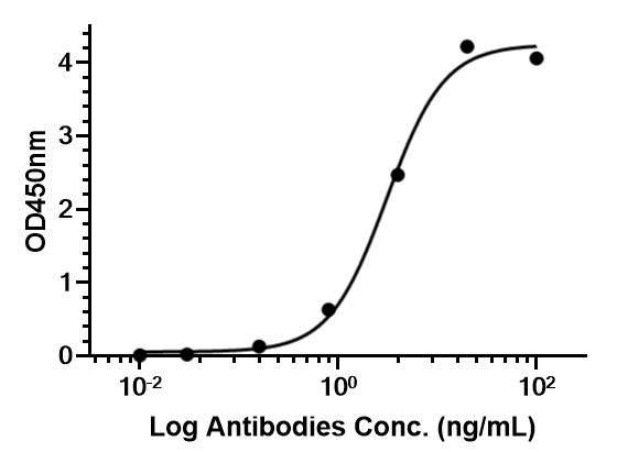 Immobilized VEGFR-2/Flk-1/KDR Fc Chimera Protein, Mouse can bind Anti-Mouse VEGFR-2 Antibody (DC101). The EC50 for this effect is 177.8 ng/mL.


Chemical Information
-
Appearance Liquid
-
Molecular Weight 150 kDa
-
Color Colorless to light yellow
-
SMILES
[Anti-Mouse VEGFR-2 Antibody (DC101)]
-
Formulation
Please refer to the lot-specific COA for specific buffer information.
-
Storage
Please store the product under the recommended conditions in the Certificate of Analysis.

Protocols
-
Multiplex immunofluorescence IHC
Multiplex immunofluorescence IHC detects multiple protein biomarkers in one tissue section by sequential antibody staining, HRP-mediated tyramide fluorophore deposition, heat-mediated antibody stripping, nuclear counterstaining, multispectral imaging, spectral unmixing, and digital cell phenotyping; TSA deposits fluorophore near the antigen so the fluorescence signal remains after primary and secondary antibodies are removed, enabling repeated staining cycles, including with antibodies from the same host species. Classic FFPE tumor immune-profiling applications use panels such as CD3, CD8, CD68/CD163, FOXP3, PD-1, PD-L1, pancytokeratin, Ki67, and DAPI to identify tumor cells, immune-cell subsets, checkpoint-marker expression, co-expression phenotypes, cell density, and spatial relationships in the tumor microenvironment.
-
Human pluripotent stem cell endothelial-cell differentiation
Human pluripotent stem cell endothelial differentiation is based on stepwise developmental patterning: early activation of WNT/GSK3β inhibition promotes mesodermal or vascular progenitor entry, followed by endothelial specification using VEGF-related signaling, BMP4, FGF2, Notch modulation, or cAMP depending on the published protocol. Endothelial differentiation is read out by acquisition of CD31, CD34, VE-cadherin/CD144, KDR/VEGFR2, vWF, Tie2, NOS3, acetylated LDL uptake, tube/network formation, barrier function, and in vivo vessel-forming capacity where tested.
-
Apoptosis
Apoptosis, also called programmed cell death, is generally characterized by distinct morphological characteristics.
-
TUNEL staining for apoptotic DNA fragmentation
TUNEL staining detects DNA strand breaks by using terminal deoxynucleotidyl transferase to add labeled nucleotides to exposed 3′-OH DNA termini, generating either microscopic staining in fixed cells or tissue sections, or fluorescence/cytometric signal in cell suspensions. TUNEL positivity reflects DNA fragmentation but should not be interpreted alone as definitive apoptosis, because TUNEL can also label necrotic, autolytic, mechanically damaged, or DNA-repair-associated DNA breaks.
-
Vascular/Branching Fractal Analysis
Vascular/branching fractal analysis quantifies the geometric complexity of vessel trees or vascular networks from segmented 2D images, commonly by converting vessels into binary and/or skeletonized maps and estimating fractal dimension using box-counting or related approaches. Fractal dimension is interpreted as an image-derived readout of vascular branching complexity, space filling, or density, and has been applied to retinal photographs, fluorescein angiography, OCT angiography, capillary perfusion maps, and in vitro Matrigel angiogenesis networks. The assay readout is generated from vessel-positive pixels after image preprocessing, vessel segmentation, binarization, and optional skeletonization; reported outputs include fractal dimension, vessel density, branchpoint density, endpoint density, vessel length density, tortuosity, and generation-based branching metrics when VESGEN-style analysis is used. The biological interpretation is limited to quantitative vascular patterning and s
-
Annexin V plus membrane-impermeant dye apoptosis staining
Annexin V-based apoptosis assays rely on the detection of phosphatidylserine (PS) externalization from the inner leaflet of the plasma membrane to the outer leaflet, an early biochemical hallmark of apoptosis. Fluorescently labeled Annexin V binds PS in a calcium-dependent manner, enabling identification of early apoptotic cells by flow cytometry or fluorescence microscopy. When combined with a membrane-impermeant DNA-binding dye (e. g. , propidium iodide), this approach allows discrimination between viable (Annexin V−/dye−), early apoptotic (Annexin V+/dye−), and late apoptotic or necrotic (Annexin V+/dye+) cell populations by assessing membrane integrity and PS exposure.
-
Endothelial Tube Formation Assay
Endothelial tube formation assay evaluates the ability of endothelial cells to attach, migrate, align, and organize into capillary-like networks when cultured on gelled basement membrane extract or Matrigel; the readout is the morphology and quantity of tube-like networks, which reflects an in vitro endothelial morphogenesis step related to angiogenesis. Basement membrane extract/Matrigel provides laminin-rich extracellular matrix cues that support endothelial differentiation into capillary-like structures, but it can contain biologically active growth factors, so growth-factor-reduced matrix is preferred when testing defined angiogenic stimulators or inhibitors.
-
Multiplex immunohistochemistry
Multiplex immunohistochemistry (mIHC), also known as tyramide dignal amplification (TSA), is an enzymatic detection method that uses horseradish peroxidase (HRP) to perform high-density in-situ labeling of target proteins or nucleic acids.
-
Apoptosis Solutions
Apoptosis is a regulated, generally non-lytic cell-death pathway that removes unwanted, damaged, infected, or abnormal cells through coordinated morphological changes, caspase activation, DNA fragmentation, and membrane remodeling. The intrinsic apoptosis pathway is controlled mainly by mitochondrial outer membrane permeabilization, BCL-2 family proteins, cytochrome c release, apoptosome formation, caspase-9 activation, and downstream executioner caspase-3/7 activation. The extrinsic apoptosis pathway is initiated by death receptors such as Fas, TNFR, and TRAIL receptors, which recruit adaptor proteins and activate caspase-8 before engaging executioner caspases or mitochondrial amplification through BID cleavage. Apoptosis is linked to many phenotypes, including cancer cell killing, tissue homeostasis, immune regulation, neurodegeneration, infection response, and treatment-induced cytotoxicity; unresolved questions include how apoptosis interacts with necroptosis, pyroptosis, ferroptos
-
Breast Cancer Modeling
Breast cancer is a heterogeneous cancer, and it has been distinguished into four subtypes: luminal A, luminal B, HER2-positive and basal-like. Molecular mutations, epigenetic alterations, hormone exposure and immune microenvironment are related to the progression of breast cancer.
-
Endothelial Cell Migration/Angiogenic Sprouting Assay
Endothelial cell migration and angiogenic sprouting assays are in vitro (and partially ex vivo-adapted) functional models that quantify the ability of endothelial cells to undergo coordinated migration, extracellular matrix invasion, and multicellular organization into capillary-like sprouts in response to pro-angiogenic stimuli such as VEGF, bFGF, or conditioned microenvironments. These assays are used to model early angiogenic events including tip-cell formation, directional migration, and lumen-like sprout extension, which collectively reflect angiogenic activation and vascular morphogenesis processes observed in vivo.
-
Research Protocol for Cancer Immunology
Cancer immunology studies how the immune system recognizes, suppresses, edits, or fails to eliminate malignant cells through tumor antigen release, antigen presentation, T-cell priming, immune trafficking, tumor-cell killing, and feedback inhibition in the tumor microenvironment. The cancer-immunity cycle links tumor antigenicity, dendritic-cell priming, CD8+ T-cell infiltration, cytotoxic function, and immune-checkpoint regulation to tumor rejection or immune escape. Immune-checkpoint pathways such as PD-1/PD-L1 and CTLA-4 suppress antitumor T-cell activity and can be therapeutically blocked, but many tumors remain resistant because of poor antigen presentation, weak T-cell infiltration, suppressive myeloid cells, regulatory T cells, and tumor-intrinsic immune-exclusion programs. Unresolved questions include which immune-cell states predict response, how tumor-intrinsic pathways exclude immune cells, how myeloid suppression limits checkpoint blockade, and which combination strategies


Purity & Documentation
-
Data Sheet (268 KB)
-
SDS (251 KB)
- English - EN (251 KB)
- Français - FR (251 KB)
- Deutsch - DE (251 KB)
- Norwegian - NO (251 KB)
- Español - ES (251 KB)
- Swedish - SV (251 KB)
- Italian - IT (251 KB)
- Korean - KR (251 KB)
- Portuguese - PT (251 KB)
-
Inhibitory Antibodies User Guide (603 KB)
References
[1]. Inoue K, et al. Treatment of human metastatic transitional cell carcinoma of the bladder in a murine model with the anti-vascular endothelial growth factor receptor monoclonal antibody DC101 and paclitaxel. Clin Cancer Res. 2000 Jul;6(7):2635-43. [Content Brief]
[2]. Bao X, et al. Enhanced anti-PD-1 therapy in hepatocellular carcinoma by tumor vascular disruption and normalization dependent on combretastatin A4 nanoparticles and DC101. Theranostics. 2021 Apr 3;11(12):5955-5969. [Content Brief]
[3]. Wang Z, et al. DC101, an anti-VEGFR2 agent, promotes high-endothelial venule formation and immune infiltration versus SAR131675 and fruquintinib. Biochem Biophys Res Commun. 2023 Jun 18;661:10-20. [Content Brief]
[4]. Ding X, et al. Distinct functions of epidermal and myeloid-derived VEGF-A in skin tumorigenesis mediated by HPV8. Cancer Res. 2015 Jan 15;75(2):330-43. [Content Brief]
[5]. Manning EA, et al. A vascular endothelial growth factor receptor-2 inhibitor enhances antitumor immunity through an immune-based mechanism. Clin Cancer Res. 2007 Jul 1;13(13):3951-9. [Content Brief]
[6]. Prewett M, et al. Antivascular endothelial growth factor receptor (fetal liver kinase 1) monoclonal antibody inhibits tumor angiogenesis and growth of several mouse and human tumors. Cancer Res. 1999 Oct 15;59(20):5209-18. [Content Brief]

Calculators
Concentration (start) × Volume (start) = Concentration (final) × Volume (final)


Anti-Mouse VEGFR-2 Antibody (DC101) Related Classifications
HY-P990106 Related Classifications
 Powered by Bioz