



ERK5 Antibody (YA451)
(Synonyms: BMK1, ERK5, PRKM7, MAPK7, Mitogen-activated protein kinase 7, MAP kinase 7, MAPK 7, Big MAP kinase 1, Extracellular signal-regulated kinase 5, BMK-1, ERK-5)
Based on 1 Customer Validation
ERK5 Antibody (YA451) is a Rabbit-derived and non-conjugated IgG monoclonal antibody, targeting to ERK5.
-
Host:
Rabbit
-
Isotype:
IgG
-
Application:
WB, ICC/IF, IP, FC
-
Reactivity :
Human, Mouse
-
Formulation:
Supplied in 1*TBS (pH7.4), 0.05% BSA and 40% Glycerol. Preservative: 0.05% Sodium Azide.
-
Conjugation:
Non-conjugated


To place orders, for customer services and technical support, please contact: MedChemExpress USA
Tel: 609-228-6898 E-mail: [email protected] [email protected]





Applications
| Application |
WB

WB: Western Blot
|
ICC/IF
ICC/IF: Immunocytochemistry/
Immunofluorescence |
FC
FC: Flow Cytometry
|
IP
IP: Immunoprecipitation
|
|---|---|---|---|---|
| Dilution Ratio | 1:500-1:2000 | 1:100-1:500 | 1:50-1:100 | Use at an assay dependent concentration. |


Product Details
ERK5 Antibody (YA451) is a Rabbit-derived and non-conjugated IgG monoclonal antibody, targeting to ERK5.
-
Host Rabbit

-
Clonality Recombinant,Monoclonal

-
Species ReactivityHuman, Mouse
-
Observed Molecular WeightObserved band size: 120 kDa
Note: Due to possible protein modifications or aggregation, the molecular weight should be confirmed by actual measurement, and the predicted value is for reference only.

-
Calculated Molecular Weight Predicted band size: 115 kDa

Entrez Gene: 5598 Human ; 23939 Mouse ;
SwissProt: Q13164 Human ; Q9WVS8 Mouse ; P0C865 Rat
OMIM: 602521 Human
Synthetic peptide corresponding to Human ERK5.AA range:767-816.
Endogenous
Protein A affinity purified.
Non-conjugated
Unmodified
IgG


Product Properties
-
Appearance
Solution
-
Formulation
Supplied in 1*TBS (pH7.4), 0.05% BSA and 40% Glycerol. Preservative: 0.05% Sodium Azide.
-
Concentration
Batch-dependent, Please check the COA for the concentration of each lot. Check Lot Concentration
-
Storage & Stability
Stored at -20°C for 1 year. Avoid repeated freeze / thaw cycles.
-
Shipping
Shipping with blue ice.


Verification Images
-
 Western blot analysis was performed on protein extracts (25 μg) from Hela (lane 2), NIH/3T3 (lane 3), 293T (lane 4), A549 (lane 5), Ramos (lane 6) and Mouse brain (lane 7) using ERK5 antibody. Proteins were transferred onto a 0.45 μm PVDF membrane using the Trans-Blot® Turbo™ system for 13 min. The membrane was then blocked with 5% nonfat milk in TBST (HY-K1025) for 1 h at room temperature. The primary antibody (1:1000) and loading control antibody GAPDH Antibody (HRP) (HY-P80954A) (1:5000) were diluted in 5% nonfat milk in TBST and incubated with the membrane overnight at 4°C. After washing, the membrane of primary antibody was incubated with HRP-conjugated goat anti-rabbit/mouse IgG secondary antibody (HY-P8001/HY-P8004) (1:5000) diluted in 5% nonfat milk in TBST for 1 h at room temperature. Protein bands were visualized using an Ultra High Sensitivity ECL detection kit (HY-K1005).
 -
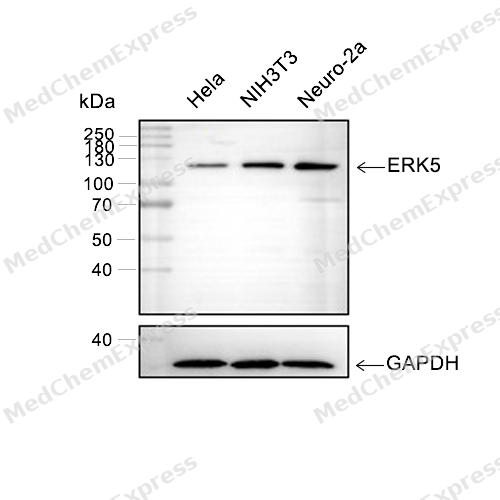 Western blot analysis of extracts from Hela(lane 2(20μg), NIH3T3(lane 3(20μg), Neuro-2a(lane 4(20μg) ) using ERK5 (HY-P80122) Rabbit mAb. Proteins were transferred to a PVDF membrane and blocked with 5% BSA in TBST for 2 hour at room temperature. The primary antibody (HY-P80122, 1/1000) and Loading control antibody (GAPDH, HY-P80954, 1/3000) was used in 5% BSA in TBST at 4°C overnight. Goat Anti-Mouse/Rabbit IgG-HRP Secondary Antibody (HY-P8004/HY-P8001, 1/10,000) was used for 1 hour at room temperature.
 -
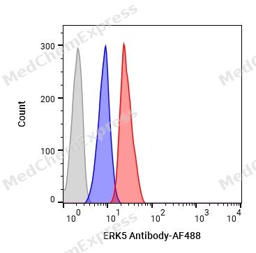 Flow cytometric analysis of 1X10⁶ HeLa cells labeling ERK5 Antibody(HY-P80122, red). Cells were fixed with 4% paraformaldehyde and permeabilised with 90% methanol. Then stained with the primary antibody at 1/100 dilution for an hour at 4℃. AF488-conjugated Goat Anti-Rabbit IgG H&L (HY-P8002) was used as the secondary antibody at 1/1,000 dilution for 30 minutes at 4℃. Rabbit IgG Isotype Control (HY-P80879, blue) was used as the isotype control, cells without incubation with primary antibody were used as the unlabeled control (black).


Background
-
Function
ERK5 (MAPK7), also known as big mitogen-activated protein kinase 1 (BMK1), is a distinct member of the mitogen-activated protein kinase (MAPK) family that integrates extracellular signals to regulate proliferation, differentiation, survival, transcriptional control, and tissue homeostasis[1][2]. Mechanistically, ERK5 is uniquely activated by MEK5 and contains an N-terminal kinase domain together with a large C-terminal transcriptional activation domain, enabling coordinated regulation of kinase-dependent and transcriptional responses[1][3]. Through the MEK5/ERK5 signaling axis, ERK5 participates in endothelial adaptation to laminar shear stress, promotes cell survival, and contributes to cardiovascular homeostasis by regulating protective transcriptional programs such as KLF2 expression[2]. In disease settings, aberrant ERK5 signaling has been associated with cancer progression, including enhanced migration, invasion, epithelial-mesenchymal transition, and metastatic behavior in multiple tumor models[1][4]. Compared with related ERK isoforms, ERK5 is distinguished by its unusually large C-terminal region containing transcriptional activation and nuclear localization elements, which confer functions beyond classical MAPK catalytic signaling[1][3]. Alternative ERK5 isoforms further diversify its biological activity, and a short isoform lacking the C-terminal transcriptional domain and nuclear localization signal has been linked to altered nucleocytoplasmic trafficking, increased cell migration, epithelial-mesenchymal transition, and poor outcomes in breast cancer models[5]. For experimental applications, selective ERK5 inhibitors and chemical probes have been developed to investigate ERK5-dependent signaling networks and disease mechanisms, supporting its continued use as a pharmacological target in cancer and vascular biology research[3].
-
Subcellular Localization
Cytoplasm; Nucleus; Nucleus, PML body
-
Expression
Tissue_specificity:It is expressed in many adult tissues. It is abundant in the heart, placenta, lungs, kidneys, and skeletal muscle. It is undetectable in the liver. -
Subunit
Interacts with MAP2K5.
-
SwissProt ID
-
Synonyms
BMK1, ERK5, PRKM7, MAPK7, Mitogen-activated protein kinase 7, MAP kinase 7, MAPK 7, Big MAP kinase 1, Extracellular signal-regulated kinase 5, BMK-1, ERK-5
-
Research Field
Neuroscience


Documentation
-
Data Sheet (262 KB)
-
SDS (252 KB)
- English - EN (252 KB)
- Français - FR (252 KB)
- Deutsch - DE (252 KB)
- Norwegian - NO (252 KB)
- Español - ES (252 KB)
- Swedish - SV (252 KB)
- Italian - IT (252 KB)
- Korean - KR (252 KB)
- Portuguese - PT (252 KB)
-
User Guide for Antibodies (1077 KB)
References
[1]. Le NT. The significance of ERK5 catalytic-independent functions in disease pathways. Front Cell Dev Biol. 2023 Aug 4;11:1235217. doi: 10.3389/fcell.2023.1235217. PMID: 37601096; PMCID: PMC10436230. et al. The significance of ERK5 catalytic-independent functions in disease pathways. Front Cell Dev Biol. 2023 Aug 4;11:1235217. [Content Brief]
[2]. Paudel R, et al. The MEK5/ERK5 Pathway in Health and Disease. Int J Mol Sci. 2021 Jul 15;22(14):7594. [Content Brief]
[3]. Elkins JM, et al. X-ray crystal structure of ERK5 (MAPK7) in complex with a specific inhibitor. J Med Chem. 2013 Jun 13;56(11):4413-21. [Content Brief]
[4]. Villalba M, et al. Is the MAPK ERK5 the nexus from FAO to NK cell-mediated metastasis immune surveillance? Front Immunol. 2025 Oct 8;16:1641865. [Content Brief]

 Powered by Bioz
