



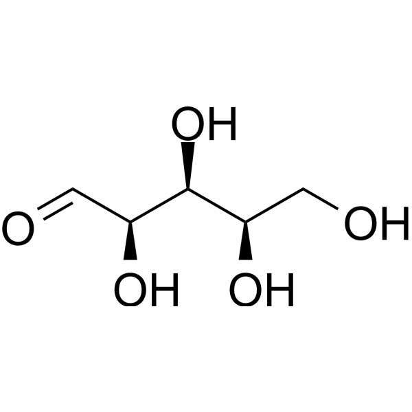
Arabinose
Based on 2 publication(s) in Google Scholar
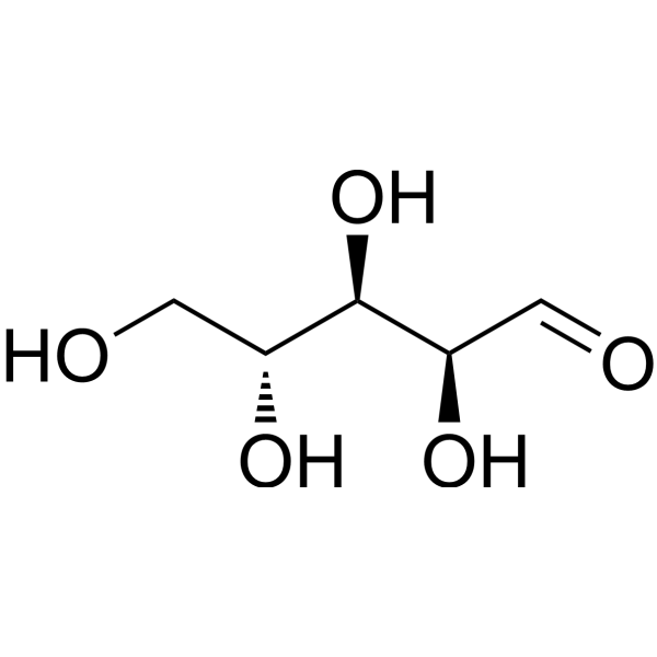
Arabinose is a pentose sugar commonly found in plants. Arabinose alleviates immune dysregulation and inflammation by promoting balanced immune responses and reducing inflammation. Arabinose induces cytotoxicity, autophagy (Autophagy), and cell cycle arrest in breast cancer cells through the p38-MAPK signaling pathway. Arabinose activates the ACSS2-PPARγ/TFEB-AMPK axis in neuroblastoma cells, thereby exerting neuromodulatory/antidepressant effects. Arabinose can also be used as an intermediate in compound synthesis. Arabinose may be applied in research related to immune inflammation, depression, breast cancer, and other diseases.
For research use only. We do not sell to patients.
- Purity : 99.87%
- CAS No.: 147-81-9
- Formula: C5H10O5
- Molecular Weight:150.13
-
Storage:
4°C, stored under nitrogen, away from moisture
* In solvent : -80°C, 6 months; -20°C, 1 month (stored under nitrogen, away from moisture)

To place orders, for customer services and technical support, please contact: MedChemExpress USA
Tel: 609-228-6898 E-mail: [email protected] [email protected]


-
 Biological Activity
-
Chemical Information
-
Solvent & Solubility
- Protocols
- Purity & Documentation
- References
-
 Help & FAQs


Publications Citing Use of MedChemExpress (MCE) Arabinose
MoreAll Endogenous Metabolite Isoforms
More

Biological Activity
Description
IC50 & Target
|
Human Endogenous Metabolite |
In Vitro
Arabinose (50 mM) induces cytotoxicity, autophagy, and cell cycle arrest in breast cancer cells via the p38-MAPK signaling pathway[1].
Arabinose induces CRTC1 expression and activates the ACSS2-PPARγ/TFEB-AMPK axis in SH-SY5Y human neuroblastoma cells, exerting neuromodulatory/antidepressant effects[1].
MedChemExpress (MCE) has not independently confirmed the accuracy of these methods. They are for reference only.
In Vivo
MedChemExpress (MCE) has not independently confirmed the accuracy of these methods. They are for reference only.
- Cell Cytotoxicity Assay
- Mammalian live/dead viability and cytotoxicity staining
- Flow cytometric DNA-content cell-cycle staining
- BrdU Incorporation Assay
- Autophagy
- Protocol for Cell Cycle
- Research Protocol for Inflammation-related Diseases
- Lysosome and acidic-vesicle live-cell staining
- Macroautophagy Solutions
- Breast Cancer Modeling
- Cell Viability Determination by MTT Colorimetric Assay
Clinical Trial
| NCT Number | Sponsor | Condition | Start Date |
Phase

|
|---|---|---|---|---|
| NCT01329991 | 2011-05 | PHASE1 |


Chemical Information
-
CAS No. 147-81-9
-
Appearance Solid
-
Molecular Weight 150.13
-
Formula C5H10O5
-
Color White to off-white
-
SMILES
OC[C@H]([C@H]([C@@H](C=O)O)O)O
-
Synonyms
(±)-Arabinose; DL-Arabinose; dl-Arabinose
-
Structure Classification
-
Initial Source
-
Shipping
Room temperature in continental US; may vary elsewhere.
-
Storage
4°C, stored under nitrogen, away from moisture
* In solvent : -80°C, 6 months; -20°C, 1 month (stored under nitrogen, away from moisture)


Publications (2)
-
Journal Impact Factor
-
Most Recent
-
Ultrason Sonochem
Ultrasonic complexation with Lycium barbarum polysaccharide significantly enhances the aqueous solubility and bioavailability of curcumin. [Abstract]2025 Dec:123:107673. PMID: 41223460 -


Solvent & Solubility
In Vitro:
H2O : 100 mg/mL (666.09 mM; ultrasonic and warming and heat to 60°C)
DMSO : 20 mg/mL (133.22 mM; ultrasonic and warming and heat to 60°C; Hygroscopic DMSO has a significant impact on the solubility of product, please use newly opened DMSO)
Please refer to the solubility information to select the appropriate solvent. Once prepared, please aliquot and store the solution to prevent product inactivation from repeated freeze-thaw cycles.
Storage method and period of stock solution: -80°C, 6 months; -20°C, 1 month (stored under nitrogen, away from moisture). When stored at -80°C, please use it within 6 months. When stored at -20°C, please use it within 1 month.
* Note: If you choose water as the stock solution, please dilute it to the working solution, then filter and sterilize it with a 0.22 μm filter before use.
Please refer to the solubility information to select the appropriate solvent. Once prepared, please aliquot and store the solution to prevent product inactivation from repeated freeze-thaw cycles.
Storage method and period of stock solution: -80°C, 6 months; -20°C, 1 month (stored under nitrogen, away from moisture). When stored at -80°C, please use it within 6 months. When stored at -20°C, please use it within 1 month.
* Note: If you choose water as the stock solution, please dilute it to the working solution, then filter and sterilize it with a 0.22 μm filter before use.
Concentration (start) × Volume (start) = Concentration (final) × Volume (final)
In Vivo:
Select the appropriate dissolution method based on your experimental animal and administration route.
- For the following dissolution methods, please ensure to first prepare a clear stock solution using an In Vitro approach and then sequentially add co-solvents:
- To ensure reliable experimental results, the clarified stock solution can be appropriately stored based on storage conditions. As for the working solution for In Vivo experiments, it is recommended to prepare freshly and use it on the same day.
- The percentages shown for the solvents indicate their volumetric ratio in the final prepared solution. If precipitation or phase separation occurs during preparation, heat and/or sonication can be used to aid dissolution.
Add each solvent one by one: 10% DMSO 40% PEG300 5% Tween-80 45% Saline
Solubility: ≥ 2 mg/mL (13.32 mM); Clear solution
This protocol yields a clear solution of ≥ 2 mg/mL (saturation unknown).
Taking 1 mL working solution as an example, add 100 μL DMSO stock solution (20.0 mg/mL) to 400 μL PEG300, and mix evenly; then add 50 μL Tween-80 and mix evenly; then add 450 μL Saline to adjust the volume to 1 mL.
Preparation of Saline: Dissolve 0.9 g sodium chloride in ddH₂O and dilute to 100 mL to obtain a clear Saline solution.
Add each solvent one by one: 10% DMSO 90% (20% SBE-β-CD in Saline)
Solubility: ≥ 2 mg/mL (13.32 mM); Clear solution
This protocol yields a clear solution of ≥ 2 mg/mL (saturation unknown).
Taking 1 mL working solution as an example, add 100 μL DMSO stock solution (20.0 mg/mL) to 900 μL 20% SBE-β-CD in Saline, and mix evenly.
Preparation of 20% SBE-β-CD in Saline (4°C, storage for one week): 2 g SBE-β-CD powder is dissolved in 10 mL Saline, completely dissolve until clear.
In Vivo Dissolution Calculator
Please enter the basic information of animal experiments:
-
-
-
-
Recommended: Prepare an additional quantity of animals to account for potential losses during experiments.
Working solution concentration: 0.22 mg/mL
This product has good water solubility, please refer to the measured solubility data in water/PBS/Saline for details.


Purity & Documentation
-
Data Sheet (274 KB)
-
SDS (396 KB)
- English - EN (396 KB)
- Français - FR (396 KB)
- Deutsch - DE (396 KB)
- Norwegian - NO (396 KB)
- Español - ES (396 KB)
- Swedish - SV (396 KB)
- Italian - IT (396 KB)
- Korean - KR (396 KB)
- Portuguese - PT (396 KB)
-
Handling Instructions (2659 KB)
References
[1]. Newman PP, Schmitt BL, Maurmann RM, Pence BD. Polysaccharides with Arabinose: Key Players in Reducing Chronic Inflammation and Enhancing Immune Health in Aging. Molecules. 2025 Mar 6;30(5):1178. [Content Brief]
[2]. Newman PP, et al. Polysaccharides with Arabinose: Key Players in Reducing Chronic Inflammation and Enhancing Immune Health in Aging. Molecules. 2025;30(5):1178. Published 2025 Mar 6. [Content Brief]
[3]. Tyson B, et al. Crystallization Behavior and Crystallographic Properties of dl-Arabinose and dl-Xylose Diastereomer Sugars. Cryst Growth Des. 2022;22(2):1371-1383. [Content Brief]


Complete Stock Solution Preparation Table
Please refer to the solubility information to select the appropriate solvent. Once prepared, please aliquot and store the solution to prevent product inactivation from repeated freeze-thaw cycles.
Storage method and period of stock solution: -80°C, 6 months; -20°C, 1 month (stored under nitrogen, away from moisture). When stored at -80°C, please use it within 6 months. When stored at -20°C, please use it within 1 month.
| Optional Solvent | Concentration Solvent Mass | 1 mg | 5 mg | 10 mg | 25 mg |
|---|---|---|---|---|---|
| DMSO / H2O | 1 mM | 6.6609 mL | 33.3045 mL | 66.6089 mL | 166.5223 mL |
| 5 mM | 1.3322 mL | 6.6609 mL | 13.3218 mL | 33.3045 mL | |
| 10 mM | 0.6661 mL | 3.3304 mL | 6.6609 mL | 16.6522 mL | |
| 15 mM | 0.4441 mL | 2.2203 mL | 4.4406 mL | 11.1015 mL | |
| 20 mM | 0.3330 mL | 1.6652 mL | 3.3304 mL | 8.3261 mL | |
| 25 mM | 0.2664 mL | 1.3322 mL | 2.6644 mL | 6.6609 mL | |
| 30 mM | 0.2220 mL | 1.1101 mL | 2.2203 mL | 5.5507 mL | |
| 40 mM | 0.1665 mL | 0.8326 mL | 1.6652 mL | 4.1631 mL | |
| 50 mM | 0.1332 mL | 0.6661 mL | 1.3322 mL | 3.3304 mL | |
| 60 mM | 0.1110 mL | 0.5551 mL | 1.1101 mL | 2.7754 mL | |
| 80 mM | 0.0833 mL | 0.4163 mL | 0.8326 mL | 2.0815 mL | |
| 100 mM | 0.0666 mL | 0.3330 mL | 0.6661 mL | 1.6652 mL |
* Note: If you choose water as the stock solution, please dilute it to the working solution, then filter and sterilize it with a 0.22 μm filter before use.


Arabinose Related Classifications
HY-N2353 Related Classifications
 Powered by Bioz