



Trifluridine
Based on 4 publication(s) in Google Scholar
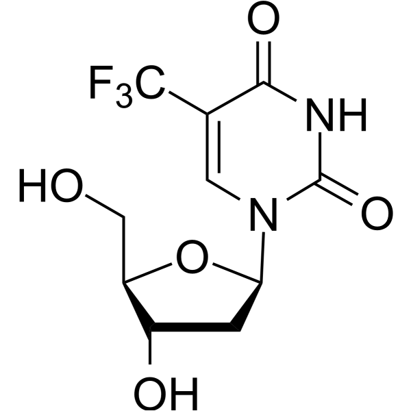
Trifluridine (Trifluorothymidine) is an irreversible and orally active thymidylate synthase inhibitor, and thereby suppressing DNA synthesis. Trifluridine is an antiviral molecule used for research of HSV, rhabdovirus and orthopoxvirus infection. Trifluridine induces cell apoptosis and autophagy. Trifluridine is also an anticancer agent used in studies of metastatic colorectal cancer, gastrointestinal tumors.
Nur für Forschungszwecke. Wir verkaufen nicht an Patienten.
- Reinheit : 99.96%
- CAS. Nr.: 70-00-8
- Formel: C10H11F3N2O5
- Molecular Weight:296.20
-
Speicherung:Powder -20°C, 3 years , 4°C, 2 years ; In solvent -80°C, 2 years , -20°C, 1 year

To place orders, for customer services and technical support, please contact: MedChemExpress USA
Tel: 609-228-6898 E-mail: [email protected] [email protected]


-
 Biologische Aktivität
-
Chemical Information
-
Lösungsmittel & Löslichkeit
- Protokoll
- Reinheit & Dokumentation
- Verweise
-
 Help & FAQs


-
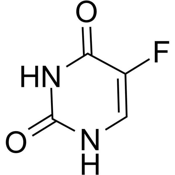
HY-90006
5-FluorouracilThymidylate Synthase Inhibitor -
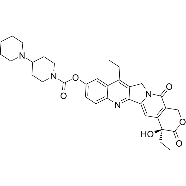
HY-16562
IrinotecanTopoisomerase I Inhibitor -
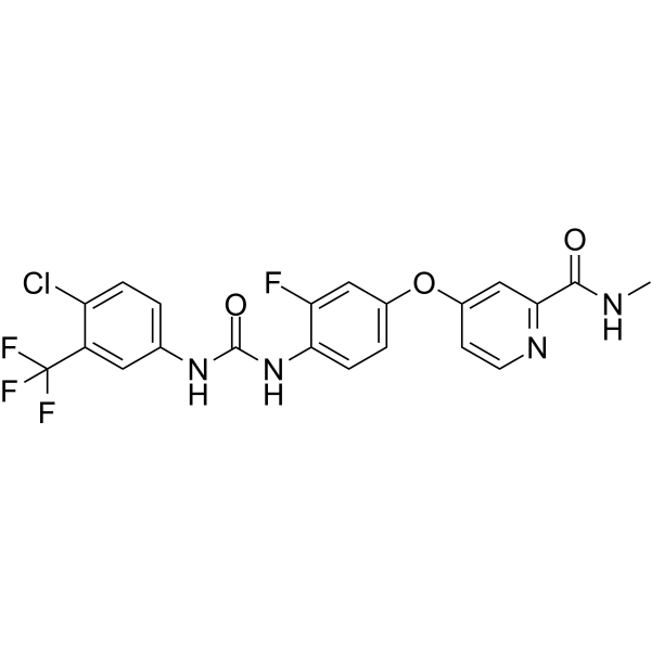
HY-10331
RegorafenibMulti-kinase Inhibitor -
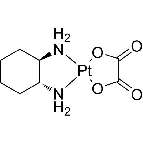
HY-17371
OxaliplatinDNA Synthesis Inhibitor -
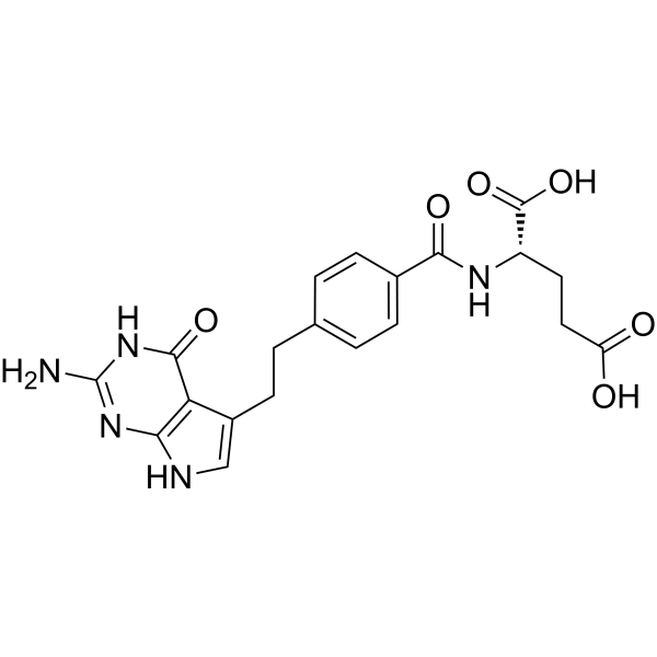
HY-10820
Pemetrexed -
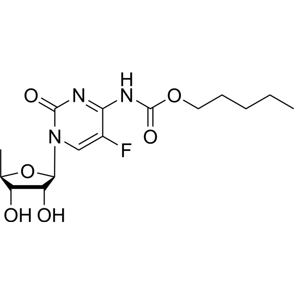
HY-B0016
CapecitabineDNA/RNA Synthesis Inhibitor -
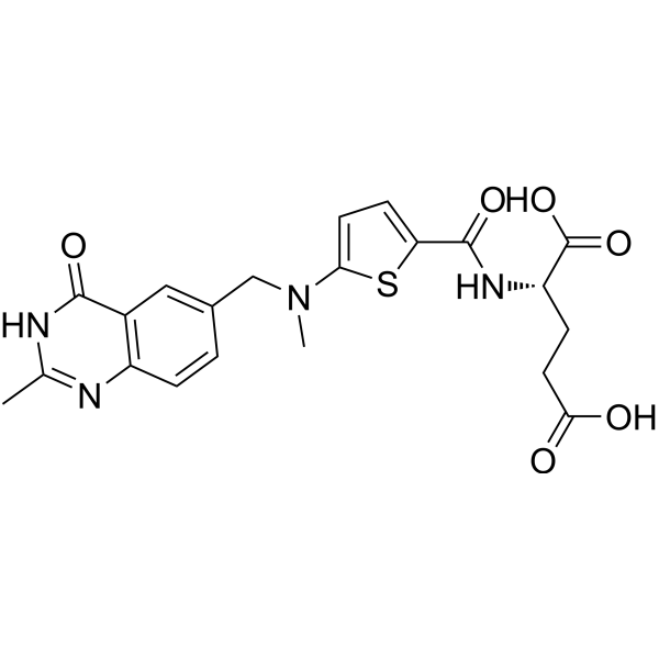
HY-10821
RaltitrexedThymidylate Synthase Inhibitor -
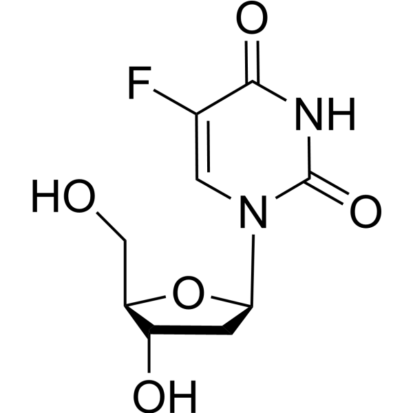
HY-B0097
FloxuridineOncology Antimetabolite -
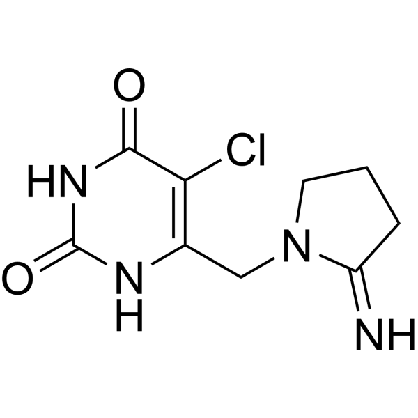
HY-A0063A
TipiracilTPase Inhibitor -
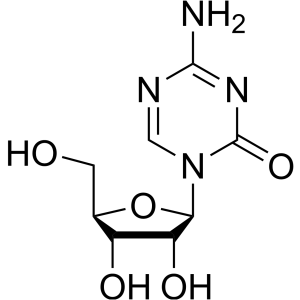
HY-10586
5-AzacytidineDNMT Inhibitor -
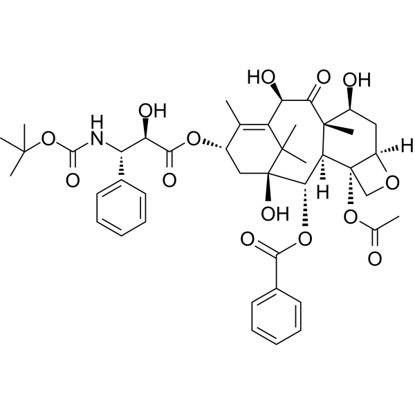
HY-B0011
DocetaxelTubulin Stabilizer -
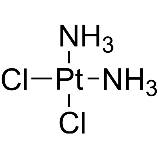
HY-17394
CisplatinDNA Alkylator
Publications Citing Use of MedChemExpress (MCE) Trifluridine
MoreAlle DNA/RNA Synthesis Isoform-spezifische Produkte anzeigen
More

Biologische Aktivität
Beschreibung
|
Thymidylate Synthase |
Nucleoside Antimetabolite/Analog |
HSV-1 |
HSV-2 |
Cellular Effect
|
Cell Line

|
Type | Value | Description | References |
|---|---|---|---|---|
| Hepatocyte | EC50 |
3.37 μM
Compound: 25
|
Antiviral activity against DHBV infected in duck hepatocytes assessed as inhibition of viral DNA replication
Antiviral activity against DHBV infected in duck hepatocytes assessed as inhibition of viral DNA replication
|
[PMID: 20857959] |
| HepG2 2.2.15 | EC50 |
10 μg/mL
Compound: 2
|
Antiviral activity against HBV infected in human 2.2.15 cell at 10 ug/ml
Antiviral activity against HBV infected in human 2.2.15 cell at 10 ug/ml
|
[PMID: 20863701] |
| Huh-7 | CC50 |
>675 μM
Compound: 25
|
Cytotoxicity against HuH7 cells
Cytotoxicity against HuH7 cells
|
[PMID: 20857959] |
| Huh-7 | CC50 |
>200 μg/mL
Compound: 2
|
Cytotoxicity against human HuH7 cells
Cytotoxicity against human HuH7 cells
|
[PMID: 20863701] |
| Vero | CC50 |
50.6 μM
Compound: TFT
|
Cytotoxicity against african green monkey Vero cells after 2 days
Cytotoxicity against african green monkey Vero cells after 2 days
|
[PMID: 17438061] |
| Vero | EC50 |
<1.7 μM
Compound: TFT
|
Antiviral activity against Herpes simplex virus 1 F infected in african green monkey Vero cells assessed as plaque reduction after 36 to 48 hrs
Antiviral activity against Herpes simplex virus 1 F infected in african green monkey Vero cells assessed as plaque reduction after 36 to 48 hrs
|
[PMID: 17438061] |
| Vero | EC50 |
1.7 μM
Compound: TFT
|
Antiviral activity against Herpes B virus 24105 infected in african green monkey Vero cells assessed as plaque reduction after 36 to 48 hrs
Antiviral activity against Herpes B virus 24105 infected in african green monkey Vero cells assessed as plaque reduction after 36 to 48 hrs
|
[PMID: 17438061] |
| Vero | EC50 |
3.4 μM
Compound: TFT
|
Antiviral activity against Herpes B virus 32425 infected in african green monkey Vero cells assessed as plaque reduction after 36 to 48 hrs
Antiviral activity against Herpes B virus 32425 infected in african green monkey Vero cells assessed as plaque reduction after 36 to 48 hrs
|
[PMID: 17438061] |
In Vitro
Trifluridine (0.5 μΜ, 3 days) induces cellular senescence in HUVECs[2].
Trifluridine (0.5 μΜ, 3 days) inhibits autophagy and autophagy flux via the mTOR pathway in HUVECs [2].
Trifluridine (0-5 μΜ, 3 days) inhibits HUVEC cells viability in a concentration-dependent way[2].
Trifluridine (5 µM-20 µM, 24 h–72 h) inhibits the proliferation in MCF-7, MDA-MB-231, BT-549 and Hs578T[4].
Trifluridine (0 µM-20 µM, 48 h) selectively induces apoptosis in MCF-7, MDA-MB-231, BT-549 and Hs578T cells [4].
Trifluridine (10 µM, 72 h) induces DNA double-strand break in MCF-10A, MCF-7, MDA-MB-231 and BT-549 cells[4].
MedChemExpress (MCE) has not independently confirmed the accuracy of these methods. They are for reference only.
-
Cell Line:HUVEC cells
-
Concentration:0-5 μM
-
Incubation Time:3 days
-
Result:Cell viability dropped sharply at 3 μM and 5 μM.
-
Cell Line:HUVEC cells
-
Concentration:0-5 μM
-
Incubation Time:3 days
-
Result:Induced cellular senescence by inhibiting autophagy flux.
-
Cell Line:HUVEC cells
-
Concentration:0-5 μM
-
Incubation Time:3 days
-
Result:Increased protein levels of senescence markers: p53, p16, SASP (IL-1, IL-6, TNF-α, p21).
-
Cell Line:MCF-7, MDA-MB-231, BT-549, Hs578T and MCF-10A(control non-tumor) cells
-
Concentration:5 µM-20 µM
-
Incubation Time:24 h–72 h
-
Result:Inhibited the proliferation of MDA-MB231, BT549 and Hs578T cells at 10µM and 20µM, significantly.
-
Cell Line:MDA-MB-231, BT-549, and Hs578T cells
-
Concentration:10µM or 20 µM
-
Incubation Time:48 h
-
Result:Increased the percentages of apoptotic TNBC cells (MDA-MB-231, BT-549, and Hs578Tcells) at 10µM and 20µM, significantly.
In Vivo
Trifluridine (75 or 150 mg/kg, oral gavage, once daily for 5 consecutive days) inhibits the growth in implanted mouse TNBC tumors [4].
MedChemExpress (MCE) has not independently confirmed the accuracy of these methods. They are for reference only.
-
Animal Model:Nude mice (human colorectal intraperitoneal xenograft model)[3]
-
Dosage:200 mg/kg(trifluridine/tipiracil)
-
Administration:Oral administration; Once a day; 5 days followed by 2 drug-free days, a total of 6 weeks
-
Result:Exhibited a significantly longer survival time compared with untreated mice.
-
Animal Model:Mouse model of breast cancer[4]
-
Dosage:75 or 150 mg/kg
-
Administration:Oral gavage; daily for 10 days
-
Result:Treatment with different doses inhibited the growth of TNBC tumors in mouse in a dose-dependent manner
-
Animal Model:The human gastric MKN45 intraperitoneal xenograft model[3]
-
Dosage:200 mg/kg (Trifluridine/tipiracil)
-
Administration:twice daily for 5 consecutive days followed by 2 drug‑free days for 6 weeks
-
Result:Prolonged the survival of mice compared with untreated mice.
- EdU Incorporation Assay (Click Chemistry-Based DNA Synthesis Measurement)
- Research Protocol for Infectious Diseases
- Detection of Apoptosis
- TUNEL staining for apoptotic DNA fragmentation
- Annexin V plus membrane-impermeant dye apoptosis staining
- BrdU Incorporation Assay
- Autophagy
- Apoptosis Solutions
- Lysosome and acidic-vesicle live-cell staining
- Macroautophagy Solutions
Clinical Trial
| NCT Number | Sponsor | Condition | Start Date |
Phase
|
|---|---|---|---|---|
| NCT01329991 | 2011-05 | PHASE1 |


Chemical Information
-
CAS. Nr. 70-00-8
-
Appearance Solid
-
Molecular Weight 296.20
-
Formel C10H11F3N2O5
-
Color White to off-white
-
SMILES
O[C@H]1C[C@H](N2C(NC(C(C(F)(F)F)=C2)=O)=O)O[C@@H]1CO
-
Synonyms
Trifluorothymidine; 5-Trifluorothymidine; TFT
-
Versand
Room temperature in continental US; may vary elsewhere.
-
Speicherung
Powder -20°C 3 years 4°C 2 years In solvent -80°C 2 years -20°C 1 year


Publications (4)
-
Journal Impact Factor
-
Most Recent
-
Signal Transduct Target Ther
Selective depletion of tumor-associated SAMHD1 enhances chemotherapeutic efficacy and antitumor immune responses. [Abstract]2025 Dec 15;10(1):406. PMID: 41392286 -
-
J Mol Med (Berl)
2019 Aug;97(8):1183-1193. PMID: 31201471 -
PLoS Pathog
Anti-orthopoxvirus drugs inhibit lumpy skin disease virus replication by targeting viral DNA polymerase. [Abstract]2026 Jan 26;22(1):e1013903. PMID: 41587213


Lösungsmittel & Löslichkeit
In Vitro:
DMSO : ≥ 100 mg/mL (337.61 mM; Hygroscopic DMSO has a significant impact on the solubility of product, please use newly opened DMSO)
H2O : 33.33 mg/mL (112.53 mM; ultrasonic and warming and heat to 60°C)
* "≥" means soluble, but saturation unknown.
Please refer to the solubility information to select the appropriate solvent. Once prepared, please aliquot and store the solution to prevent product inactivation from repeated freeze-thaw cycles.
Storage method and period of stock solution: -80°C, 2 years; -20°C, 1 year. When stored at -80°C, please use it within 2 years. When stored at -20°C, please use it within 1 year.
* Note: If you choose water as the stock solution, please dilute it to the working solution, then filter and sterilize it with a 0.22 μm filter before use.
Please refer to the solubility information to select the appropriate solvent. Once prepared, please aliquot and store the solution to prevent product inactivation from repeated freeze-thaw cycles.
Storage method and period of stock solution: -80°C, 2 years; -20°C, 1 year. When stored at -80°C, please use it within 2 years. When stored at -20°C, please use it within 1 year.
* Note: If you choose water as the stock solution, please dilute it to the working solution, then filter and sterilize it with a 0.22 μm filter before use.
Konzentration (Stammlösung) × Volumen (Stammlösung) = Konzentration (Ziellösung) × Volumen (Ziellösung)
In Vivo:
Select the appropriate dissolution method based on your experimental animal and administration route.
- For the following dissolution methods, please ensure to first prepare a clear stock solution using an In Vitro approach and then sequentially add co-solvents:
- To ensure reliable experimental results, the clarified stock solution can be appropriately stored based on storage conditions. As for the working solution for In Vivo experiments, it is recommended to prepare freshly and use it on the same day.
- The percentages shown for the solvents indicate their volumetric ratio in the final prepared solution. If precipitation or phase separation occurs during preparation, heat and/or sonication can be used to aid dissolution.
Add each solvent one by one: 10% DMSO 40% PEG300 5% Tween-80 45% Saline
Solubility: ≥ 2.5 mg/mL (8.44 mM); Clear solution
This protocol yields a clear solution of ≥ 2.5 mg/mL (saturation unknown).
Taking 1 mL working solution as an example, add 100 μL DMSO stock solution (25.0 mg/mL) to 400 μL PEG300, and mix evenly; then add 50 μL Tween-80 and mix evenly; then add 450 μL Saline to adjust the volume to 1 mL.
Preparation of Saline: Dissolve 0.9 g sodium chloride in ddH₂O and dilute to 100 mL to obtain a clear Saline solution.
Add each solvent one by one: 10% DMSO 90% (20% SBE-β-CD in Saline)
Solubility: ≥ 2.5 mg/mL (8.44 mM); Clear solution
This protocol yields a clear solution of ≥ 2.5 mg/mL (saturation unknown).
Taking 1 mL working solution as an example, add 100 μL DMSO stock solution (25.0 mg/mL) to 900 μL 20% SBE-β-CD in Saline, and mix evenly.
Preparation of 20% SBE-β-CD in Saline (4°C, storage for one week): 2 g SBE-β-CD powder is dissolved in 10 mL Saline, completely dissolve until clear.
In Vivo Dissolution Calculator
Please enter the basic information of animal experiments:
-
-
-
-
Recommended: Prepare an additional quantity of animals to account for potential losses during experiments.
Working solution concentration: 0.22 mg/mL
This product has good water solubility, please refer to the measured solubility data in water/PBS/Saline for details.


Reinheit & Dokumentation
-
Data Sheet (286 KB)
-
SDS (419 KB)
- English - EN (419 KB)
- Français - FR (419 KB)
- Deutsch - DE (419 KB)
- Norwegian - NO (419 KB)
- Español - ES (419 KB)
- Swedish - SV (419 KB)
- Italian - IT (419 KB)
- Korean - KR (419 KB)
- Portuguese - PT (419 KB)
-
Handling Instructions (2659 KB)
Verweise
[1]. Okayama T, et al. Involvement of concentrative nucleoside transporter 1 in intestinal absorption of trifluorothymidine, a novel antitumor nucleoside, in rats. J Pharmacol Exp Ther. 2012 Feb;340(2):457-62. [Content Brief]
[2]. Jia HJ, et al. Trifluridine induces HUVECs senescence by inhibiting mTOR-dependent autophagy. Biochem Biophys Res Commun. 2022 Jun 25;610:119-126. [Content Brief]
[3]. Suzuki N,et al. Trifluridine/tipiracil increases survival rates in peritoneal dissemination mouse models of human colorectal and gastric cancer. Oncol Lett. 2017 Jul;14(1):639-646. [Content Brief]
[4]. Li J, et al. Trifluridine selectively inhibits cell growth and induces cell apoptosis of triple-negative breast cancer. Am J Cancer Res. 2020 Feb 1;10(2):507-522. [Content Brief]


Complete Stock Solution Preparation Table
Please refer to the solubility information to select the appropriate solvent. Once prepared, please aliquot and store the solution to prevent product inactivation from repeated freeze-thaw cycles.
Storage method and period of stock solution: -80°C, 2 years; -20°C, 1 year. When stored at -80°C, please use it within 2 years. When stored at -20°C, please use it within 1 year.
| Optional Solvent | Concentration Solvent Mass | 1 mg | 5 mg | 10 mg | 25 mg |
|---|---|---|---|---|---|
| H2O / DMSO | 1 mM | 3.3761 mL | 16.8805 mL | 33.7610 mL | 84.4024 mL |
| 5 mM | 0.6752 mL | 3.3761 mL | 6.7522 mL | 16.8805 mL | |
| 10 mM | 0.3376 mL | 1.6880 mL | 3.3761 mL | 8.4402 mL | |
| 15 mM | 0.2251 mL | 1.1254 mL | 2.2507 mL | 5.6268 mL | |
| 20 mM | 0.1688 mL | 0.8440 mL | 1.6880 mL | 4.2201 mL | |
| 25 mM | 0.1350 mL | 0.6752 mL | 1.3504 mL | 3.3761 mL | |
| 30 mM | 0.1125 mL | 0.5627 mL | 1.1254 mL | 2.8134 mL | |
| 40 mM | 0.0844 mL | 0.4220 mL | 0.8440 mL | 2.1101 mL | |
| 50 mM | 0.0675 mL | 0.3376 mL | 0.6752 mL | 1.6880 mL | |
| 60 mM | 0.0563 mL | 0.2813 mL | 0.5627 mL | 1.4067 mL | |
| 80 mM | 0.0422 mL | 0.2110 mL | 0.4220 mL | 1.0550 mL | |
| 100 mM | 0.0338 mL | 0.1688 mL | 0.3376 mL | 0.8440 mL |
* Note: If you choose water as the stock solution, please dilute it to the working solution, then filter and sterilize it with a 0.22 μm filter before use.


Trifluridine Related Classifications
HY-A0061 Related Classifications
 Powered by Bioz