



LRRKtide

Based on 1 Customer Validation
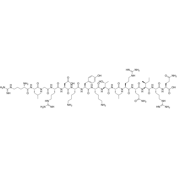
LRRKtide is a polypeptide substrate. LRRKtide is a specific phosphorylation substrate of LRRK2, a kinase associated with Parkinson's disease, and its phosphorylation site is a threonine residue. LRRKtide can be used for characterization of the catalytic properties of LRRK2, as well as studies on kinase activity and inhibition. LRRKtide is applicable to research related to Parkinson's disease.
Nur für Forschungszwecke. Wir verkaufen nicht an Patienten.
- Reinheit : 99.94%
- CAS. Nr.: 1071466-49-3
- Formel: C83H147N31O22
- Molecular Weight:1931.25
-
Speicherung:
Sealed storage, away from moisture.
Powder -80°C, 2 years , -20°C, 1 year* In solvent : -80°C, 6 months; -20°C, 1 month (sealed storage, away from moisture)

To place orders, for customer services and technical support, please contact: MedChemExpress USA
Tel: 609-228-6898 E-mail: [email protected] [email protected]


-
 Biologische Aktivität
-
Chemical Information
-
Lösungsmittel & Löslichkeit
- Protokoll
- Reinheit & Dokumentation
- Verweise
-
 Help & FAQs




Biologische Aktivität
Beschreibung
In Vitro
LRRKtide is a key tool peptide for dissecting the functions of LRRK2 and the mechanisms of Parkinson's disease[1].
MedChemExpress (MCE) has not independently confirmed the accuracy of these methods. They are for reference only.


Chemical Information
-
CAS. Nr. 1071466-49-3
-
Appearance Solid
-
Molecular Weight 1931.25
-
Formel C83H147N31O22
-
Color White to off-white
-
Sequence
Arg-Leu-Gly-Arg-Asp-Lys-Tyr-Lys-Thr-Leu-Arg-Gln-Ile-Arg-Gln
-
Sequence Shortening
RLGRDKYKTLRQIRQ
-
Versand
Room temperature in continental US; may vary elsewhere.
-
Speicherung
Sealed storage, away from moisture
Powder -80°C 2 years -20°C 1 year * In solvent : -80°C, 6 months; -20°C, 1 month (sealed storage, away from moisture)


Lösungsmittel & Löslichkeit
In Vitro:
H2O : ≥ 100 mg/mL (51.78 mM)
DMSO : 20 mg/mL (10.36 mM; Need ultrasonic; Hygroscopic DMSO has a significant impact on the solubility of product, please use newly opened DMSO)
* "≥" means soluble, but saturation unknown.
Please refer to the solubility information to select the appropriate solvent. Once prepared, please aliquot and store the solution to prevent product inactivation from repeated freeze-thaw cycles.
Storage method and period of stock solution: -80°C, 6 months; -20°C, 1 month (sealed storage, away from moisture). When stored at -80°C, please use it within 6 months. When stored at -20°C, please use it within 1 month.
* Note: If you choose water as the stock solution, please dilute it to the working solution, then filter and sterilize it with a 0.22 μm filter before use.
Please refer to the solubility information to select the appropriate solvent. Once prepared, please aliquot and store the solution to prevent product inactivation from repeated freeze-thaw cycles.
Storage method and period of stock solution: -80°C, 6 months; -20°C, 1 month (sealed storage, away from moisture). When stored at -80°C, please use it within 6 months. When stored at -20°C, please use it within 1 month.
* Note: If you choose water as the stock solution, please dilute it to the working solution, then filter and sterilize it with a 0.22 μm filter before use.
Konzentration (Stammlösung) × Volumen (Stammlösung) = Konzentration (Ziellösung) × Volumen (Ziellösung)


Reinheit & Dokumentation
-
Data Sheet (271 KB)
-
SDS (254 KB)
- English - EN (254 KB)
- Français - FR (254 KB)
- Deutsch - DE (254 KB)
- Norwegian - NO (254 KB)
- Español - ES (254 KB)
- Swedish - SV (254 KB)
- Italian - IT (254 KB)
- Korean - KR (254 KB)
- Portuguese - PT (254 KB)
-
Handling Instructions (2659 KB)
Verweise
[1]. Liu M, et al. Kinetic mechanistic studies of wild-type leucine-rich repeat kinase 2: characterization of the kinase and GTPase activities. Biochemistry. 2010;49(9):2008-2017. [Content Brief]
[2]. Ray S, et al. The Parkinson disease-linked LRRK2 protein mutation I2020T stabilizes an active state conformation leading to increased kinase activity. J Biol Chem. 2014;289(19):13042-13053. [Content Brief]


Complete Stock Solution Preparation Table
Please refer to the solubility information to select the appropriate solvent. Once prepared, please aliquot and store the solution to prevent product inactivation from repeated freeze-thaw cycles.
Storage method and period of stock solution: -80°C, 6 months; -20°C, 1 month (sealed storage, away from moisture). When stored at -80°C, please use it within 6 months. When stored at -20°C, please use it within 1 month.
| Optional Solvent | Concentration Solvent Mass | 1 mg | 5 mg | 10 mg | 25 mg |
|---|---|---|---|---|---|
| DMSO / H2O | 1 mM | 0.5178 mL | 2.5890 mL | 5.1780 mL | 12.9450 mL |
| 5 mM | 0.1036 mL | 0.5178 mL | 1.0356 mL | 2.5890 mL | |
| 10 mM | 0.0518 mL | 0.2589 mL | 0.5178 mL | 1.2945 mL | |
| H2O | 15 mM | 0.0345 mL | 0.1726 mL | 0.3452 mL | 0.8630 mL |
| 20 mM | 0.0259 mL | 0.1294 mL | 0.2589 mL | 0.6472 mL | |
| 25 mM | 0.0207 mL | 0.1036 mL | 0.2071 mL | 0.5178 mL | |
| 30 mM | 0.0173 mL | 0.0863 mL | 0.1726 mL | 0.4315 mL | |
| 40 mM | 0.0129 mL | 0.0647 mL | 0.1294 mL | 0.3236 mL | |
| 50 mM | 0.0104 mL | 0.0518 mL | 0.1036 mL | 0.2589 mL |
* Note: If you choose water as the stock solution, please dilute it to the working solution, then filter and sterilize it with a 0.22 μm filter before use.


LRRKtide Related Classifications
HY-P5979 Related Classifications
 Powered by Bioz