



5-POHSA
5-POHSA is a CD36 and Src kinase modulator acting on taste bud cells, and belongs to the fatty acid esters of hydroxy fatty acids (FAHFA) family. 5-POHSA binds to lingual CD36 to initiate intracellular signaling pathways, activate downstream Src kinase, and rapidly elevate intracellular calcium levels. 5-POHSA interacts with the tongue-brain axis by binding to CD36, so as to ameliorate taste disorders and obesity-related adverse reactions. 5-POHSA is applicable to the research of obesity.
For research use only. We do not sell to patients.
- CAS No.: 2161370-68-7
- Formula: C34H64O4
- Molecular Weight:536.87
-
Storage:
Please store the product under the recommended conditions in the Certificate of Analysis.


To place orders, for customer services and technical support, please contact: MedChemExpress USA
Tel: 609-228-6898 E-mail: [email protected] [email protected]


-
 Biological Activity
-
Chemical Information
- Protocols
- Purity & Documentation
- References
-
 Help & FAQs


-
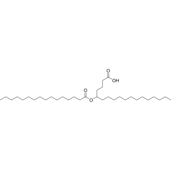
HY-116193
5-PAHSAAnti-Inflammatory Agent -
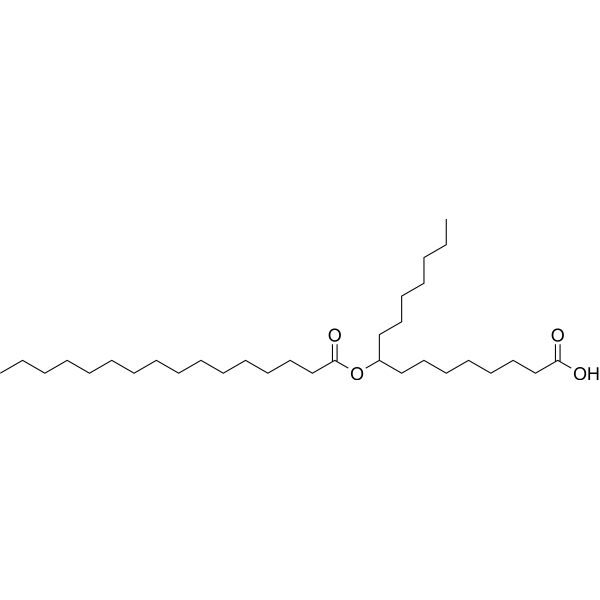
HY-117586
9-PAHPAAntidiabetic/Anti-inflammatory Agent -

HY-158584
13-OAHSAFAHFA -

HY-W709961
5-OAHSAEndogenous Metabolite -
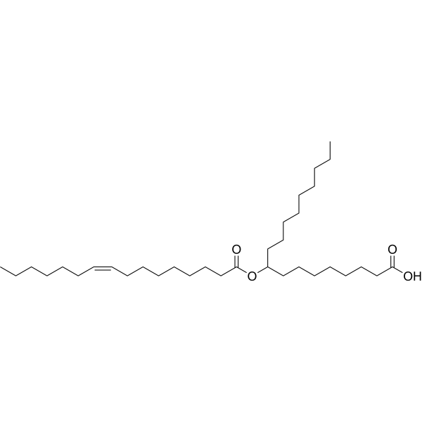
HY-131668
9-POHSAAnti-Inflammatory Agent -
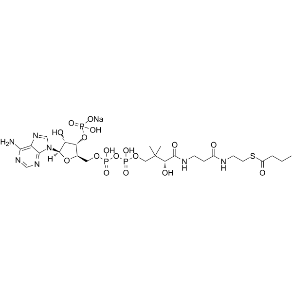
HY-138177
Butyryl-Coenzyme A sodiumIntermediate Metabolite -
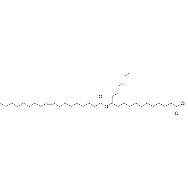
HY-113878
12-OAHSAAnti-Obese Agent -

HY-158583
13-POHSAFAHFA -
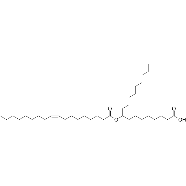
HY-131934
9-OAHSAAnti-Inflammatory reagent -
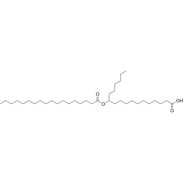
HY-130434
12-SAHSAFAHFA -

HY-116193R
5-PAHSA (Standard)Anti-Inflammatory Agent -

HY-130352
9-SAHSAFAHFA


Biological Activity
Description
In Vitro
5-POHSA (5-100 μM; 5 min) activates Src kinase in cultured mouse taste bud cells in a concentration-dependent manner[1].
5-POHSA (20 μM) significantly increases calcium influx in mouse taste bud cells cultured in vitro via a CD36-dependent mechanism; in mock-transfected cells, its combined use with Linoleic acid (HY-N0729) produces a synergistic effect on calcium signals, while this response is attenuated in CD36 siRNA-transfected cells[1].
MedChemExpress (MCE) has not independently confirmed the accuracy of these methods. They are for reference only.


Chemical Information
-
CAS No. 2161370-68-7
-
Molecular Weight 536.87
-
Formula C34H64O4
-
SMILES
CCCCCCCCCCCCCC(OC(CCCCCCC/C=C\CCCCCC)=O)CCCC(O)=O
-
Shipping
Room temperature in continental US; may vary elsewhere.
-
Storage
Please store the product under the recommended conditions in the Certificate of Analysis.


Purity & Documentation
References

Calculators
Concentration (start) × Volume (start) = Concentration (final) × Volume (final)


5-POHSA Related Classifications
HY-W769935 Related Classifications
 Powered by Bioz