



MM-206

Based on 1 Customer Validation
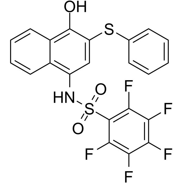
MM-206, a STAT3 activity inhibitor, potently inhibits the STAT3 SH2 domain-phosphopeptide interaction with IC50 of 1.2 μM. MM-206 demonstrates dose-dependent induction of apoptosis in acute myeloid leukemia (AML) cell lines.
For research use only. We do not sell to patients.
- Purity : 99.9%
- CAS No.: 1809581-87-0
- Formula: C22H12F5NO3S2
- Molecular Weight:497.46
-
Storage:Powder -20°C, 3 years ; In solvent -80°C, 6 months , -20°C, 1 month

To place orders, for customer services and technical support, please contact: MedChemExpress USA
Tel: 609-228-6898 E-mail: [email protected] [email protected]


-
 Biological Activity
-
Chemical Information
-
Solvent & Solubility
- Protocols
- Purity & Documentation
- References
-
 Help & FAQs


-
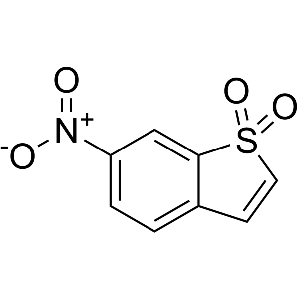
HY-13818
StatticSTAT3 Inhibitor -
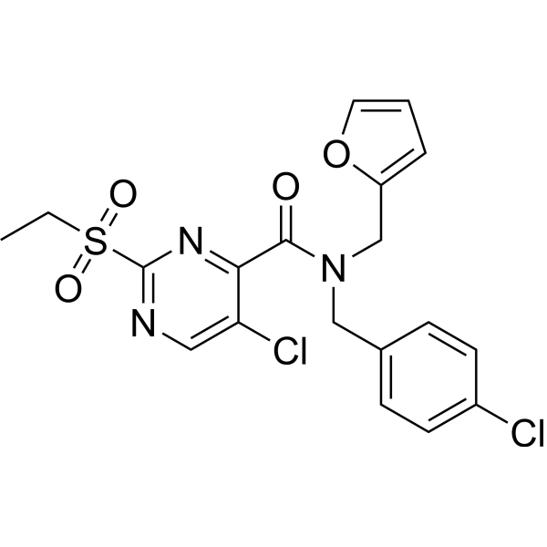
HY-136179
ZAP-180013ZAP-70 Inhibitor -
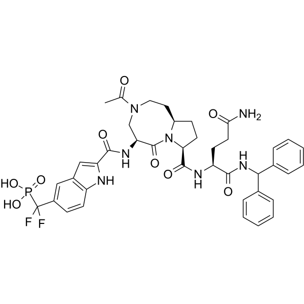
HY-129603
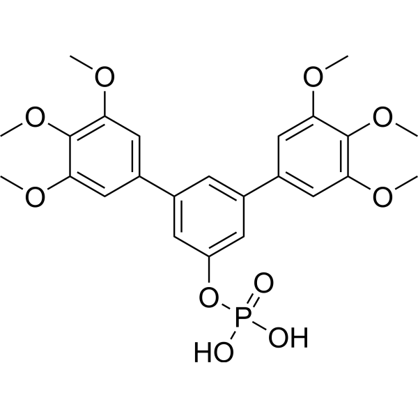
SI-109STAT3 Inhibitor -
HY-136546
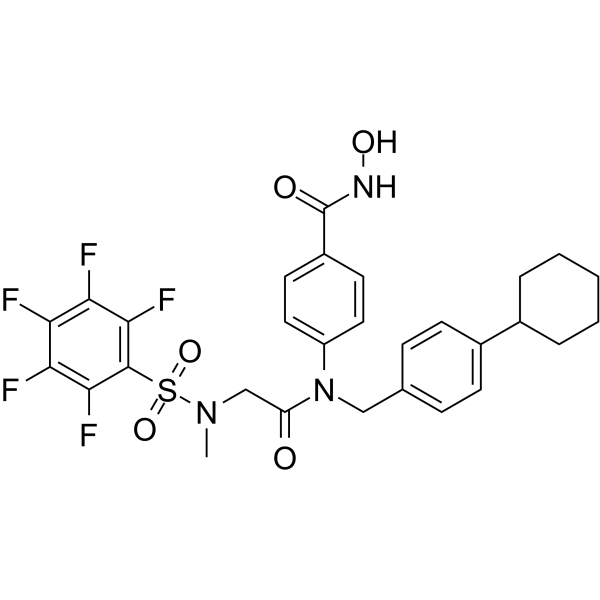
Stafia-1STAT5a Inhibitor -
HY-100494
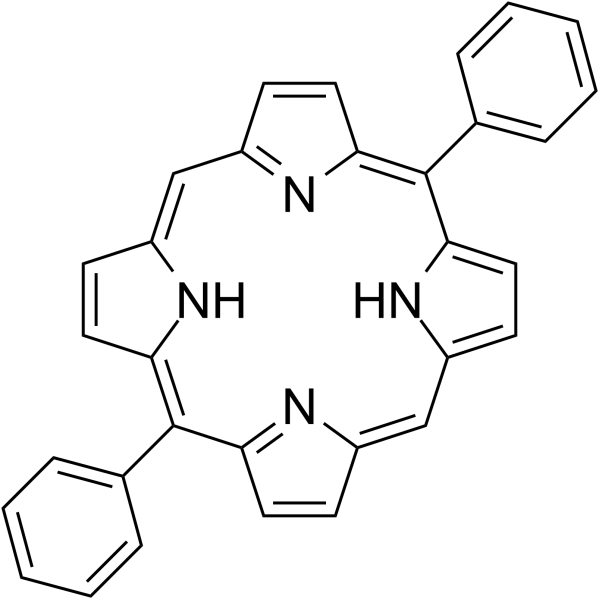
SH5-07STAT3 Inhibitor -
HY-W035137
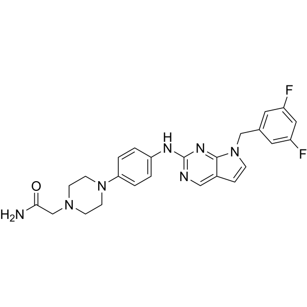
5,15-DiphenylporphyrinSTAT3 Antagonist -
HY-134772
AS1810722STAT6 Inhibitor -

HY-13818R
Stattic (Standard)STAT3 Inhibitor -
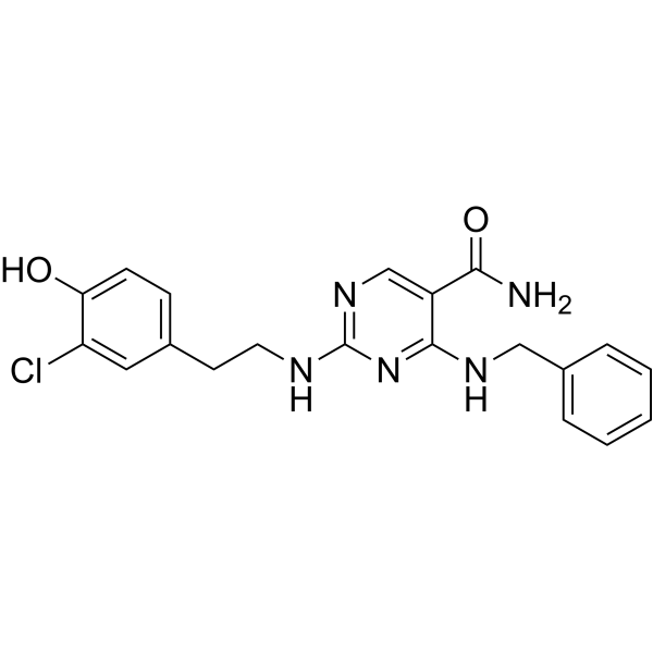
HY-100614
AS1517499STAT6 Inhibitor -
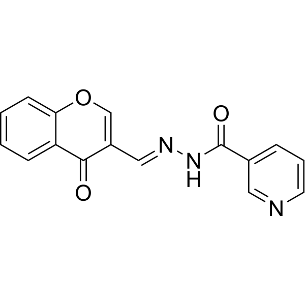
HY-101853
STAT5-IN-1STAT5 Inhibitor -
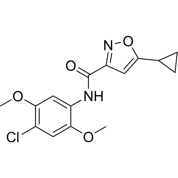
HY-111152
ML115STAT3 Agonist -
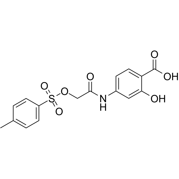
HY-15146
NSC 74859STAT3 Inhibitor


Biological Activity
Description


Chemical Information
-
CAS No. 1809581-87-0
-
Appearance Solid
-
Molecular Weight 497.46
-
Formula C22H12F5NO3S2
-
Color Off-white to light yellow
-
SMILES
O=S(C1=C(C(F)=C(C(F)=C1F)F)F)(NC2=C3C=CC=CC3=C(C(SC4=CC=CC=C4)=C2)O)=O
-
Shipping
Room temperature in continental US; may vary elsewhere.
-
Storage
Powder -20°C 3 years In solvent -80°C 6 months -20°C 1 month


Solvent & Solubility
In Vitro:
DMF : 25 mg/mL (50.26 mM; Need ultrasonic and warming)
Please refer to the solubility information to select the appropriate solvent. Once prepared, please aliquot and store the solution to prevent product inactivation from repeated freeze-thaw cycles.
Storage method and period of stock solution: -80°C, 6 months; -20°C, 1 month. When stored at -80°C, please use it within 6 months. When stored at -20°C, please use it within 1 month.
Please refer to the solubility information to select the appropriate solvent. Once prepared, please aliquot and store the solution to prevent product inactivation from repeated freeze-thaw cycles.
Storage method and period of stock solution: -80°C, 6 months; -20°C, 1 month. When stored at -80°C, please use it within 6 months. When stored at -20°C, please use it within 1 month.
Concentration (start) × Volume (start) = Concentration (final) × Volume (final)


Purity & Documentation
-
Data Sheet (275 KB)
-
SDS (251 KB)
- English - EN (251 KB)
- Français - FR (251 KB)
- Deutsch - DE (251 KB)
- Norwegian - NO (251 KB)
- Español - ES (251 KB)
- Swedish - SV (251 KB)
- Italian - IT (251 KB)
- Korean - KR (251 KB)
- Portuguese - PT (251 KB)
-
Handling Instructions (2659 KB)
References


Complete Stock Solution Preparation Table
Please refer to the solubility information to select the appropriate solvent. Once prepared, please aliquot and store the solution to prevent product inactivation from repeated freeze-thaw cycles.
Storage method and period of stock solution: -80°C, 6 months; -20°C, 1 month. When stored at -80°C, please use it within 6 months. When stored at -20°C, please use it within 1 month.
| Optional Solvent | Concentration Solvent Mass | 1 mg | 5 mg | 10 mg | 25 mg |
|---|---|---|---|---|---|
| DMF | 1 mM | 2.0102 mL | 10.0511 mL | 20.1021 mL | 50.2553 mL |
| 5 mM | 0.4020 mL | 2.0102 mL | 4.0204 mL | 10.0511 mL | |
| 10 mM | 0.2010 mL | 1.0051 mL | 2.0102 mL | 5.0255 mL | |
| 15 mM | 0.1340 mL | 0.6701 mL | 1.3401 mL | 3.3504 mL | |
| 20 mM | 0.1005 mL | 0.5026 mL | 1.0051 mL | 2.5128 mL | |
| 25 mM | 0.0804 mL | 0.4020 mL | 0.8041 mL | 2.0102 mL | |
| 30 mM | 0.0670 mL | 0.3350 mL | 0.6701 mL | 1.6752 mL | |
| 40 mM | 0.0503 mL | 0.2513 mL | 0.5026 mL | 1.2564 mL | |
| 50 mM | 0.0402 mL | 0.2010 mL | 0.4020 mL | 1.0051 mL |


MM-206 Related Classifications
HY-121725 Related Classifications
 Powered by Bioz