



BI-655064

Based on 1 Customer Validation
BI-655064, a humanised anti-CD40 antibody with has fragment crystallisable (Fc) regions with two mutations that prevent Fc-mediated antibody-dependent or complement-mediated cellular cytotoxicity and platelet activation. BI-655064 can be used for the study of autoimmune disease, such as lupus nephritis and rheumatoid arthritis (RA).
Nur für Forschungszwecke. Wir verkaufen nicht an Patienten.
- Reinheit : 98.56%
- Molecular Weight:146.74 kDa
-
Speicherung:
Please store the product under the recommended conditions in the Certificate of Analysis.


To place orders, for customer services and technical support, please contact: MedChemExpress USA
Tel: 609-228-6898 E-mail: [email protected] [email protected]


-
 Biologische Aktivität
-
Chemical Information
- Protokoll
- Reinheit & Dokumentation
- Verweise
-
 Help & FAQs


Alle TNF Receptor Isoform-spezifische Produkte anzeigen
More

Biologische Aktivität
Beschreibung
Isotype
Human IgG1 (L234A/L235A) kappa
Recommend Isotype Controls
Species Reactivity
Human
IC50 & Target
TNFRSF5/CD40
Gene ID
Accession
Conjugated
Unconjugated
Reconsititution
The product can be reconstituted/diluted with sterile PBS or saline.
Anwendung
ELISA, FACS, Functional assay
Verified Bioactivity
-
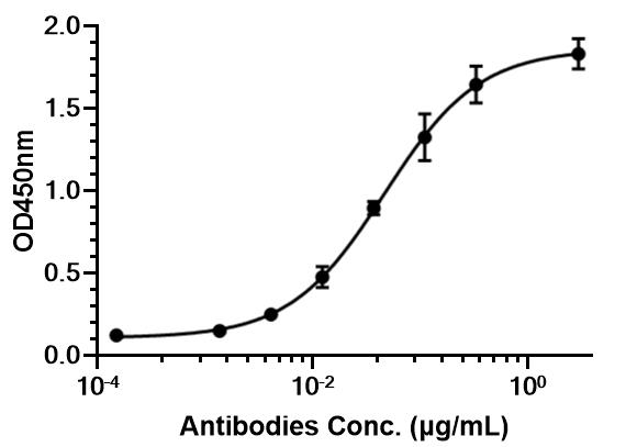 Immobilized hu-CD40-his can bind BI-655064. The EC50 for this effect is 46.74 ng/mL.


Chemical Information
-
Appearance Liquid
-
Molecular Weight 146.74 kDa
-
Color Colorless to light yellow
-
SMILES
N/A
-
Versand
Shipping with dry ice.
-
Formulation
Please refer to the lot-specific COA for specific buffer information.
-
Speicherung
Please store the product under the recommended conditions in the Certificate of Analysis.


Reinheit & Dokumentation
-
Data Sheet (255 KB)
-
SDS (251 KB)
- English - EN (251 KB)
- Français - FR (251 KB)
- Deutsch - DE (251 KB)
- Norwegian - NO (251 KB)
- Español - ES (251 KB)
- Swedish - SV (251 KB)
- Italian - IT (251 KB)
- Korean - KR (251 KB)
- Portuguese - PT (251 KB)
-
Inhibitory Antibodies User Guide (603 KB)
Verweise

Calculators
Konzentration (Stammlösung) × Volumen (Stammlösung) = Konzentration (Ziellösung) × Volumen (Ziellösung)


BI-655064 Related Classifications
HY-P991513 Related Classifications
 Powered by Bioz