



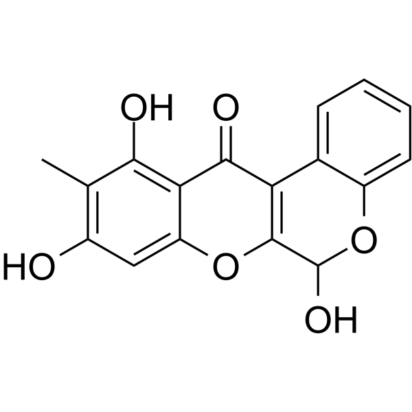
Boeravinone B

Based on 1 Customer Validation
Boeravinone B, a dual inhibitor of NorA bacterial efflux pump of Staphylococcus aureus and human P-Glycoprotein, reduces the biofilm formation and intracellular invasion of bacteria. Boeravinone B act as anti-aging and anti-apoptosis phyto-molecules during oxidative stress.
商品は「研究用試薬」です。人や動物の医療用・臨床診断用・食品用の製品ではありません。
研究用途以外に使用した場合、当社は一切の責任を負いかねます。
- 純度 : 97.59%
- CAS 番号: 114567-34-9
- 分子式: C17H12O6
- 分子量:312.27
-
保管条件:
4°C, protect from light
* In solvent : -80°C, 6 months; -20°C, 1 month (protect from light)

To place orders, for customer services and technical support, please contact: MedChemExpress USA
Tel: 609-228-6898 E-mail: [email protected] [email protected]


-
 生物活性
生物活性
-
化学情報
-
溶剤 & 溶解度
- プロトコル
- 純度とドキュメンテーション
- 参考文献
-
 Help & FAQs


-
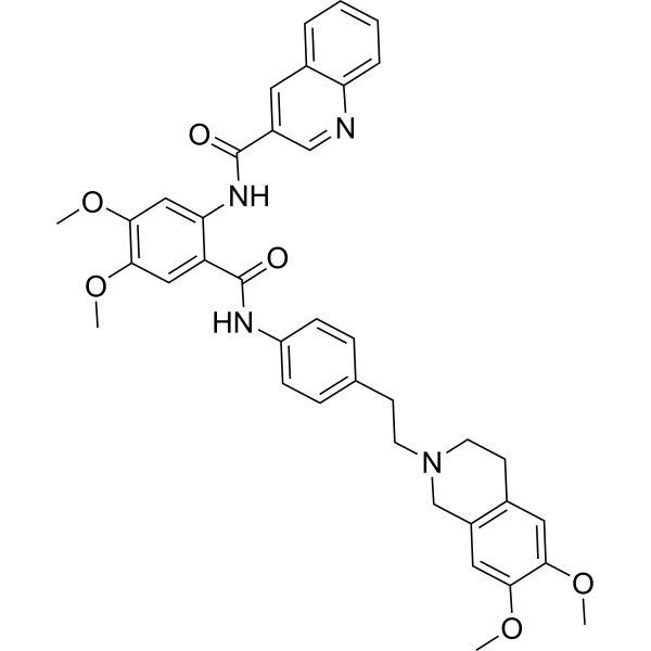
HY-10550
TariquidarP-gp Inhibitor -
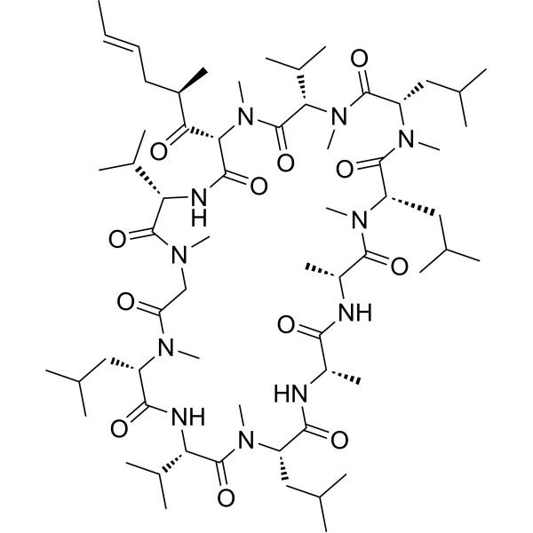
HY-17384
ValspodarP-glycoprotein Inhibitor -
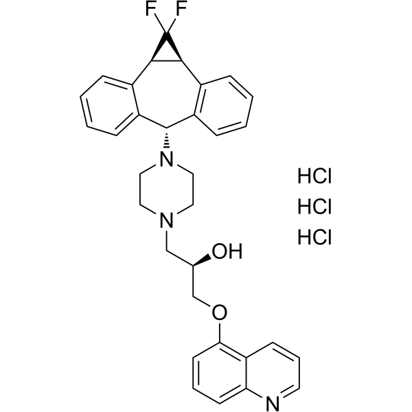
HY-50671
Zosuquidar trihydrochlorideP-gp Inhibitor -
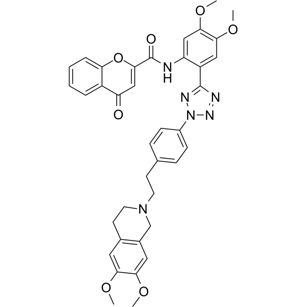
HY-13646
EncequidarP-glycoprotein Inhibitor -
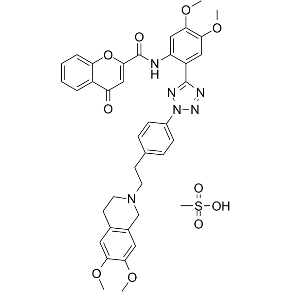
HY-13646A
Encequidar mesylateP-gp Inhibitor -
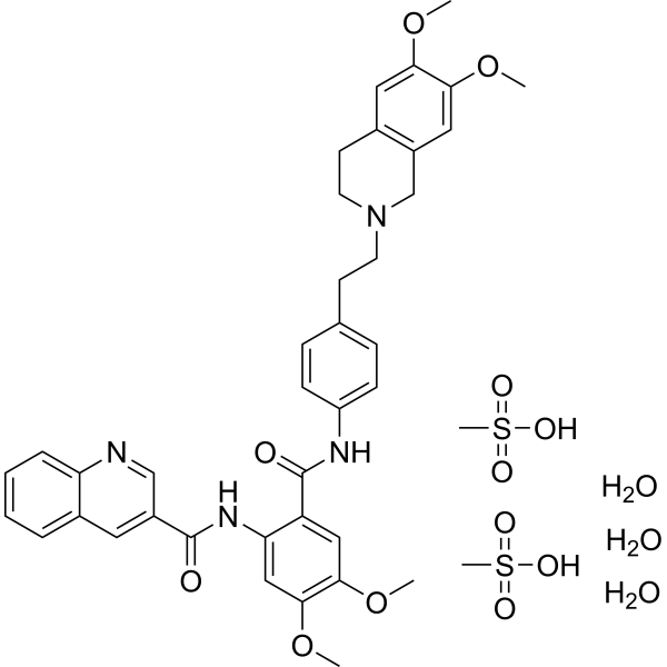
HY-10550A
Tariquidar methanesulfonate, hydrateP-gp Inhibitor -
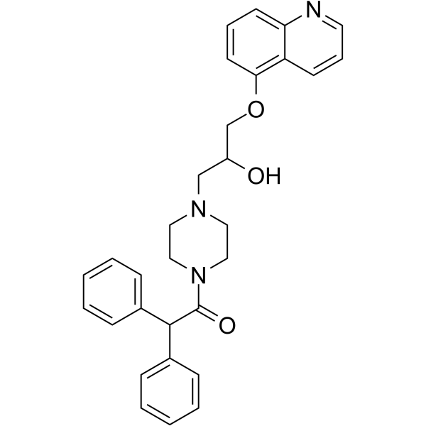
HY-17013
DofequidarP-glycoprotein Inhibitor -
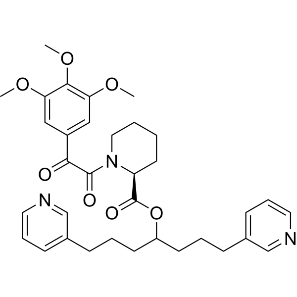
HY-13574A
BiricodarP-glycoprotein Modulator -
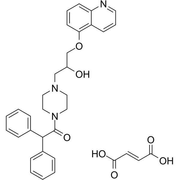
HY-17013A
Dofequidar fumarateP-glycoprotein Inhibitor -
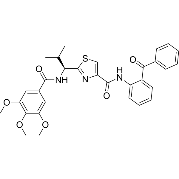
HY-101511
TTT-28ABCB1 Inhibitor -

HY-10550R
Tariquidar (Standard)P-gp Inhibitor -
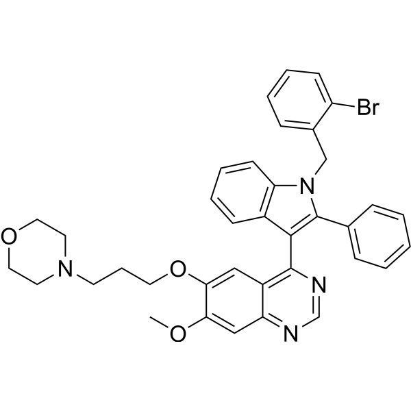
HY-132866
YS-370P-gp Inhibitor


生物活性
製品説明
体外実験
Boeravinone B is also active against a methicillin-resistant S. aureus (MRSA) strain, which showed a fourfold reduction in the ciprofloxacin MIC[1].
MedChemExpress (MCE) has not independently confirmed the accuracy of these methods. They are for reference only.


化学情報
-
CAS 番号 114567-34-9
-
性状 Solid
-
分子量 312.27
-
分子式 C17H12O6
-
Color Light yellow to yellow
-
SMILES
O=C1C2=C(C(O)OC3=CC=CC=C32)OC4=CC(O)=C(C)C(O)=C14
-
Structure Classification
-
Initial Source
-
輸送条件
Room temperature in continental US; may vary elsewhere.
-
保管条件
4°C, protect from light
* In solvent : -80°C, 6 months; -20°C, 1 month (protect from light)


溶剤 & 溶解度
体外:
DMSO : 50 mg/mL (160.12 mM; Need ultrasonic; Hygroscopic DMSO has a significant impact on the solubility of product, please use newly opened DMSO)
Please refer to the solubility information to select the appropriate solvent. Once prepared, please aliquot and store the solution to prevent product inactivation from repeated freeze-thaw cycles.
Storage method and period of stock solution: -80°C, 6 months; -20°C, 1 month (protect from light). When stored at -80°C, please use it within 6 months. When stored at -20°C, please use it within 1 month.
Please refer to the solubility information to select the appropriate solvent. Once prepared, please aliquot and store the solution to prevent product inactivation from repeated freeze-thaw cycles.
Storage method and period of stock solution: -80°C, 6 months; -20°C, 1 month (protect from light). When stored at -80°C, please use it within 6 months. When stored at -20°C, please use it within 1 month.
濃度 (開始) × 体積 (開始) = 濃度 (終了) × 体積 (終了)


純度とドキュメンテーション
-
データシート (275 KB)
-
SDS (623 KB)
- English - EN (623 KB)
- Français - FR (623 KB)
- Deutsch - DE (623 KB)
- Norwegian - NO (623 KB)
- Español - ES (623 KB)
- Swedish - SV (623 KB)
- Italian - IT (623 KB)
- Korean - KR (623 KB)
- Portuguese - PT (623 KB)
-
取扱説明書 (2659 KB)
参考文献
[1]. Samsher Singh, Boeravinone B, A Novel Dual Inhibitor of NorA Bacterial Efflux Pump of Staphylococcus aureus and Human P-Glycoprotein, Reduces the Biofilm Formation and Intracellular Invasion of Bacteria. Front Microbiol. 2017 Oct 4;8:1868. [Content Brief]
[2]. Shivtej P Biradar, et al. Chebulinic acid and Boeravinone B act as anti-aging and anti-apoptosis phyto-molecules during oxidative stress. Mitochondrion. 2019 May;46:236-246. [Content Brief]


Complete Stock Solution Preparation Table
Please refer to the solubility information to select the appropriate solvent. Once prepared, please aliquot and store the solution to prevent product inactivation from repeated freeze-thaw cycles.
Storage method and period of stock solution: -80°C, 6 months; -20°C, 1 month (protect from light). When stored at -80°C, please use it within 6 months. When stored at -20°C, please use it within 1 month.
| Optional Solvent | Concentration Solvent Mass | 1 mg | 5 mg | 10 mg | 25 mg |
|---|---|---|---|---|---|
| DMSO | 1 mM | 3.2024 mL | 16.0118 mL | 32.0236 mL | 80.0589 mL |
| 5 mM | 0.6405 mL | 3.2024 mL | 6.4047 mL | 16.0118 mL | |
| 10 mM | 0.3202 mL | 1.6012 mL | 3.2024 mL | 8.0059 mL | |
| 15 mM | 0.2135 mL | 1.0675 mL | 2.1349 mL | 5.3373 mL | |
| 20 mM | 0.1601 mL | 0.8006 mL | 1.6012 mL | 4.0029 mL | |
| 25 mM | 0.1281 mL | 0.6405 mL | 1.2809 mL | 3.2024 mL | |
| 30 mM | 0.1067 mL | 0.5337 mL | 1.0675 mL | 2.6686 mL | |
| 40 mM | 0.0801 mL | 0.4003 mL | 0.8006 mL | 2.0015 mL | |
| 50 mM | 0.0640 mL | 0.3202 mL | 0.6405 mL | 1.6012 mL | |
| 60 mM | 0.0534 mL | 0.2669 mL | 0.5337 mL | 1.3343 mL | |
| 80 mM | 0.0400 mL | 0.2001 mL | 0.4003 mL | 1.0007 mL | |
| 100 mM | 0.0320 mL | 0.1601 mL | 0.3202 mL | 0.8006 mL |


Boeravinone B 関連分類
HY-N2947 関連分類
 Powered by Bioz