



Bortezomib-d8
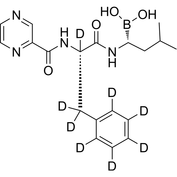
Bortezomib-d8 is the deuterium labeled Bortezomib. Bortezomib (PS-341) is a reversible and selective proteasome inhibitor, and potently inhibits 20S proteasome (Ki=0.6 nM) by targeting a threonine residue. Bortezomib disrupts the cell cycle, induces apoptosis, and inhibits NF-κB. Bortezomib is the first proteasome inhibitor anticancer agent. Bortezomib can be used for the study of multiple myeloma (MM). Bortezomib effectively inhibits TREM2 expression in tumor-associated macrophages (TAMs).
For research use only. We do not sell to patients.
- Formula: C19H17D8BN4O4
- Molecular Weight:392.29
-
Storage:
-20°C, stored under nitrogen
* In solvent : -80°C, 6 months; -20°C, 1 month (stored under nitrogen)

To place orders, for customer services and technical support, please contact: MedChemExpress USA
Tel: 609-228-6898 E-mail: [email protected] [email protected]


-
 Biological Activity
-
Chemical Information
-
Solvent & Solubility
- Protocols
- Purity & Documentation
- References
-
 Help & FAQs




Biological Activity
Description
In Vitro
Stable heavy isotopes of hydrogen, carbon, and other elements have been incorporated into drug molecules, largely as tracers for quantitation during the drug development process. Deuteration has gained attention because of its potential to affect the pharmacokinetic and metabolic profiles of drugs[1].
MedChemExpress (MCE) has not independently confirmed the accuracy of these methods. They are for reference only.
- RNA extraction experimental
- Detection of Apoptosis
- TUNEL staining for apoptotic DNA fragmentation
- Flow cytometric DNA-content cell-cycle staining
- Annexin V plus membrane-impermeant dye apoptosis staining
- BrdU Incorporation Assay
- Apoptosis Solutions
- Protocol for Cell Cycle
- Research Protocol for Inflammation-related Diseases
Application
1. This compound can be used as a tracer
2. This compound can be used as an internal standard for quantitative analysis by NMR, GC-MS, or LC-MS.


Chemical Information
-
Unlabeled CAS 179324-69-7
-
Appearance Solid
-
Molecular Weight 392.29
-
Formula C19H17D8BN4O4
-
Color White to off-white
-
SMILES
OB([C@H](CC(C)C)NC(C(NC(C1=NC=CN=C1)=O)([2H])C([2H])([2H])C2=C([2H])C([2H])=C([2H])C([2H])=C2[2H])=O)O
-
Synonyms
PS-341-d8; LDP-341-d8; NSC 681239-d8
-
Shipping
Room temperature in continental US; may vary elsewhere.
-
Storage
-20°C, stored under nitrogen
* In solvent : -80°C, 6 months; -20°C, 1 month (stored under nitrogen)


Solvent & Solubility
In Vitro:
Ethanol : 66.67 mg/mL (169.95 mM; ultrasonic and warming and heat to 60°C)
DMSO : 50 mg/mL (127.46 mM; Need ultrasonic; Hygroscopic DMSO has a significant impact on the solubility of product, please use newly opened DMSO)
Please refer to the solubility information to select the appropriate solvent. Once prepared, please aliquot and store the solution to prevent product inactivation from repeated freeze-thaw cycles.
Storage method and period of stock solution: -80°C, 6 months; -20°C, 1 month (stored under nitrogen). When stored at -80°C, please use it within 6 months. When stored at -20°C, please use it within 1 month.
Please refer to the solubility information to select the appropriate solvent. Once prepared, please aliquot and store the solution to prevent product inactivation from repeated freeze-thaw cycles.
Storage method and period of stock solution: -80°C, 6 months; -20°C, 1 month (stored under nitrogen). When stored at -80°C, please use it within 6 months. When stored at -20°C, please use it within 1 month.
Concentration (start) × Volume (start) = Concentration (final) × Volume (final)


Purity & Documentation
-
Data Sheet (280 KB)
-
SDS (252 KB)
- English - EN (252 KB)
- Français - FR (252 KB)
- Deutsch - DE (252 KB)
- Norwegian - NO (252 KB)
- Español - ES (252 KB)
- Swedish - SV (252 KB)
- Italian - IT (252 KB)
- Korean - KR (252 KB)
- Portuguese - PT (252 KB)
-
Handling Instructions (2659 KB)
References
[1]. Russak EM, et al. Impact of Deuterium Substitution on the Pharmacokinetics of Pharmaceuticals. Ann Pharmacother. 2019;53(2):211-216. [Content Brief]
[2]. Adams J, et al. Proteasome inhibitors: a novel class of potent and effective antitumor agents. Cancer Res. 1999 Jun 1;59(11):2615-22. [Content Brief]
[3]. Shahshahan MA, et al. Potential usage of proteasome inhibitor bortezomib (Velcade, PS-341) in the treatment of metastaticmelanoma: basic and clinical aspects. Am J Cancer Res. 2011;1(7):913-24. [Content Brief]
[4]. Pérez-Galán P, et al. The proteasome inhibitor bortezomib induces apoptosis in mantle-cell lymphoma through generation of ROS and Noxa activation independent of p53 status. Blood. 2006 Jan 1;107(1):257-64. [Content Brief]
[5]. Yerlikaya A, et al. Combined effects of the proteasome inhibitor bortezomib and Hsp70 inhibitors on the B16F10 melanoma cell line. Mol Med Rep. 2010 Mar-Apr;3(2):333-9. [Content Brief]
[6]. Mujtaba T, et al. Advances in the understanding of mechanisms and therapeutic use of bortezomib. Discov Med. 2011 Dec;12(67):471-80. [Content Brief]
[7]. Fernández Y, et al. Chemical blockage of the proteasome inhibitory function of bortezomib: impact on tumor cell death. J Biol Chem. 2006 Jan 13;281(2):1107-18. [Content Brief]
[8]. Wang Y, et al. TREM2-Mediated Cholesterol Efflux in Macrophages Inhibits Anti-Tumor Immunity via Limitation of CD4+ T and NK Cells. Adv Sci (Weinh). 2025 Oct 20:e06995. [Content Brief]


Complete Stock Solution Preparation Table
Please refer to the solubility information to select the appropriate solvent. Once prepared, please aliquot and store the solution to prevent product inactivation from repeated freeze-thaw cycles.
Storage method and period of stock solution: -80°C, 6 months; -20°C, 1 month (stored under nitrogen). When stored at -80°C, please use it within 6 months. When stored at -20°C, please use it within 1 month.
| Optional Solvent | Concentration Solvent Mass | 1 mg | 5 mg | 10 mg | 25 mg |
|---|---|---|---|---|---|
| DMSO / Ethanol | 1 mM | 2.5491 mL | 12.7457 mL | 25.4913 mL | 63.7284 mL |
| 5 mM | 0.5098 mL | 2.5491 mL | 5.0983 mL | 12.7457 mL | |
| 10 mM | 0.2549 mL | 1.2746 mL | 2.5491 mL | 6.3728 mL | |
| 15 mM | 0.1699 mL | 0.8497 mL | 1.6994 mL | 4.2486 mL | |
| 20 mM | 0.1275 mL | 0.6373 mL | 1.2746 mL | 3.1864 mL | |
| 25 mM | 0.1020 mL | 0.5098 mL | 1.0197 mL | 2.5491 mL | |
| 30 mM | 0.0850 mL | 0.4249 mL | 0.8497 mL | 2.1243 mL | |
| 40 mM | 0.0637 mL | 0.3186 mL | 0.6373 mL | 1.5932 mL | |
| 50 mM | 0.0510 mL | 0.2549 mL | 0.5098 mL | 1.2746 mL | |
| 60 mM | 0.0425 mL | 0.2124 mL | 0.4249 mL | 1.0621 mL | |
| 80 mM | 0.0319 mL | 0.1593 mL | 0.3186 mL | 0.7966 mL | |
| 100 mM | 0.0255 mL | 0.1275 mL | 0.2549 mL | 0.6373 mL |


Bortezomib-d8 Related Classifications
HY-10227S Related Classifications
 Powered by Bioz