



Broussonin A

Based on 1 Customer Validation
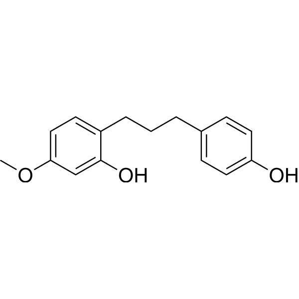
Broussonin A is a potent BChE inhibitor, with an IC50 of 4.16 µM. Broussonin A is a diarylpropane natural product that can be isolated from the bark of Broussonetia papyrifera after solid fermentation.
Nur für Forschungszwecke. Wir verkaufen nicht an Patienten.
- Reinheit : 97%
- CAS. Nr.: 73731-87-0
- Formel: C16H18O3
- Molecular Weight:258.31
-
Speicherung:
-20°C, protect from light
* In solvent : -80°C, 6 months; -20°C, 1 month (protect from light)

To place orders, for customer services and technical support, please contact: MedChemExpress USA
Tel: 609-228-6898 E-mail: [email protected] [email protected]


-
 Biologische Aktivität
-
Chemical Information
- Protokoll
- Reinheit & Dokumentation
- Verweise
-
 Help & FAQs




Biologische Aktivität
Beschreibung
IC50 & Target
|
BChE 4.16 μM (IC50) |
Cellular Effect
|
Cell Line

|
Type | Value | Description | References |
|---|---|---|---|---|
| 3T3-L1 | IC50 |
74.5 μM
Compound: 8
|
Antiadipogenic activity against mouse 3T3L1 cells assessed as inhibition of differentiation after 7 days by oil-red O staining
Antiadipogenic activity against mouse 3T3L1 cells assessed as inhibition of differentiation after 7 days by oil-red O staining
|
[PMID: 19757853] |
| CV-1 | EC50 |
0.53 μM
Compound: 1
|
Transactivation of human recombinant ERbeta expressed in african green monkey CV1 cells co-expressing ERE-Luc after 24 hrs by luciferase reporter gene assay
Transactivation of human recombinant ERbeta expressed in african green monkey CV1 cells co-expressing ERE-Luc after 24 hrs by luciferase reporter gene assay
|
[PMID: 20493686] |
| CV-1 | EC50 |
1.59 μM
Compound: 1
|
Transactivation of human recombinant ERalpha expressed in african green monkey CV1 cells co-expressing ERE-Luc after 24 hrs by luciferase reporter gene assay
Transactivation of human recombinant ERalpha expressed in african green monkey CV1 cells co-expressing ERE-Luc after 24 hrs by luciferase reporter gene assay
|
[PMID: 20493686] |
In Vitro
Broussonin A (0.1-10 µM, 30 min) suppresses VEGF-A-stimulated endothelial cell proliferation by regulating the expression of cell cycle-related proteins and the phosphorylation status of retinoblastoma protein[1].
Broussonin A (0-10 µM, 30 min)abrogates VEGF-A-stimulated angiogenic responses including endothelial cell migration, invasion, tube formation and microvessel formation from rat aortic rings[1].
MedChemExpress (MCE) has not independently confirmed the accuracy of these methods. They are for reference only. Further protocols information, click here.


Chemical Information
-
CAS. Nr. 73731-87-0
-
Appearance Solid
-
Molecular Weight 258.31
-
Formel C16H18O3
-
Color White to off-white
-
SMILES
OC1=CC(OC)=CC=C1CCCC2=CC=C(O)C=C2
-
Structure Classification
-
Versand
Room temperature in continental US; may vary elsewhere.
-
Speicherung
-20°C, protect from light
* In solvent : -80°C, 6 months; -20°C, 1 month (protect from light)

Protokoll
-
Cell Cytotoxicity Assay
Cytotoxicity assays are usually based on the assessment of cell membrane damage, which can also be indirectly detected by measuring cell viability. Detection methods include MTT assay, CKK-8 assay, LDH assay and ATP assay, etc.


Reinheit & Dokumentation
-
Data Sheet (266 KB)
-
SDS (393 KB)
- English - EN (393 KB)
- Français - FR (393 KB)
- Deutsch - DE (393 KB)
- Norwegian - NO (393 KB)
- Español - ES (393 KB)
- Swedish - SV (393 KB)
- Italian - IT (393 KB)
- Korean - KR (393 KB)
- Portuguese - PT (393 KB)
-
Handling Instructions (2659 KB)
Verweise
[1]. Kim JH, et al. Broussonin A- and B-mediated inhibition of angiogenesis by blockade of VEGFR-2 signalling pathways and integrin β1 expression. J Cell Mol Med. 2022 Feb;26(4):1194-1205. [Content Brief]
[2]. Lee JP, et al. Potent inhibition of acetylcholinesterase by sargachromanol I from Sargassum siliquastrum and by selected natural compounds. Bioorg Chem. 2019 Aug;89:103043. [Content Brief]

Calculators
Konzentration (Stammlösung) × Volumen (Stammlösung) = Konzentration (Ziellösung) × Volumen (Ziellösung)


Broussonin A Related Classifications
HY-N2960 Related Classifications
 Powered by Bioz