



Lokivetmab

Based on 1 Customer Validation
Lokivetmab (Anti-Canine IL31 Recombinant Antibody) is an anti-canine IL-31 monoclonal antibody. Lokivetmab inhibits IL-31-mediated activation of pruritogenic signals in peripheral sensory neurons and reduces TH2-weighted inflammation. Lokivetmab demonstrates long-term efficacy in controlling pruritus and improving skin lesions in dogs with canine atopic dermatitis (CAD). Lokivetmab can be used for the research of atopic dermatitis (AD) in dogs.
For research use only. We do not sell to patients.
- Purity : 99.34%
- CAS No.: 1533403-95-0
- Molecular Weight:147.51 kDa
-
Storage:
Please store the product under the recommended conditions in the Certificate of Analysis.


To place orders, for customer services and technical support, please contact: MedChemExpress USA
Tel: 609-228-6898 E-mail: [email protected] [email protected]


-
 Biological Activity
-
Chemical Information
- Protocols
- Purity & Documentation
- References
-
 Help & FAQs


-

HY-P703718
IL-31 Protein, Canine (HEK293, His) -

HY-P99045
SacituzumabAnti-TROP-2 Antibody -

HY-138298A
Trastuzumab deruxtecanHER2 ADC -

HY-P99557
TirnovetmabIL-31 Neutralizer -

HY-P99297
BedinvetmabAnti-Canine NGF mAb -

HY-P99380
ZanidatamabAnti- HER2 Antibody -

HY-P99042
PolatuzumabAnti-CD79b mAb -

HY-141601
Telisotuzumab vedotinAnti-c-MET ADC


Biological Activity
Description
Isotype
Canine IgG2 kappa
Recommend Isotype Controls
Species Reactivity
Canine
IC50 & Target
[1]|
IL-31 |
In Vivo
MedChemExpress (MCE) has not independently confirmed the accuracy of these methods. They are for reference only.
-
Animal Model:Canine atopic dermatitis (CAD) dog model[1]
-
Dosage:2 mg/kg
-
Administration:i.v. injection, every 4-8 weeks, 12 months
-
Result:Achieved pruritus control with 87% (64/75) of dogs maintaining PVAS below baseline.
Improved skin lesions with mean VetVAS (skin lesion score).
Gene ID
256353430 [NCBI]
Accession
BAH97742
Conjugated
Unconjugated
Reconsititution
The product can be reconstituted/diluted with sterile PBS or saline.
Format
-
IgG2-kappa
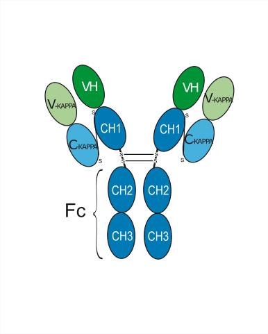
Application
ELISA, FACS, Functional assay
Verified Bioactivity
-
 Immobilized IL-31 Protein, Canine (HEK293, His, HY-P703718) can bind Lokivetmab. The EC50 for this effect is 59.37 ng/mL.


Chemical Information
-
CAS No. 1533403-95-0
-
Appearance Liquid
-
Molecular Weight 147.51 kDa
-
Color Colorless to light yellow
-
SMILES
[Lokivetmab]
-
Synonyms
Anti-Canine IL31 Recombinant Antibody; CAN34D3-65; PF-06443537
-
Shipping
Shipping with dry ice.
-
Formulation
Please refer to the lot-specific COA for specific buffer information.
-
Storage
Please store the product under the recommended conditions in the Certificate of Analysis.

Protocols
-
Naïve CD4+ T-cell subset differentiation/polarization
Naïve CD4+ T-cell subset differentiation/polarization is an in vitro assay in which purified naïve CD4+ T cells are activated through TCR and CD28 costimulation and cultured with defined cytokines and neutralizing antibodies to generate Th0, Th1, Th2, Th17, or induced Treg-like populations. Differentiation is detected by subset-associated cytokines and transcription factors: IFN-γ/T-bet for Th1, IL-4/GATA3 for Th2, IL-17A/RORγt for Th17, and Foxp3 for induced Treg cells. The assay readout is usually generated by intracellular cytokine staining after restimulation, transcription-factor staining by flow cytometry, ELISA of secreted cytokines, or gene-expression analysis. The result reflects cytokine-directed lineage commitment or polarization rather than antigen-specific immune protection by itself.
-
TPA/Croton Oil Ear Edema and Dermatitis
The TPA (12-O-tetradecanoylphorbol-13-acetate) and croton oil-induced mouse ear edema model is a well-established acute cutaneous inflammation system used to evaluate topical anti-inflammatory activity by measuring edema formation, neutrophil infiltration, vascular permeability, and cytokine-mediated skin responses in vivo. The inflammatory response is triggered by topical application of phorbol esters (TPA) or croton oil constituents, leading to rapid activation of protein kinase C signaling, leukocyte recruitment, and increased vascular permeability, which can be quantified by ear thickness, weight, dye extravasation, and biochemical markers such as myeloperoxidase (MPO) activity and pro-inflammatory mediators in ear tissue homogenates. This model is widely used for screening anti-inflammatory agents, where reductions in edema and inflammatory biomarkers reflect suppression of acute dermal inflammation and immune cell infiltration. Histological evaluation typically confirms epidermal
-
Research Protocol for Inflammation-related Diseases
The NLRP3 inflammasome is a cytosolic innate immune signaling platform that integrates priming signals and danger-signal activation to promote caspase-1 activation, maturation of IL-1β and IL-18, and gasdermin D-mediated pyroptotic cell death. The core experimental logic is to determine whether inflammatory disease phenotypes are driven by increased NLRP3 expression, ASC-containing inflammasome assembly, caspase-1 cleavage, GSDMD cleavage, and extracellular release of IL-1β/IL-18 rather than by nonspecific cell injury alone. The pathway is strongly linked to inflammation-related disease phenotypes because monosodium urate crystals activate NALP3/NLRP3 inflammasome signaling in gout-like crystal inflammation, cholesterol crystals activate NLRP3 inflammasomes in atherogenesis models, and DSS-induced intestinal inflammation has been reported to involve NLRP3 inflammasome activity. However, experimental colitis studies also show context-dependent protective effects of NLRP3 inflammasome co
-
Recombinant antibody expression and Protein A/G purification
Recombinant antibody expression produces antibody chains from cloned antibody genes in mammalian cells, commonly HEK293-derived cells, so that secreted IgG or Fc-fusion antibodies can be collected from culture supernatant. Protein A/G purification is affinity chromatography in which immobilized Protein A, Protein G, or Protein A/G binds the antibody Fc region; nonbound culture components are washed away, and bound antibody is recovered by changing buffer conditions, commonly acidic elution followed by neutralization.


Purity & Documentation
-
Data Sheet (256 KB)
-
SDS (252 KB)
- English - EN (252 KB)
- Français - FR (252 KB)
- Deutsch - DE (252 KB)
- Norwegian - NO (252 KB)
- Español - ES (252 KB)
- Swedish - SV (252 KB)
- Italian - IT (252 KB)
- Korean - KR (252 KB)
- Portuguese - PT (252 KB)
-
Inhibitory Antibodies User Guide (603 KB)
References

Calculators
Concentration (start) × Volume (start) = Concentration (final) × Volume (final)


Lokivetmab Related Classifications
HY-P99301 Related Classifications
 Powered by Bioz