



Cephaeline hydrochloride
Based on 2 publication(s) in Google Scholar
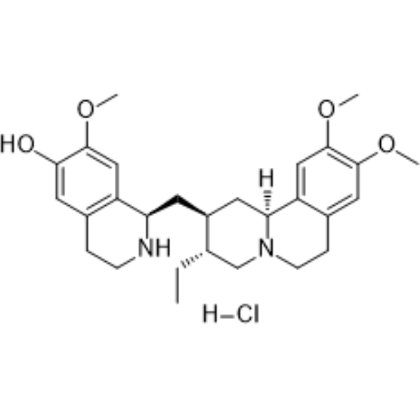
Cephaeline hydrochloride ((-)-Cephaeline (hydrochloride); NSC 32944 (monohydrochloride)) is a ferroptosis inducer, with broad-spectrum anticancer and antiviral activities. Cephaeline hydrochloride induces Ferroptosis by upregulating p53, inhibiting NRF2, activating ULK3, downregulating the expressions of SLC7A11 and GPX4 in a p53-dependent manner, reducing GSH and mitochondrial membrane potential, and increasing lipid peroxidation and iron accumulation. Cephaeline hydrochloride inhibits cancer cell proliferation, migration and tumor growth. Cephaeline hydrochloride inhibits Ebola virus (EBOV) VLP entry and infection, with IC50 values of 3.27 μM and 22.18 μM respectively; it also inhibits Zika virus (ZIKV) NS5 RdRp activity (IC50 = 976 nM), and binds to severe acute respiratory syndrome coronavirus 2 (SARS-CoV-2) RdRp and N protein, with Kd values of 8.9 μM and 53.8 μM respectively. Cephaeline hydrochloride can be used in studies related to breast cancer, lung cancer, COVID-19, EBOV and ZIKV infections.
For research use only. We do not sell to patients.
- Purity : 98.08%
- CAS No.: 3738-70-3
- Formula: C28H39ClN2O4
- Molecular Weight:503.07
-
Storage:Powder -20°C, 3 years , 4°C, 2 years ; In solvent -80°C, 6 months , -20°C, 1 month

To place orders, for customer services and technical support, please contact: MedChemExpress USA
Tel: 609-228-6898 E-mail: [email protected] [email protected]


-
 Biological Activity
-
Chemical Information
-
Solvent & Solubility
- Protocols
- Purity & Documentation
- References
-
 Help & FAQs


Publications Citing Use of MedChemExpress (MCE) Cephaeline hydrochloride
More Customer Validation & Images

-
Cell Proliferation/Viability Assay
-
Cell Migration/Invasion Assay
-
WB


Biological Activity
Description
IC50 & Target
|
EBOV 22.18 μM (IC50) |
ZIKV NS5 RdRp 976 nM (IC50) |
SARS-CoV-2 RdRp 8.9 μM (Kd) |
SARS-CoV-2 N protein 53.8 μM (Kd) |
GPX4 |
In Vitro
Cephaeline hydrochloride (2.5-320 nM; 72 h) potently inhibits the proliferation of 4T1 and MDA-MB-231 breast cancer cells, with IC50 values of 38.89 nM and 50.29 nM, respectively[2].
Cephaeline hydrochloride (80 nM; 24 h) inhibits the migration of 4T1 and MDA-MB-231 breast cancer cells[2].
Cephaeline (14 day) hydrochloride inhibits colony formation of 4T1 and MDA-MB-231 breast cancer cells, and this effect is attenuated by p53 siRNA knockdown[2].
Cephaeline hydrochloride (5-400 nM; 24-72 h) potently inhibits the viability of H460 and A549 human lung cancer cells in a time-dependent manner, with IC50 values ranging from 35 nM to 89 nM[1].
Cephaeline hydrochloride (0.01-30 μM; 48 h) inhibits the cell viability of mucoepidermoid carcinoma cell lines UM-HMC-1, UM-HMC-2 and UM-HMC-3A, with IC50 values of 0.16 μM, 2.08 μM and 0.02 μM, respectively[4].
Cephaeline hydrochloride (at the corresponding IC50 concentration; 72 h) inhibits the cell growth of UM-HMC-1 and UM-HMC-2 mucoepidermoid carcinoma cell lines, while the growth inhibitory effect on UM-HMC-3A is not statistically significant[4].
Cephaeline hydrochloride (at respective IC50 concentrations) inhibits cell migration of the mucoepidermoid carcinoma cell lines UM-HMC-1, UM-HMC-2, and UM-HMC-3A, with the onset time of inhibition varying among different cell lines[4].
Cephaeline hydrochloride (at the corresponding IC50 concentration; 24-48 h) increases the acetylation level of histone H3 lysine 9 in the mucoepidermoid carcinoma cell lines UM-HMC-1, UM-HMC-2 and UM-HMC-3A[4].
Cephaeline hydrochloride (at the corresponding IC50 concentration; 24 h) reduces the level of ALDH+ cancer stem cells in UM-HMC-2 mucoepidermoid carcinoma cells, but increases the ALDH+ level in UM-HMC-1 and UM-HMC-3A cells[4].
Cephaeline hydrochloride (at the corresponding IC50 concentration; 5 days) completely inhibits tumor sphere formation in UM-HMC-1 and UM-HMC-2 mucoepidermoid carcinoma cells, and reduces tumor sphere formation in UM-HMC-3A cells[4].
Cephaeline hydrochloride (20-80 nM; 72 h) reduces intracellular GSH levels in a concentration-dependent manner and increases intracellular MDA levels in 4T1 and MDA-MB-231 breast cancer cells[2].
Cephaeline hydrochloride (20-80 nM; 72 h) increases intracellular ROS levels in 4T1 and MDA-MB-231 breast cancer cells in a concentration-dependent manner[2].
Cephaeline hydrochloride (20-80 nM; 72 h) increases the levels of total iron and Fe2+ in 4T1 and MDA-MB-231 breast cancer cells in a concentration-dependent manner[2].
Cephaeline hydrochloride (20-80 nM; 72 h) downregulates the protein expression of SLC7A11 and GPX4 in a concentration-dependent manner, and upregulates the protein expression of p53 in 4T1 and MDA-MB-231 breast cancer cells[2].
The ferroptosis and cell death induced in human lung cancer cells H460 and A549 by cephaeline hydrochloride (100 nM; 24 h) are mediated by targeting NRF2, as pretreatment with the NRF2 agonist TBHQ reverses these effects[1].
Cephaeline hydrochloride (80 nM; 72 h) induces ferroptosis-related changes (decreased GSH levels, increased MDA and ROS levels, downregulated expression of SLC7A11 and GPX4) in 4T1 and MDA-MB-231 breast cancer cells, and these effects are blocked by p53 siRNA knockdown[2].
Cephaeline hydrochloride (25-100 nM; 24 h) increases intracellular iron levels and reactive oxygen species levels in H460 and A549 human lung cancer cells[1].
Cephaeline hydrochloride (25-100 nM; 24 h) regulates the expression of ferroptosis- and iron metabolism-related genes in H460 and A549 human lung cancer cells, downregulates the expression of GPX4, SLC7A11 and SLC40A1, and upregulates the expression of transferrin[1].
Cephaeline hydrochloride (25-100 nM; 24 h) downregulates the protein levels of GPX4, SLC7A11, SLC40A1 and NRF2, while upregulates the protein expression of transferrin in human lung cancer H460 and A549 cells[1].
Cephaeline hydrochloride (25-100 nM; 24 h) induces lipid peroxidation in human lung cancer cells H460 and A549, which can be detected by increased levels of malondialdehyde[1].
Cephaeline hydrochloride (25-100 nM; 24 h) reduces glutathione levels in H460 and A549 human lung cancer cells, thereby impairing their antioxidant capacity[1].
Cephaeline hydrochloride (1.950-50 μM; 90 s injection, 180 s dissociation) binds to purified SARS-CoV-2 nsp12 with a KD value of 19.6 μM[3].
Cephaeline hydrochloride (1.301-50 μM; 90 s injection, 180 s dissociation) binds to the SARS-CoV-2 nsp12-nsp7-nsp8-RNA complex with a KD value of 8.94 μM[3].
Cephaeline hydrochloride (1.301-50 μM; 90 s injection, 180 s dissociation) binds to purified SARS-CoV-2 N protein with a KD value of 58.24 μM[3].
Cephaeline hydrochloride (24 h) potently inhibits the replication of SARS-CoV-2 in Vero E6 cells, with an EC50 of 0.0123 μM[3].
Cephaeline (1 h) hydrochloride inhibits ZIKV NS5 RdRp polymerase activity in HEK293 cells, with an IC50 value of 976 nM[5].
Cephaeline (72 h) hydrochloride inhibits Ebola virus VLP entry into HeLa cells with an IC50 value of 3.27 μM, and suppresses Ebola virus infection in Vero E6 cells with an IC50 value of 22.18 nM[5].
Cephaeline (24 h) hydrochloride exhibits cytotoxicity in Vero E6, Huh-7 and HEK293T cells, with CC50 values of 49.048, 3.035 and 1.995 μM, respectively[3].
MedChemExpress (MCE) has not independently confirmed the accuracy of these methods. They are for reference only.
-
Cell Line:H460 and A549 human lung cancer cell lines
-
Concentration:25, 50, 100 nM
-
Incubation Time:24 h
-
Result:Significantly downregulated the expression of antioxidant-related genes GPX4 and SLC7A11 in both H460 and A549 cells.
Upregulated the iron influx-promoting gene transferrin in both H460 and A549 cells.
Downregulated the iron efflux-regulating gene SLC40A1 in both H460 and A549 cells.
-
Cell Line:H460 and A549 human lung cancer cell lines
-
Concentration:5, 15, 25, 50, 100, 200, 400 nM
-
Incubation Time:24 h; 48 h; 72 h
-
Result:Exhibited concentration- and time-dependent inhibitory effects on H460 and A549 cell viability.
Reduced H460 cell viability with IC50 values of 88 nM at 24 h, 58 nM at 48 h, and 35 nM at 72 h.
Reduced A549 cell viability with IC50 values of 89 nM at 24 h, 65 nM at 48 h, and 43 nM at 72 h.
-
Cell Line:H460 and A549 human lung cancer cell lines
-
Concentration:25, 50, 100 nM
-
Incubation Time:24 h
-
Result:Significantly reduced protein levels of GPX4, SLC7A11, SLC40A1, and the antioxidant regulatory protein NRF2 in both H460 and A549 cells.
Increased transferrin protein levels in both H460 and A549 cells.
Produced results consistent with RT-qPCR gene expression findings.
-
Cell Line:H460 and A549 human lung cancer cell lines
-
Concentration:100 nM
-
Incubation Time:24 h
-
Result:Induced cell death in both H460 and A549 cells.
Induced increases in lipid ROS and lipid peroxidation in both H460 and A549 cells.
Reduced intracellular GSH levels in both H460 and A549 cells.
Induced LDH release in both H460 and A549 cells.
Downregulated GPX4 and SLC7A11 proteins in both H460 and A549 cells.
Had all above effects significantly reversed by pre-treatment with NRF2 agonist TBHQ.
-
Cell Line:murine 4T1 breast cancer cells, human MDA-MB-231 breast cancer cells
-
Concentration:2.5, 5, 10, 20, 40, 80, 160 and 320 nM
-
Incubation Time:72 h
-
Result:Inhibited cell viability and proliferation in a dose-dependent manner in both cell lines.
Exhibited an IC50 value of 38.89 nM for 4T1 cells.
Exhibited an IC50 value of 50.29 nM for MDA-MB-231 cells.
-
Cell Line:murine 4T1 breast cancer cells, human MDA-MB-231 breast cancer cells
-
Concentration:80 nM
-
Incubation Time:24 h
-
Result:Significantly reduced the migratory capacity of both 4T1 and MDA-MB-231 cells, as measured by decreased wound closure relative to untreated controls.
-
Cell Line:murine 4T1 breast cancer cells, human MDA-MB-231 breast cancer cells
-
Concentration:20, 40 and 80 nM
-
Incubation Time:72 h
-
Result:Caused a concentration-dependent decrease in intracellular GSH levels in both 4T1 and MDA-MB-231 cells.
Caused a corresponding concentration-dependent increase in intracellular MDA levels in both 4T1 and MDA-MB-231 cells.
-
Cell Line:murine 4T1 breast cancer cells, human MDA-MB-231 breast cancer cells
-
Concentration:20, 40 and 80 nM
-
Incubation Time:72 h
-
Result:Increased ROS levels in 4T1 and MDA-MB-231 breast cancer cells in a concentration-dependent manner.
-
Cell Line:murine 4T1 breast cancer cells, human MDA-MB-231 breast cancer cells
-
Concentration:20, 40 and 80 nM
-
Incubation Time:72 h
-
Result:Caused a concentration-dependent downregulation of SLC7A11 protein expression in both 4T1 and MDA-MB-231 cells.
Caused a concentration-dependent downregulation of GPX4 protein expression in both 4T1 and MDA-MB-231 cells.
Caused a concentration-dependent upregulation of p53 protein expression in both 4T1 and MDA-MB-231 cells.
Increased nuclear p53 fluorescence intensity in both cell lines via immunofluorescence staining.
-
Cell Line:human mucoepidermoid carcinoma cell lines UM-HMC-1, UM-HMC-2, UM-HMC-3A
-
Concentration:0.16 μM (UM-HMC-1); 2.08 μM (UM-HMC-2); 0.02 μM (UM-HMC-3A)
-
Incubation Time:24 h, 48 h, 72 h
-
Result:Inhibited cell growth in UM-HMC-1 and UM-HMC-2, with the largest difference between treated and control groups observed at 72h.
Inhibited growth in UM-HMC-3A but did not reach statistical significance.
-
Cell Line:human mucoepidermoid carcinoma cell lines UM-HMC-1, UM-HMC-2, UM-HMC-3A
-
Concentration:0.16 μM (UM-HMC-1); 2.08 μM (UM-HMC-2); 0.02 μM (UM-HMC-3A)
-
Incubation Time:6 h, 12 h, 24 h, 48 h
-
Result:Reduced cell migration in all three cell lines.
Induced significant inhibition in UM-HMC-1 at 48h and 60h.
Induced reduced migration in UM-HMC-2 and UM-HMC-3A as early as 24h after treatment.
-
Cell Line:human mucoepidermoid carcinoma cell lines UM-HMC-1, UM-HMC-2, UM-HMC-3A
-
Concentration:0.16 μM (UM-HMC-1); 2.08 μM (UM-HMC-2); 0.02 μM (UM-HMC-3A)
-
Incubation Time:24 h, 48 h
-
Result:Induced significant H3K9ac acetylation in all three cell lines as early as 24h after treatment, and acetylation levels remained elevated through 48h.
In Vivo
MedChemExpress (MCE) has not independently confirmed the accuracy of these methods. They are for reference only.
-
Animal Model:BALB/c-nu (5-week-old female) were injected subcutaneously in the right back with 1 × 10⁶ H460 lung cancer cells (dissolved in 0.1 mL PBS) to establish a subcutaneous tumor model[1]
-
Dosage:5 mg/kg; 10 mg/kg
-
Administration:i.p.; daily; 12 days
-
Result:Exerted significant in vivo antitumour effects at 5 mg/kg and 10 mg/kg doses.
Achieved an ED50 of 3 mg/kg and minimum effective concentration of 2.5 mg/kg in the subcutaneous tumor xenograft model.
Demonstrated equivalent anti-lung cancer efficacy to erastin at 10 mg/kg dose.
Caused no significant differences in body weight compared with control mice.
Induced changes in ferroptosis-related protein expression in tumor tissue consistent with in vitro findings.
- Cell migration
- Somatic Cell Culture
- Breast Cancer Modeling
- Fluorescent plasma-membrane potential dye assay
- Mitochondrial membrane-potential and mitochondrial mass staining
- Ferroptosis Solutions
- MTT Cell Proliferation Assay
- Research Protocol for Infectious Diseases
- Mitochondrial membrane-potential fluorescent assay
- BrdU Incorporation Assay
- CFSE Dye Dilution Proliferation Assay
- Dye-dilution cell tracking and proliferation staining
- Patient-Derived Orthotopic Xenograft (PDOX)
- CCK-8/WST-8 Cell Proliferation Assay
- Protocol for Cell Cycle
- Cell Counting-Based Growth Curve Assay
- Colony Formation (Clonogenic) Assay
- Cell Viability Determination by MTT Colorimetric Assay
- EdU Incorporation Assay (Click Chemistry-Based DNA Synthesis Measurement)
- Ki-67 Immunostaining Proliferation Assay
- PCNA Immunodetection Proliferation Assay
- Protocol for Cell Counting and Cell Density Analysis
- Patient-Derived Xenograft (PDX)
- Subcutaneous Cell-Line-Derived Xenograft
- Orthotopic Cell-Line Xenograft


Chemical Information
-
CAS No. 3738-70-3
-
Appearance Solid
-
Molecular Weight 503.07
-
Formula C28H39ClN2O4
-
Color White to light yellow
-
SMILES
OC1=CC2=C([C@@H](C[C@@H]3[C@@H](CC)CN4CCC5=CC(OC)=C(OC)C=C5[C@]4([H])C3)NCC2)C=C1OC.[H]Cl
-
Synonyms
(-)-Cephaeline hydrochloride; NSC 32944 monohydrochloride
-
Structure Classification
-
Initial Source
-
Shipping
Room temperature in continental US; may vary elsewhere.
-
Storage
Powder -20°C 3 years 4°C 2 years In solvent -80°C 6 months -20°C 1 month


Publications (2)
-
Journal Impact Factor
-
Most Recent
-
J Cancer Res Clin Oncol
Prognostic value of amino acid metabolism-related gene expression in invasive breast carcinoma. [Abstract]2023 Oct;149(13):11117-11133. PMID: 37340191
Cephaeline hydrochloride purchased from MedChemExpress. Usage Cited in: J Cancer Res Clin Oncol. 2023 Oct;149(13):11117-11133. [Abstract]
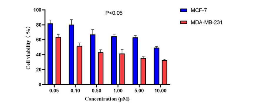
MDA-MB-231 and MCF-7 cells were treated with different dose of Cephaeline (0.1, 0.5, 1, 5, 10 μM) and cell viability were calculated by Cell Counting Kit-8.
Cephaeline hydrochloride purchased from MedChemExpress. Usage Cited in: J Cancer Res Clin Oncol. 2023 Oct;149(13):11117-11133. [Abstract]
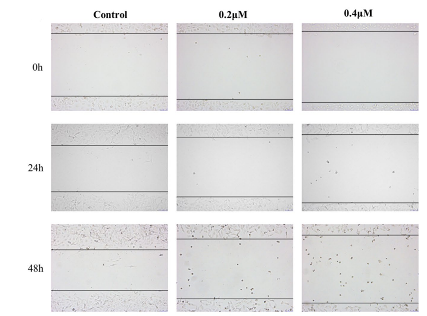
Representative images of cell scratch-wound healing test taken by a microscope at × 100 magnification treated with Cephaeline (0.2, 0.4 μM).
Cephaeline hydrochloride purchased from MedChemExpress. Usage Cited in: J Cancer Res Clin Oncol. 2023 Oct;149(13):11117-11133. [Abstract]
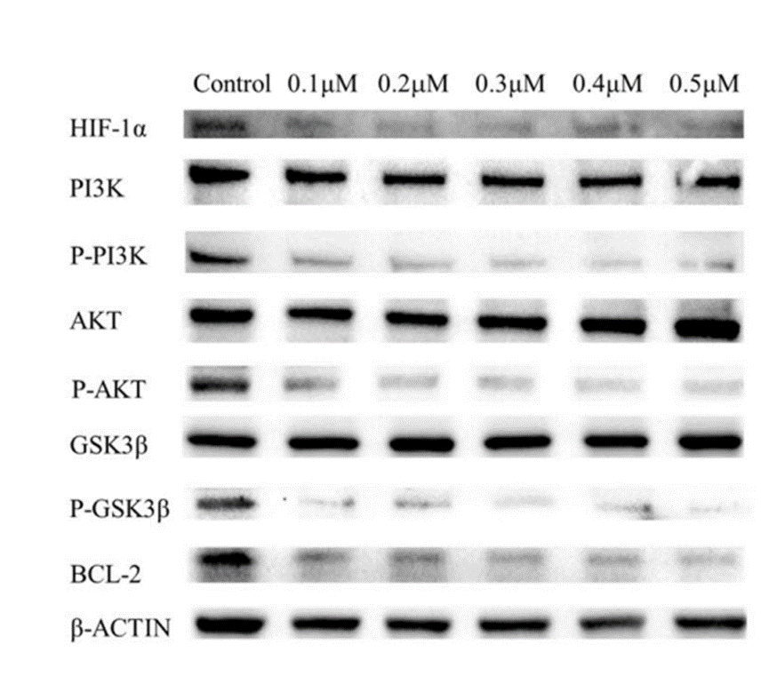
Representative western blots for PI3K/AKT signal pathway and HIF-1α under different doses of Cephaeline (0.1, 0.2, 0.3, 0.4, 0.5 μM).
-
Daru
2026 Jun 19;34(2):38. PMID: 42319734


Solvent & Solubility
In Vitro:
DMSO : 50 mg/mL (99.39 mM; Need ultrasonic; Hygroscopic DMSO has a significant impact on the solubility of product, please use newly opened DMSO)
Please refer to the solubility information to select the appropriate solvent. Once prepared, please aliquot and store the solution to prevent product inactivation from repeated freeze-thaw cycles.
Storage method and period of stock solution: -80°C, 6 months; -20°C, 1 month. When stored at -80°C, please use it within 6 months. When stored at -20°C, please use it within 1 month.
Please refer to the solubility information to select the appropriate solvent. Once prepared, please aliquot and store the solution to prevent product inactivation from repeated freeze-thaw cycles.
Storage method and period of stock solution: -80°C, 6 months; -20°C, 1 month. When stored at -80°C, please use it within 6 months. When stored at -20°C, please use it within 1 month.
Concentration (start) × Volume (start) = Concentration (final) × Volume (final)
In Vivo:
Select the appropriate dissolution method based on your experimental animal and administration route.
- For the following dissolution methods, please ensure to first prepare a clear stock solution using an In Vitro approach and then sequentially add co-solvents:
- To ensure reliable experimental results, the clarified stock solution can be appropriately stored based on storage conditions. As for the working solution for In Vivo experiments, it is recommended to prepare freshly and use it on the same day.
- The percentages shown for the solvents indicate their volumetric ratio in the final prepared solution. If precipitation or phase separation occurs during preparation, heat and/or sonication can be used to aid dissolution.
Add each solvent one by one: 10% DMSO 40% PEG300 5% Tween-80 45% Saline
Solubility: ≥ 1.25 mg/mL (2.48 mM); Clear solution
This protocol yields a clear solution of ≥ 1.25 mg/mL (saturation unknown).
Taking 1 mL working solution as an example, add 100 μL DMSO stock solution (12.5 mg/mL) to 400 μL PEG300, and mix evenly; then add 50 μL Tween-80 and mix evenly; then add 450 μL Saline to adjust the volume to 1 mL.
Preparation of Saline: Dissolve 0.9 g sodium chloride in ddH₂O and dilute to 100 mL to obtain a clear Saline solution.
Add each solvent one by one: 10% DMSO 90% (20% SBE-β-CD in Saline)
Solubility: ≥ 1.25 mg/mL (2.48 mM); Clear solution
This protocol yields a clear solution of ≥ 1.25 mg/mL (saturation unknown).
Taking 1 mL working solution as an example, add 100 μL DMSO stock solution (12.5 mg/mL) to 900 μL 20% SBE-β-CD in Saline, and mix evenly.
Preparation of 20% SBE-β-CD in Saline (4°C, storage for one week): 2 g SBE-β-CD powder is dissolved in 10 mL Saline, completely dissolve until clear.
In Vivo Dissolution Calculator
Please enter the basic information of animal experiments:
-
-
-
-
Recommended: Prepare an additional quantity of animals to account for potential losses during experiments.
Please enter your animal formula composition:
-
%DMSO +
Recommended: Keep the proportion of DMSO in working solution below 2% if your animal is weak.
-
%+
-
+%Tween-80 + +
-
%Saline +
The co-solvents required include: DMSO, . All of co-solvents are available by MedChemExpress (MCE). , Tween 80. All of co-solvents are available by MedChemExpress (MCE).
Working solution concentration: 0.22 mg/mL
Method for preparing stock solution: mg drug dissolved in μL DMSO. Stock solution concentration: mg/mL.
1. Take μL DMSO stock solution;
2. Add μL .
μL , mix evenly;
3. Then add μL Tween 80, mix evenly;
4. Then add μL
Please ensure that the stock solution in the first step is dissolved to a clear state, and add co-solvents in sequence. You can use ultrasonic heating (ultrasonic cleaner, recommended frequency 20-40 kHz), vortexing, etc. to assist dissolution.


Purity & Documentation
-
Data Sheet (307 KB)
-
SDS (254 KB)
- English - EN (254 KB)
- Français - FR (254 KB)
- Deutsch - DE (254 KB)
- Norwegian - NO (254 KB)
- Español - ES (254 KB)
- Swedish - SV (254 KB)
- Italian - IT (254 KB)
- Korean - KR (254 KB)
- Portuguese - PT (254 KB)
-
Handling Instructions (2659 KB)
References
[1]. Chen P, et al. Cephaeline promotes ferroptosis by targeting NRF2 to exert anti-lung cancer efficacy. Pharmaceutical biology. 2024 Dec;62(1):195-206. [Content Brief]
[2]. Li X, et al. Cephaeline promotes ferroptosis in breast cancer via p53/SLC7A11/GPX4 axis. Daru : journal of Faculty of Pharmacy, Tehran University of Medical Sciences. 2026 Jun 19;34(2):38. [Content Brief]
[3]. Ren PX, et al. A multi-targeting drug design strategy for identifying potent anti-SARS-CoV-2 inhibitors. Acta pharmacologica Sinica. 2022 Feb;43(2):483-493. [Content Brief]
[4]. Silva LC, et al. Cephaeline is an inductor of histone H3 acetylation and inhibitor of mucoepidermoid carcinoma cancer stem cells. Journal of oral pathology & medicine : official publication of the International Association of Oral Pathologists and the American Academy of Oral Pathology. 2022 Jul;51(6):553-562. [Content Brief]


Complete Stock Solution Preparation Table
Please refer to the solubility information to select the appropriate solvent. Once prepared, please aliquot and store the solution to prevent product inactivation from repeated freeze-thaw cycles.
Storage method and period of stock solution: -80°C, 6 months; -20°C, 1 month. When stored at -80°C, please use it within 6 months. When stored at -20°C, please use it within 1 month.
| Optional Solvent | Concentration Solvent Mass | 1 mg | 5 mg | 10 mg | 25 mg |
|---|---|---|---|---|---|
| DMSO | 1 mM | 1.9878 mL | 9.9390 mL | 19.8779 mL | 49.6949 mL |
| 5 mM | 0.3976 mL | 1.9878 mL | 3.9756 mL | 9.9390 mL | |
| 10 mM | 0.1988 mL | 0.9939 mL | 1.9878 mL | 4.9695 mL | |
| 15 mM | 0.1325 mL | 0.6626 mL | 1.3252 mL | 3.3130 mL | |
| 20 mM | 0.0994 mL | 0.4969 mL | 0.9939 mL | 2.4847 mL | |
| 25 mM | 0.0795 mL | 0.3976 mL | 0.7951 mL | 1.9878 mL | |
| 30 mM | 0.0663 mL | 0.3313 mL | 0.6626 mL | 1.6565 mL | |
| 40 mM | 0.0497 mL | 0.2485 mL | 0.4969 mL | 1.2424 mL | |
| 50 mM | 0.0398 mL | 0.1988 mL | 0.3976 mL | 0.9939 mL | |
| 60 mM | 0.0331 mL | 0.1656 mL | 0.3313 mL | 0.8282 mL | |
| 80 mM | 0.0248 mL | 0.1242 mL | 0.2485 mL | 0.6212 mL |


Cephaeline hydrochloride Related Classifications
HY-N2076 Related Classifications
 Powered by Bioz