



m-PEG2-azide

Based on 1 Customer Validation
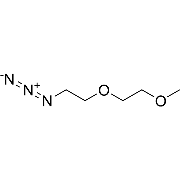
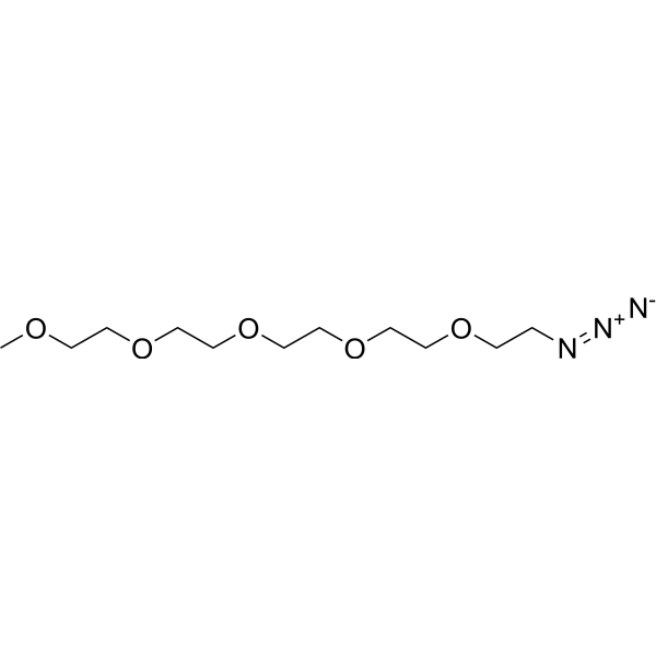
m-PEG2-azide is a PEG-based PROTAC linker can be used in the synthesis of PROTACs. m-PEG2-azide is a click chemistry reagent, it contains an Azide group and can undergo copper-catalyzed azide-alkyne cycloaddition reaction (CuAAc) with molecules containing Alkyne groups. It can also undergo strain-promoted alkyne-azide cycloaddition (SPAAC) reactions with molecules containing DBCO or BCN groups.
Nur für Forschungszwecke. Wir verkaufen nicht an Patienten.
- Reinheit : 97.0%
- CAS. Nr.: 215181-61-6
- Formel: C5H11N3O2
- Molecular Weight:145.16
-
Speicherung:Pure form -20°C, 3 years , 4°C, 2 years ; In solvent -80°C, 6 months , -20°C, 1 month

To place orders, for customer services and technical support, please contact: MedChemExpress USA
Tel: 609-228-6898 E-mail: [email protected] [email protected]


-
 Biologische Aktivität
-
Chemical Information
- Protokoll
- Reinheit & Dokumentation
- Verweise
-
 Help & FAQs


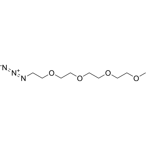
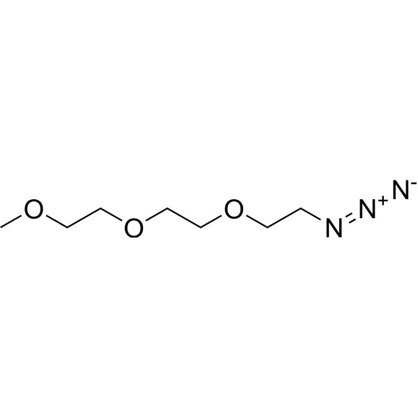
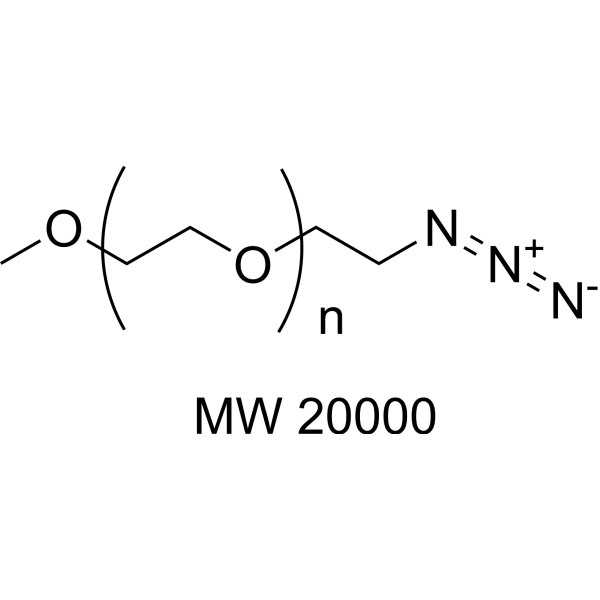

Alle PROTAC Linkers Isoform-spezifische Produkte anzeigen
More

Biologische Aktivität
Beschreibung
In Vitro
PROTACs contain two different ligands connected by a linker; one is a ligand for an E3 ubiquitin ligase and the other is for the target protein. PROTACs exploit the intracellular ubiquitin-proteasome system to selectively degrade target proteins.
MedChemExpress (MCE) has not independently confirmed the accuracy of these methods. They are for reference only.


Chemical Information
-
CAS. Nr. 215181-61-6
-
Appearance Liquid
-
Molecular Weight 145.16
-
Formel C5H11N3O2
-
Color Colorless to light yellow
-
SMILES
COCCOCCN=[N+]=[N-]
-
Versand
Room temperature in continental US; may vary elsewhere.
-
Speicherung
Pure form -20°C 3 years 4°C 2 years In solvent -80°C 6 months -20°C 1 month


Reinheit & Dokumentation
-
Data Sheet (262 KB)
-
SDS (467 KB)
- English - EN (467 KB)
- Français - FR (467 KB)
- Deutsch - DE (467 KB)
- Norwegian - NO (467 KB)
- Español - ES (467 KB)
- Swedish - SV (467 KB)
- Italian - IT (467 KB)
- Korean - KR (467 KB)
- Portuguese - PT (467 KB)
-
Handling Instructions (2659 KB)
Verweise

Calculators
Konzentration (Stammlösung) × Volumen (Stammlösung) = Konzentration (Ziellösung) × Volumen (Ziellösung)


m-PEG2-azide Related Classifications
HY-130578 Related Classifications
 Powered by Bioz