
Pulchinenoside E2
Based on 1 Customer Validation
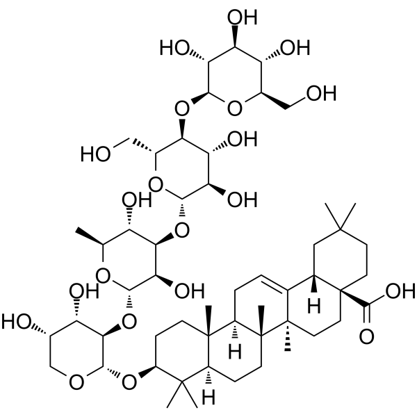
Pulchinenoside E2 is a triterpenoid saponin. Pulchinenoside E2 inhibits the phosphorylation of STAT3 and JAK2, blocks the nuclear translocation and transcriptional activity of STAT3, and suppresses STAT3-dependent mitochondrial oxidative phosphorylation. Pulchinenoside E2 inhibits invasion, migration and autophagy of triple-negative breast cancer cells. Pulchinenoside E2 can be used in research related to triple-negative breast cancer.
Nur für Forschungszwecke. Wir verkaufen nicht an Patienten.
- Reinheit : 99.28%
- CAS. Nr.: 244202-36-6
- Formel: C53H86O21
- Molecular Weight:1059.24
-
Speicherung:
-20°C, sealed storage, away from moisture
* In solvent : -80°C, 6 months; -20°C, 1 month (sealed storage, away from moisture)
Biologische Aktivität
Beschreibung
IC50 & Target
[1]|
JAK2 |
STAT3 |
Cellular Effect
|
Cell Line
|
Type | Value | Description | References |
|---|---|---|---|---|
| A-375 | IC50 |
5.9 μM
Compound: 9
|
Cytotoxicity against human A375 cells after 72 hrs by sulforhodamine B assay
|
[PMID: 23639650] |
| A549 | IC50 |
7.9 μM
Compound: 9
|
Cytotoxicity against human A549 cells after 72 hrs by sulforhodamine B assay
|
[PMID: 23639650] |
| HL-60 | IC50 |
2.6 μg/mL
Compound: 8
|
Cytotoxicity against human HL-60 cells after 72 hrs by MTT assay
|
[PMID: 10514313] |
| HL-60 | IC50 |
3.5 μM
Compound: 9
|
Cytotoxicity against human HL60 cells after 72 hrs by MTT assay
|
[PMID: 23639650] |
In Vitro
Pulchinenoside E2 (0.75-1.5 μM; 24 h) induces lysosomal dysfunction in HS-578T and MDA-MB-231 TNBC cells, characterized by suppressed proteolytic activity, time-dependent degradation of mature CTSD, and lysosomal alkalinization[1].
Pulchinenoside E2 (0.75-1.5 μM; 24 h) dose-dependently increases GFP-LC3B puncta formation and impairs autophagic flux in HS-578T and MDA-MB-231 TNBC cells[1].
MedChemExpress (MCE) has not independently confirmed the accuracy of these methods. They are for reference only. Further protocols information, click here.
-
Cell Line:HS-578T, MDA-MB-231 TNBC cells
-
Concentration:0.75, 1.5 μM
-
Incubation Time:24 h
-
Result:Dose-dependently increased the number of autophagic puncta and fluorescence intensity of GFP-LC3B in both cell lines.
Dose-dependently increased the LC3II/LC3I ratio and accumulation of the autophagy substrate p62.
Parmacokinetics
| Species | Dose | Route | Tmax | Cmax | T1/2 | AUC0-t | MRTlast |
|---|---|---|---|---|---|---|---|
| Mice[1] | 5 mg/kg | i.p. | 6.30 h | 35.70 μg/mL | >12 h | 338.80 μg·h/mL | 6.67 h |
In Vivo
MedChemExpress (MCE) has not independently confirmed the accuracy of these methods. They are for reference only.
-
Animal Model:M-NSG mice (female, 4-6 weeks old, tail vein injection of MDA-MB-231 cells to induce metastases)[2]
-
Dosage:5 mg/kg; 10 mg/kg
-
Administration:i.p.; alternate days; 30 days
-
Result:Markedly reduced pulmonary and hepatic metastatic lesion numbers and tumor volumes in mice treated with 10 mg/kg compared to controls.
Significantly decreased phosphorylated STAT3 (Tyr705/Ser727) expression in metastatic tumors via immunohistochemical staining.
Accumulated LC3B and p62 in metastatic tumors, consistent with autophagic flux blockade.
Maintained serum AST, ALT, CRE, and BUN levels comparable to controls.
Showed no significant histopathological damage in heart and kidney tissues.
Maintained stable body weight throughout treatment, indicating no overt toxicity.
Chemical Information
-
CAS. Nr. 244202-36-6
-
Appearance Solid
-
Molecular Weight 1059.24
-
Formel C53H86O21
-
Color White to off-white
-
SMILES
CC1(C)[C@@H](O[C@H]2[C@H](O[C@H]3[C@H](O)[C@H](O[C@@H]4O[C@H](CO)[C@@H](O[C@@H]5O[C@H](CO)[C@@H](O)[C@H](O)[C@H]5O)[C@H](O)[C@H]4O)[C@@H](O)[C@H](C)O3)[C@@H](O)[C@@H](O)CO2)CC[C@@]6(C)[C@@]1([H])CC[C@]7(C)[C@]6([H])CC=C8[C@@]7(C)CC[C@]9(C(O)=O)[C@@]8([H])CC(C)(C)CC9
-
Structure Classification
-
Initial Source
-
Versand
Room temperature in continental US; may vary elsewhere.
-
Speicherung
-20°C, sealed storage, away from moisture
* In solvent : -80°C, 6 months; -20°C, 1 month (sealed storage, away from moisture)
Protokoll
-
Kinase activity and phosphorylation assays
Kinase activity assays measure the ability of kinases to transfer phosphate groups from ATP to specific substrates, while phosphorylation assays detect the presence and levels of phosphorylated proteins. Common methods include radiolabeled ATP incorporation (e. g. ,), ADP release detection via bioluminescence (e. g. ,[3]), enzyme-linked immunosorbent assays (ELISA) for phospho-specific epitopes (e. g. ,[6]), and microtiter-based formats for high-throughput screening (e. g. ,[8]). The ADP-Glo assay quantifies kinase activity by measuring ADP produced during phosphorylation using a luciferase-based system. Radiometric assays involve autoradiography or scintillation counting after incorporation of 32P-labeled ATP into substrate proteins. ELISA-based approaches rely on phospho-specific antibodies to detect activated kinases in cell lysates or purified samples.
-
Western Blot
Western blotting (WB) is a commonly used experimental method in molecular biology, biochemistry, and immunogenetics for identifying and quantifying target proteins. It combines gel electrophoresis with immunoassay, enabling researchers to analyze protein expression, post-translational modifications, and molecular weight.
-
Cytoplasmic-Nuclear Fractionated Protein Extraction
Cytoplasmic-nuclear fractionated protein extraction separates soluble cytoplasmic proteins from nuclear-enriched proteins by mild plasma-membrane permeabilization, differential centrifugation, washing of nuclei, and extraction of nuclear proteins for downstream immunoblotting or related molecular analysis. The readout is the relative abundance of a protein in cytoplasmic and nuclear fractions, commonly assessed by western blotting together with compartment markers such as tubulin or pyruvate kinase for cytoplasm and lamin, nucleoporin, hnRNP, H2AX, or Lamin B for nuclear fractions.
-
Dual Luciferin reporter gene assay
Luciferin reporter gene assay is a reporting system to detect the activity of Firefly Luciferase using luciferin as a substrate, which is often used in the research of miRNA target gene verification and promoter transcriptive activity regulation. Dual luciferase usually refers to Firefly luciferase and Renilla luciferase.
-
Cell migration
Cell migration is a method that plays an important role in wound healing, cell differentiation, embryonic development, etc.
-
Autophagy
Autophagy is a process in which eukaryotic cells use lysosomes to degrade their own cytoplasmic proteins and damaged organelles under the regulation of autophagy related gene (Atg). Microtubule-associated proteins light chain 3 (LC3) is recognized as autophagy marker, which transfers from cytoplasmic LC3 (LC3-I) to membrane type (LC3-II). LC3-II/I ratio could be detected by Western Blot and fluorescence microscopy.
-
Cell invasion
Cell invasion is the ability of cells to migrate from one area to another via the extracellular matrix. Cell invasion is the response of normal and cancer cells to chemical and mechanical stimuli. Before migrating to a new region, the extracellular matrix is degraded by proteases within the cell. Cell invasion often occurs during wound repair, vascularization and inflammation, abnormal tissue invasion, and tumor cell metastasis.
-
Protocol for Kinase activity and phosphorylation assays
Kinase activity assays measure transfer of phosphate from ATP to a protein or peptide substrate, generating phosphorylated substrate, ADP, or incorporated radiolabeled phosphate as the readout; phosphorylation assays measure site-specific phosphorylation in cells or tissues as a proxy for kinase-pathway activation, inhibition, or substrate regulation. Phosphorylation can be detected by phospho-specific Western blot, immunoprecipitation kinase assay, phospho-immunofluorescence, phospho-flow cytometry, luminescent ADP detection, radiolabeled ATP incorporation, or reporter-based pathway assays, and these readouts can be applied to cancer cells, primary neurons, mouse tumors, organoids, inflammatory macrophages, ferroptosis studies, and mitophagy studies when the kinase target is biologically relevant.
-
Lysosome and acidic-vesicle live-cell staining
Lysosome and acidic-vesicle live-cell staining detects acidic intracellular compartments by using membrane-permeant acidotropic probes that accumulate in low-pH vesicles, including lysosomes, late endosomes, autolysosomes, and acidic phagosomes. LysoTracker staining is commonly used as an intensity-based readout of acidic lysosomal compartment abundance or enlargement, while acridine orange produces green fluorescence in less concentrated compartments and red fluorescence after concentration-dependent accumulation in acidic vesicular organelles. Loss or reduction of acridine-orange red signal can be used as a readout of lysosomal membrane permeabilization or reduced acidic-vesicle integrity. This protocol is designed for live cultured cells and can be adapted for fluorescence microscopy, high-content imaging, plate-reader readout, or flow cytometry when the selected literature supports the readout. Because these dyes report acidotropic accumulation rather than lysosome identity alone,
-
Macroautophagy Solutions
Macroautophagy is a conserved lysosome-dependent degradation pathway in which cytoplasmic material is sequestered into double-membrane autophagosomes and delivered to lysosomes for degradation and recycling. The pathway supports cellular homeostasis during nutrient limitation, organelle stress, protein-aggregate accumulation, infection, differentiation, and tissue remodeling by coupling cargo sequestration, autophagosome maturation, lysosomal fusion, and degradation of cargo-derived macromolecules. The core molecular sequence includes initiation by nutrient- and stress-regulated autophagy machinery, autophagosome nucleation, LC3/ATG8-family conjugation to autophagosomal membranes, cargo selection through receptors such as SQSTM1/p62, autophagosome-lysosome fusion, and lysosomal degradation. LC3 was identified as a mammalian homolog of yeast Atg8 that localizes to autophagosomal membranes after processing, and p62/SQSTM1 was shown to connect ubiquitinated cargo with autophagic degradati
-
Breast Cancer Modeling
Breast cancer is a heterogeneous cancer, and it has been distinguished into four subtypes: luminal A, luminal B, HER2-positive and basal-like. Molecular mutations, epigenetic alterations, hormone exposure and immune microenvironment are related to the progression of breast cancer.
Reinheit & Dokumentation
-
Data Sheet (283 KB)
-
SDS (252 KB)
- English - EN (252 KB)
- Français - FR (252 KB)
- Deutsch - DE (252 KB)
- Norwegian - NO (252 KB)
- Español - ES (252 KB)
- Swedish - SV (252 KB)
- Italian - IT (252 KB)
- Korean - KR (252 KB)
- Portuguese - PT (252 KB)
-
Handling Instructions (2659 KB)
Verweise
Calculators
Konzentration (Stammlösung) × Volumen (Stammlösung) = Konzentration (Ziellösung) × Volumen (Ziellösung)

