



Physalin D

Based on 1 Customer Validation
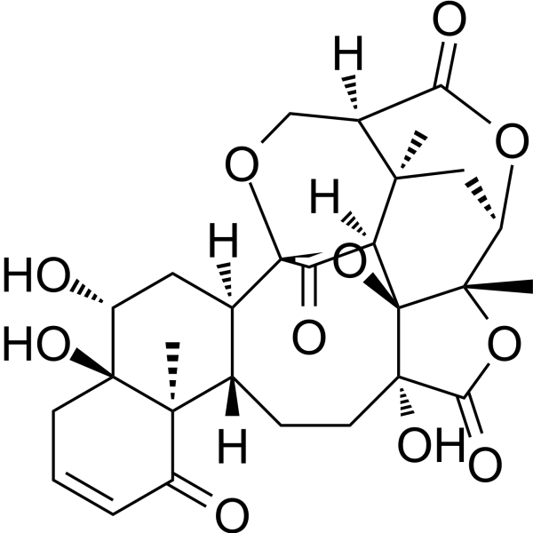
Physalin D is isolated from the aerial parts of Physalis angulate. Physalin D displays considerable cytotoxicity against several cancer cell lines, showing IC50 values in the range of d 0.28 to 2.43 microg/mL. Physalin D has the potential for the cancer disease.
For research use only. We do not sell to patients.
- Purity : 98.21%
- CAS No.: 54980-22-2
- Formula: C28H32O11
- Molecular Weight:544.55
-
Storage:
-20°C, sealed storage, away from moisture
* In solvent : -80°C, 6 months; -20°C, 1 month (sealed storage, away from moisture)

To place orders, for customer services and technical support, please contact: MedChemExpress USA
Tel: 609-228-6898 E-mail: [email protected] [email protected]


-
 Biological Activity
-
Chemical Information
- Protocols
- Purity & Documentation
- References
-
 Help & FAQs




Biological Activity
Description
Cellular Effect
|
Cell Line

|
Type | Value | Description | References |
|---|---|---|---|---|
| A549 | IC50 |
>50 μM
Compound: 20
|
Antiproliferative activity against human A549 cells assessed as reduction in cell viability incubated for 48 hrs by CCK8 assay
Antiproliferative activity against human A549 cells assessed as reduction in cell viability incubated for 48 hrs by CCK8 assay
|
[PMID: 35608269] |
| HepG2 | IC50 |
>50 μM
Compound: 20
|
Antiproliferative activity against human HepG2 cells assessed as reduction in cell viability incubated for 48 hrs by CCK8 assay
Antiproliferative activity against human HepG2 cells assessed as reduction in cell viability incubated for 48 hrs by CCK8 assay
|
[PMID: 35608269] |
| L02 | IC50 |
>50 μM
Compound: 20
|
Cytotoxicity against human L02 cells assessed as reduction in cell viability incubated for 48 hrs by CCK8 assay
Cytotoxicity against human L02 cells assessed as reduction in cell viability incubated for 48 hrs by CCK8 assay
|
[PMID: 35608269] |
| MCF7 | IC50 |
>50 μM
Compound: 20
|
Antiproliferative activity against human MCF7 cells assessed as reduction in cell viability incubated for 48 hrs by CCK8 assay
Antiproliferative activity against human MCF7 cells assessed as reduction in cell viability incubated for 48 hrs by CCK8 assay
|
[PMID: 35608269] |
| RAW264.7 | IC50 |
>50 μM
Compound: 20
|
Cytotoxicity against mouse RAW264.7 cells assessed as reduction in cell viability incubated for 48 hrs by CCK8 assay
Cytotoxicity against mouse RAW264.7 cells assessed as reduction in cell viability incubated for 48 hrs by CCK8 assay
|
[PMID: 35608269] |
| RAW264.7 | IC50 |
49.8 μM
Compound: 20
|
Antiinflammatory activity in mouse RAW264.7 assessed as inhibition of LPS-stimulated NO production pretreated with LPS followed by compund addition by Griess reagent based microplate reader assay
Antiinflammatory activity in mouse RAW264.7 assessed as inhibition of LPS-stimulated NO production pretreated with LPS followed by compund addition by Griess reagent based microplate reader assay
|
[PMID: 35608269] |
| SGC-7901 | IC50 |
>50 μM
Compound: 20
|
Antiproliferative activity against human SGC-7901 cells assessed as reduction in cell viability incubated for 48 hrs by CCK8 assay
Antiproliferative activity against human SGC-7901 cells assessed as reduction in cell viability incubated for 48 hrs by CCK8 assay
|
[PMID: 35608269] |


Chemical Information
-
CAS No. 54980-22-2
-
Appearance Solid
-
Molecular Weight 544.55
-
Formula C28H32O11
-
Color White to off-white
-
SMILES
C[C@]12[C@]34[C@]5([H])[C@]6([C@@](C(O[C@@H]2C6)=O)([H])CO[C@](C5=O)([C@@]7([H])[C@]([C@@]8([C@@]([C@@H](C7)O)(CC=CC8=O)O)C)([H])CC[C@@]4(C(O1)=O)O)O3)C
-
Structure Classification
-
Initial Source
-
Shipping
Room temperature in continental US; may vary elsewhere.
-
Storage
-20°C, sealed storage, away from moisture
* In solvent : -80°C, 6 months; -20°C, 1 month (sealed storage, away from moisture)

Protocols
-
Cell Cytotoxicity Assay
Cytotoxicity assays are usually based on the assessment of cell membrane damage, which can also be indirectly detected by measuring cell viability. Detection methods include MTT assay, CKK-8 assay, LDH assay and ATP assay, etc.
-
Mammalian live/dead viability and cytotoxicity staining
Live/dead viability and cytotoxicity staining assays are based on the simultaneous detection of intracellular esterase activity in metabolically active (viable) cells and membrane integrity loss in non-viable cells. In commonly used dual-staining approaches, membrane-permeant fluorogenic substrates are converted by intracellular esterases into fluorescent products in live cells, while impermeant DNA-binding dyes selectively enter cells with compromised plasma membranes and label nucleic acids in dead or dying cells, enabling discrimination between viable and non-viable populations by fluorescence microscopy or flow cytometry.
-
Cell Viability Determination by MTT Colorimetric Assay
The following protocol uses the MTT colorimetric assay as a classic literature-established method for assessing cell viability/metabolic activity in cultured mammalian cells. MTT[3-(4,5-dimethylthiazol-2-yl)-2,5-diphenyltetrazolium bromide] is reduced by metabolically active cells to a colored formazan product; the amount of formazan is quantified spectrophotometrically and provides an indirect measure of metabolically active viable cells. Importantly, MTT reduction reflects cellular oxidoreductase/metabolic activity rather than an absolute direct count of living cells, so changes in cellular metabolism can alter the signal independently of cell number.


Purity & Documentation
-
Data Sheet (284 KB)
-
SDS (252 KB)
- English - EN (252 KB)
- Français - FR (252 KB)
- Deutsch - DE (252 KB)
- Norwegian - NO (252 KB)
- Español - ES (252 KB)
- Swedish - SV (252 KB)
- Italian - IT (252 KB)
- Korean - KR (252 KB)
- Portuguese - PT (252 KB)
-
Handling Instructions (2659 KB)
References
[1]. Magalhães HI, et al. In-vitro and in-vivo antitumour activity of physalins B and D from Physalis angulata. J Pharm Pharmacol. 2006;58(2):235-241. [Content Brief]
[2]. Xiang D, Zou J, Zhu X, et al. Physalin D attenuates hepatic stellate cell activation and liver fibrosis by blocking TGF-β/Smad and YAP signaling. Phytomedicine. 2020;78:153294. [Content Brief]
[3]. Ding N, Lu Y, Cui H, et al. Physalin D inhibits RANKL-induced osteoclastogenesis and bone loss via regulating calcium signaling. BMB Rep. 2020;53(3):154-159. [Content Brief]
[4]. Ding N, Wang Y, Dou C, et al. Physalin D regulates macrophage M1/M2 polarization via the STAT1/6 pathway. J Cell Physiol. 2019;234(6):8788-8796. [Content Brief]

Calculators
Concentration (start) × Volume (start) = Concentration (final) × Volume (final)


Physalin D Related Classifications
HY-N9912 Related Classifications
 Powered by Bioz