



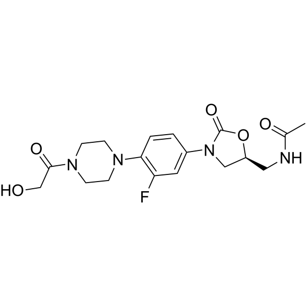
Eperezolid

Based on 1 Customer Validation
Eperezolid (PNU-100592) is an orally active protein synthesis inhibitor that targets the bacterial 50S ribosomal subunit. Eperezolid competitively binds to a specific site on the ribosomal 50S subunit (overlapping with the binding sites of chloramphenicol (HY-B0239) and lincomycin (HY-117660)) to inhibit the translation initiation stage and exert antibacterial activity. Eperezolid can induce host cell autophagy to enhance the clearance of intracellular mycobacteria, and its MIC90 for Staphylococcus aureus and Enterococcus is 1-4 μg/mL. Eperezolid is mainly used for antibacterial research on infections with Gram-positive bacteria such as methicillin-resistant (HY-121544) Staphylococci and vancomycin-resistant (HY-B0671) Enterococci, as well as infections with intracellular bacteria such as Mycobacterium tuberculosis.
Nos produits utilisent uniquement pour la recherche. Nous ne vendons pas aux patients.
- Pureté : 96.55%
- CAS No.: 165800-04-4
- Formule: C18H23FN4O5
- Masse moléculaire:394.40
-
Stockage:Powder -20°C, 3 years , 4°C, 2 years ; In solvent -80°C, 2 years , -20°C, 1 year

To place orders, for customer services and technical support, please contact: MedChemExpress USA
Tel: 609-228-6898 E-mail: [email protected] [email protected]


-
 Activité biologique
-
Chemical Information
-
Solvant et solubilité
- Protocole
- Pureté et documentation
- Références
-
 Help & FAQs


Voir tous les produits spécifiques à Isoform Antibiotic
More

Activité biologique
Description
IC50 & Target
|
Oxazolidinone |
In Vitro
1. Antibacterial activity test: Eperezolid (1×MIC-4×MIC; 24 h) shows antibacterial activity in vitro against Methicillin-sensitive/resistant Staphylococcus aureus, coagulase-negative Staphylococci and Vancomycin-sensitive/resistant Enterococci with MIC90 of 1-4 μg/mL. Eperezolid exhibits an antibacterial effect and the post-antibiotic effect (PAE) against Enterococcus faecalis and Enterococcus faecium was 0.1-1.2 h[1].
2. Autophagy induction assay:
Eperezolid (1 μM; 24 h) induces autophagy in dTHP-1 cells, as evidenced by increased conversion of LC3-I to LC3-II, increased formation of LC3 puncta, and a significant increase in the number of autophagic vacuoles as confirmed by MDC staining. It also inhibits the survival of intracellular Mycobacterium smegmatis[1].
3. Ribosome binding assay:
Eperezolid (1-100 μM; 10 min) specifically binds to the 50S ribosomal subunit of Escherichia coli with a Kd of approximately 20 μM. Its binding can be competitively inhibited by chloramphenicol and lincomycin, but does not affect peptidyl transferase activity or translation termination[1].
MedChemExpress (MCE) has not independently confirmed the accuracy of these methods. They are for reference only. Further protocols information, click here.
-
Cell Line:Staphylococcus aureus, Staphylococcus epidermidis, Enterococcus faecalis, Enterococcus faecium
-
Concentration:1×MIC (1-4 μg/mL), 4×MIC
-
Incubation Time:24 h
-
Result:Showed bacteriostatic activity, with no significant reduction in bacterial counts (>1 log10 CFU/mL) over 24 h.
PAE was measured at 0.8 h against staphylococci and 0.1-0.8 h against enterococci, with higher PAE observed at 4×MIC.
-
Cell Line:dTHP-1 human monocyte-derived macrophages
-
Concentration:1 μM
-
Incubation Time:24 h
-
Result:Showed a significant increase in LC3-II/LC3-I ratio, and confocal microscopy revealed enhanced LC3 puncta formation.
MDC staining indicated a 2-fold increase in autophagic vacuoles compared to control.
Intracellular M. smegmatis viability was reduced by 50% compared to untreated controls.
In Vivo
MedChemExpress (MCE) has not independently confirmed the accuracy of these methods. They are for reference only.
-
Animal Model:Sprague-Dawley rats (male, 175-250 g, 6-8 weeks old) with intra-abdominal abscess induced by Enterococcus faecalis 1310 or Vancomycin-resistant Enterococcus faecium A1221[3]
-
Dosage:25 mg/kg
-
Administration:Intravenous infusion or oral gavage, twice daily (q12h), for 4.5 days
-
Result:For E. faecalis infection, had no significantly reduction in bacterial density in abscesses compared to untreated controls (mean viable bacteria: 8.3 log10 CFU/g vs. 8.6 log10 CFU/g).
For vancomycin-resistant E. faecium infection, i.v. eperezolid reduced bacterial density to 6.5 log10 CFU/g (vs. 8.25 log10 CFU/g in controls), while oral administration showed a modest reduction to 7.8 log10 CFU/g.
No sterile abscesses were observed in any treatment group.


Chemical Information
-
CAS No. 165800-04-4
-
Appearance Solid
-
Masse moléculaire 394.40
-
Formule C18H23FN4O5
-
Color White to off-white
-
SMILES
O=C(O[C@H]1CNC(C)=O)N(C1)C2=CC(F)=C(C=C2)N3CCN(CC3)C(CO)=O
-
Synonyms
PNU-100592
-
Livraison
Room temperature in continental US; may vary elsewhere.
-
Stockage
Powder -20°C 3 years 4°C 2 years In solvent -80°C 2 years -20°C 1 year


Solvant et solubilité
In Vitro:
DMSO : 40 mg/mL (101.42 mM; Need ultrasonic; Hygroscopic DMSO has a significant impact on the solubility of product, please use newly opened DMSO)
Please refer to the solubility information to select the appropriate solvent. Once prepared, please aliquot and store the solution to prevent product inactivation from repeated freeze-thaw cycles.
Storage method and period of stock solution: -80°C, 2 years; -20°C, 1 year. When stored at -80°C, please use it within 2 years. When stored at -20°C, please use it within 1 year.
Please refer to the solubility information to select the appropriate solvent. Once prepared, please aliquot and store the solution to prevent product inactivation from repeated freeze-thaw cycles.
Storage method and period of stock solution: -80°C, 2 years; -20°C, 1 year. When stored at -80°C, please use it within 2 years. When stored at -20°C, please use it within 1 year.
Concentration (start) × Volume (start) = Concentration (final) × Volume (final)
In Vivo:
Select the appropriate dissolution method based on your experimental animal and administration route.
- For the following dissolution methods, please ensure to first prepare a clear stock solution using an In Vitro approach and then sequentially add co-solvents:
- To ensure reliable experimental results, the clarified stock solution can be appropriately stored based on storage conditions. As for the working solution for In Vivo experiments, it is recommended to prepare freshly and use it on the same day.
- The percentages shown for the solvents indicate their volumetric ratio in the final prepared solution. If precipitation or phase separation occurs during preparation, heat and/or sonication can be used to aid dissolution.
Add each solvent one by one: 10% DMSO 40% PEG300 5% Tween-80 45% Saline
Solubility: ≥ 2.5 mg/mL (6.34 mM); Clear solution
This protocol yields a clear solution of ≥ 2.5 mg/mL (saturation unknown).
Taking 1 mL working solution as an example, add 100 μL DMSO stock solution (25.0 mg/mL) to 400 μL PEG300, and mix evenly; then add 50 μL Tween-80 and mix evenly; then add 450 μL Saline to adjust the volume to 1 mL.
Preparation of Saline: Dissolve 0.9 g sodium chloride in ddH₂O and dilute to 100 mL to obtain a clear Saline solution.
Add each solvent one by one: 10% DMSO 90% (20% SBE-β-CD in Saline)
Solubility: ≥ 2.5 mg/mL (6.34 mM); Clear solution
This protocol yields a clear solution of ≥ 2.5 mg/mL (saturation unknown).
Taking 1 mL working solution as an example, add 100 μL DMSO stock solution (25.0 mg/mL) to 900 μL 20% SBE-β-CD in Saline, and mix evenly.
Preparation of 20% SBE-β-CD in Saline (4°C, storage for one week): 2 g SBE-β-CD powder is dissolved in 10 mL Saline, completely dissolve until clear.
In Vivo Dissolution Calculator
Please enter the basic information of animal experiments:
-
-
-
-
Recommended: Prepare an additional quantity of animals to account for potential losses during experiments.
Please enter your animal formula composition:
-
%DMSO +
Recommended: Keep the proportion of DMSO in working solution below 2% if your animal is weak.
-
%+
-
+%Tween-80 + +
-
%Saline +
The co-solvents required include: DMSO, . All of co-solvents are available by MedChemExpress (MCE). , Tween 80. All of co-solvents are available by MedChemExpress (MCE).
Working solution concentration: 0.22 mg/mL
Method for preparing stock solution: mg drug dissolved in μL DMSO. Stock solution concentration: mg/mL.
1. Take μL DMSO stock solution;
2. Add μL .
μL , mix evenly;
3. Then add μL Tween 80, mix evenly;
4. Then add μL
Please ensure that the stock solution in the first step is dissolved to a clear state, and add co-solvents in sequence. You can use ultrasonic heating (ultrasonic cleaner, recommended frequency 20-40 kHz), vortexing, etc. to assist dissolution.

Protocole
-
Research Protocol for Infectious Diseases
Infectious-disease experiments test how pathogens interact with host barriers, innate immune receptors, inflammatory signaling, pathogen replication, and tissue injury; pattern-recognition receptors such as TLRs, RIG-I-like receptors, NOD-like receptors, and inflammasomes detect microbial molecules and activate NF-κB, interferon, and cytokine responses. The central hypothesis is that infection severity reflects the balance between pathogen burden and host response: protective inflammation restricts pathogen growth, whereas excessive or mislocalized inflammation contributes to tissue damage and disease phenotype. Unresolved questions include which host pathways are protective versus pathogenic, why some infection models fail to translate to human disease, and which combined readouts best predict clinically relevant infection outcomes.
-
Autophagy
Autophagy is a process in which eukaryotic cells use lysosomes to degrade their own cytoplasmic proteins and damaged organelles under the regulation of autophagy related gene (Atg). Microtubule-associated proteins light chain 3 (LC3) is recognized as autophagy marker, which transfers from cytoplasmic LC3 (LC3-I) to membrane type (LC3-II). LC3-II/I ratio could be detected by Western Blot and fluorescence microscopy.
-
Protocol for Pharmacokinetic Study
Pharmacokinetic studies quantify how an organism handles a drug over time through absorption, distribution, metabolism, and excretion, and the core experimental readout is the concentration-time profile of parent drug and, when relevant, metabolites in biological matrices such as plasma, whole blood, urine, bile, or tissue. Pharmacokinetic analysis links dose, route, exposure, clearance, half-life, distribution, bioavailability, and systemic exposure to drug efficacy and toxicity hypotheses rather than measuring a signaling pathway directly. The literature links pharmacokinetics to drug-development phenotypes by showing that drug metabolism and pharmacokinetics influence compound progression, exposure-response interpretation, safety margins, dosing strategy, and failure risk during discovery and development. DMPK science contributes to compound optimization by integrating physicochemical properties, in vitro metabolism, transporter behavior, in vivo exposure, and pharmacodynamic contex
-
Lysosome and acidic-vesicle live-cell staining
Lysosome and acidic-vesicle live-cell staining detects acidic intracellular compartments by using membrane-permeant acidotropic probes that accumulate in low-pH vesicles, including lysosomes, late endosomes, autolysosomes, and acidic phagosomes. LysoTracker staining is commonly used as an intensity-based readout of acidic lysosomal compartment abundance or enlargement, while acridine orange produces green fluorescence in less concentrated compartments and red fluorescence after concentration-dependent accumulation in acidic vesicular organelles. Loss or reduction of acridine-orange red signal can be used as a readout of lysosomal membrane permeabilization or reduced acidic-vesicle integrity. This protocol is designed for live cultured cells and can be adapted for fluorescence microscopy, high-content imaging, plate-reader readout, or flow cytometry when the selected literature supports the readout. Because these dyes report acidotropic accumulation rather than lysosome identity alone,
-
Gram Staining of Tissue Sections
Gram staining of tissue sections is a histochemical technique used to differentiate Gram-positive and Gram-negative bacteria within histological specimens based on differences in bacterial cell wall structure and dye retention, adapted from classical bacteriological Gram staining into tissue-compatible “histological Gram stain” variants. In tissue applications, modifications of the Brown-Hopps and Brown-Brenn methods are commonly used to improve differentiation of microorganisms embedded within host connective tissue and to reduce overstaining or loss of Gram-negative signal, which are known limitations of earlier approaches. The principle relies on crystal violet-iodine complex retention in Gram-positive organisms and subsequent decolorization and counterstaining steps that allow contrast visualization of Gram-negative organisms against tissue background.
-
Bacterial live/dead nucleic-acid viability staining
The LIVE/DEAD bacterial viability staining method is based on differential permeability of nucleic-acid-binding fluorescent dyes, most commonly SYTO 9 and propidium iodide (PI), which enables discrimination of bacterial populations with intact versus compromised cytoplasmic membranes. SYTO 9 penetrates both intact and damaged bacterial membranes and binds nucleic acids to produce green fluorescence, whereas propidium iodide penetrates only cells with compromised membranes and fluoresces red while also reducing SYTO 9 signal through competitive binding and fluorescence interactions. The resulting fluorescence pattern is interpreted as a proxy for membrane integrity, which is widely used as an indicator of bacterial viability in microscopy, flow cytometry, and spectroscopic platforms. However, mechanistic studies show that SYTO 9 and PI interactions involve displacement and fluorescence resonance energy transfer effects, which can influence signal interpretation depending on dye ratios a
-
Macroautophagy Solutions
Macroautophagy is a conserved lysosome-dependent degradation pathway in which cytoplasmic material is sequestered into double-membrane autophagosomes and delivered to lysosomes for degradation and recycling. The pathway supports cellular homeostasis during nutrient limitation, organelle stress, protein-aggregate accumulation, infection, differentiation, and tissue remodeling by coupling cargo sequestration, autophagosome maturation, lysosomal fusion, and degradation of cargo-derived macromolecules. The core molecular sequence includes initiation by nutrient- and stress-regulated autophagy machinery, autophagosome nucleation, LC3/ATG8-family conjugation to autophagosomal membranes, cargo selection through receptors such as SQSTM1/p62, autophagosome-lysosome fusion, and lysosomal degradation. LC3 was identified as a mammalian homolog of yeast Atg8 that localizes to autophagosomal membranes after processing, and p62/SQSTM1 was shown to connect ubiquitinated cargo with autophagic degradati


Pureté et documentation
-
Fiche technique (278 KB)
-
SDS (393 KB)
- English - EN (393 KB)
- Français - FR (393 KB)
- Deutsch - DE (393 KB)
- Norwegian - NO (393 KB)
- Español - ES (393 KB)
- Swedish - SV (393 KB)
- Italian - IT (393 KB)
- Korean - KR (393 KB)
- Portuguese - PT (393 KB)
-
Instruction de manipulation (2659 KB)
Références
[1]. Rybak MJ, et al. Comparative in vitro activities and postantibiotic effects of the oxazolidinone compounds eperezolid (PNU-100592) and linezolid (PNU-100766) versus vancomycin against Staphylococcus aureus, coagulase-negative staphylococci, Enterococcus faecalis, and Enterococcus faecium. Antimicrob Agents Chemother. 1998 Mar;42(3):721-4. [Content Brief]
[3]. Schülin T, et al. Activities of the oxazolidinones linezolid and eperezolid in experimental intra-abdominal abscess due to Enterococcus faecalis or vancomycin-resistant Enterococcus faecium. Antimicrob Agents Chemother. 1999 Dec;43(12):2873-6. [Content Brief]
[4]. Lin AH, et al. The oxazolidinone eperezolid binds to the 50S ribosomal subunit and competes with binding of chloramphenicol and lincomycin. Antimicrob Agents Chemother. 1997 Oct;41(10):2127-31. [Content Brief]


Complete Stock Solution Preparation Table
Please refer to the solubility information to select the appropriate solvent. Once prepared, please aliquot and store the solution to prevent product inactivation from repeated freeze-thaw cycles.
Storage method and period of stock solution: -80°C, 2 years; -20°C, 1 year. When stored at -80°C, please use it within 2 years. When stored at -20°C, please use it within 1 year.
| Optional Solvent | Concentration Solvent Mass | 1 mg | 5 mg | 10 mg | 25 mg |
|---|---|---|---|---|---|
| DMSO | 1 mM | 2.5355 mL | 12.6775 mL | 25.3550 mL | 63.3874 mL |
| 5 mM | 0.5071 mL | 2.5355 mL | 5.0710 mL | 12.6775 mL | |
| 10 mM | 0.2535 mL | 1.2677 mL | 2.5355 mL | 6.3387 mL | |
| 15 mM | 0.1690 mL | 0.8452 mL | 1.6903 mL | 4.2258 mL | |
| 20 mM | 0.1268 mL | 0.6339 mL | 1.2677 mL | 3.1694 mL | |
| 25 mM | 0.1014 mL | 0.5071 mL | 1.0142 mL | 2.5355 mL | |
| 30 mM | 0.0845 mL | 0.4226 mL | 0.8452 mL | 2.1129 mL | |
| 40 mM | 0.0634 mL | 0.3169 mL | 0.6339 mL | 1.5847 mL | |
| 50 mM | 0.0507 mL | 0.2535 mL | 0.5071 mL | 1.2677 mL | |
| 60 mM | 0.0423 mL | 0.2113 mL | 0.4226 mL | 1.0565 mL | |
| 80 mM | 0.0317 mL | 0.1585 mL | 0.3169 mL | 0.7923 mL | |
| 100 mM | 0.0254 mL | 0.1268 mL | 0.2535 mL | 0.6339 mL |


Eperezolid Related Classifications
HY-10393 Related Classifications
 Powered by Bioz