



cis-4-Hydroxy-L-proline

Based on 1 Customer Validation
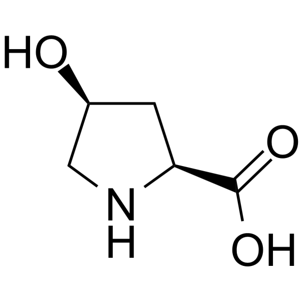
cis-4-Hydroxy-L-proline is an orally active proline analog and collagen production inhibitor. cis-4-Hydroxy-L-proline inhibits cell growth by preventing the deposition of triple helical collagen on the cell layer. cis-4-Hydroxy-L-proline inhibits N-nitrosomethylurea-induced breast tumor growth. cis-4-Hydroxy-L-proline improves lung compliance and reduces prostate weight. cis-4-Hydroxy-L-proline can be used in the study of hypoxic pulmonary hypertension.
For research use only. We do not sell to patients.
- Purity : 98.0%
- CAS No.: 618-27-9
- Formula: C5H9NO3
- Molecular Weight:131.13
-
Storage:Powder -20°C, 3 years , 4°C, 2 years ; In solvent -80°C, 6 months , -20°C, 1 month

To place orders, for customer services and technical support, please contact: MedChemExpress USA
Tel: 609-228-6898 E-mail: [email protected] [email protected]


-
 Biological Activity
-
Chemical Information
-
Solvent & Solubility
- Protocols
- Purity & Documentation
- References
-
 Help & FAQs


-
HY-Y0252
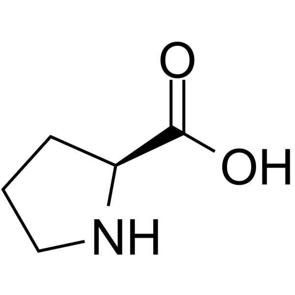
L-Proline -
HY-W008129
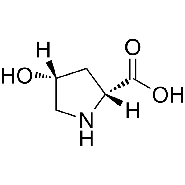
H-D-cis-Hyp-OHProline Derivative -
HY-N0304
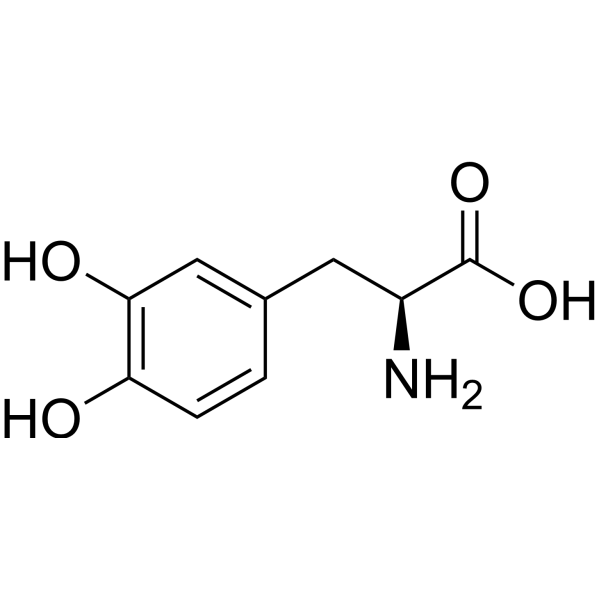
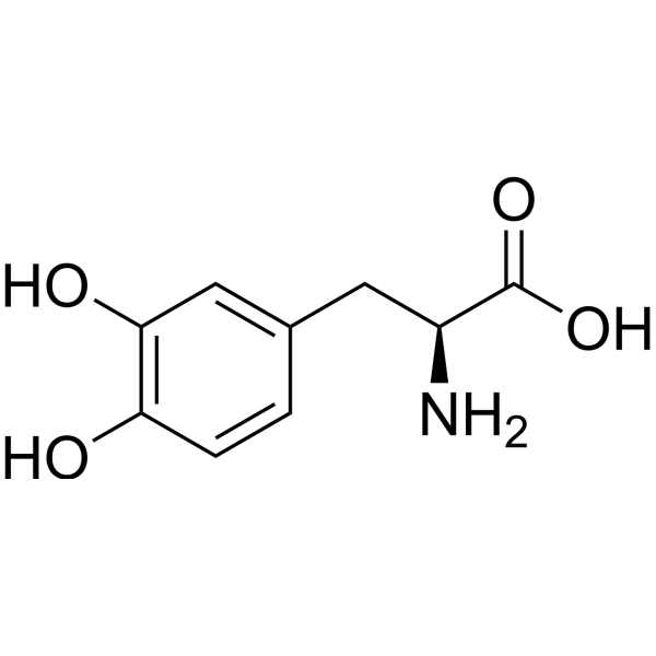
L-DOPADopamine Precursor -
HY-N0304R
L-DOPA (Standard)Dopamine Precursor -
HY-Y0588
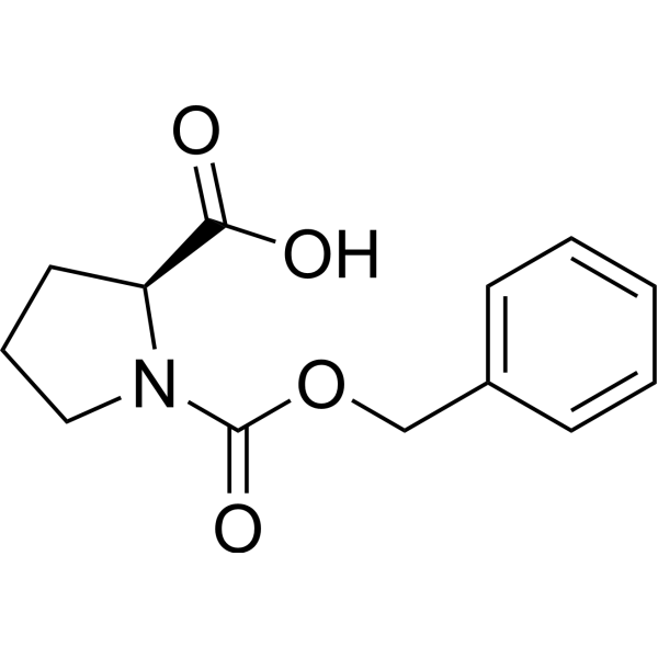
CarbobenzoxyprolineProlidase Inhibitor -
HY-Y0754
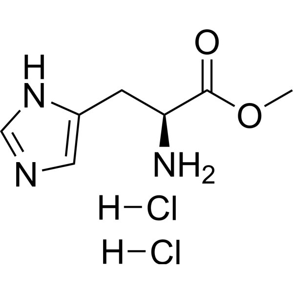
Methyl L-histidinate dihydrochlorideAmino Acid Derivative -
HY-W040821
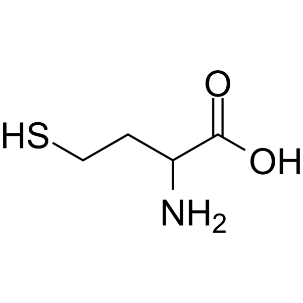
DL-HomocysteineNeurotoxin -

HY-40136R
cis-4-Hydroxy-L-proline (Standard)Collagen Production Inhibitor -

HY-W008129R
H-D-cis-Hyp-OH (Standard)Amino Acid Derivative -
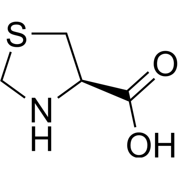
HY-W012676
L-ThioprolineAmino Acid Derivative -
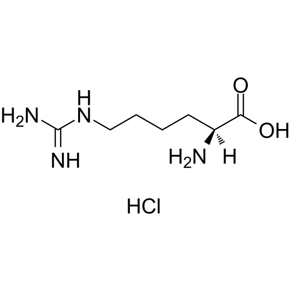
HY-W012340
H-HomoArg-OH hydrochlorideTNAP Inhibitor -
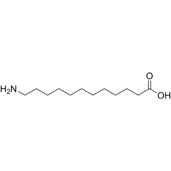
HY-W105740
12-Aminododecanoic acidPROTAC Linker
All Endogenous Metabolite Isoforms
More

Biological Activity
Description
In Vitro
cis-4-Hydroxy-L-proline (100-200 μg/mL; 1 week) inhibits the accumulation of collagenous and noncollagenous proteins in the culture medium in the in vitro co-culture experiment of rat Schwann cells (SC) and dorsal root ganglion neurons[1].
cis-4-Hydroxy-L-proline (200 μg/mL; 6 h) reduces the proportion of collagen synthesis in total protein synthesis and increases the degradation proportion of newly synthesized collagen in freshly isolated chick tendon fibroblasts[2].
cis-4-Hydroxy-L-proline (3200-32000 μg/mL; 24 h) significantly reduces the production of albumin and urea by hepatocytes[3].
MedChemExpress (MCE) has not independently confirmed the accuracy of these methods. They are for reference only. Further protocols information, click here.
In Vivo
cis-4-Hydroxy-L-proline (200 mg/kg; s.c.; twice daily; 30 days) partially prevents the accumulation of collagen in the lungs and partially improves the decrease in lung compliance in the Bleomycin (HY-108345)-induced pulmonary fibrosis model experiment of hamsters[4].
cis-4-Hydroxy-L-proline (200 mg/kg; s.c.; injected alone for 3 days first and then co-injected with dihydrotestosterone for 10 days) significantly reduces the wet weight of the prostate, inhibits the proliferation and differentiation of prostatic glandular cells in response to androgen stimulation in Androgen-induced prostate growth model in prepubertal castrated guinea pigs[5].
CHOP-PEG (polymeric prodrug of cis-4-Hydroxy-L-proline, 0.8 mg by continuous s.c.; 10 mg by single s.c.; 40 mg by single i.v.; 90 mg by single intratracheal injection; 7 days) can prevent and reverse chronic hypoxic pulmonary hypertension in the rat hypoxic pulmonary hypertension model[6].
MedChemExpress (MCE) has not independently confirmed the accuracy of these methods. They are for reference only.
-
Animal Model:Rats[3]
-
Dosage:285 mg/kg, 507 mg/kg, 903 mg/kg
-
Administration:Oral gavage (p.o.), 28 days
-
Result:Increased ALAT and ASAT at a dose of 903 mg/kg.
Increased albumin at 507 mg/kg.
Increased blood urea nitrogen at 903 mg/kg CHP.
Revealed a pale appearance of the liver due to drug-induced anemia.


Chemical Information
-
CAS No. 618-27-9
-
Appearance Solid
-
Molecular Weight 131.13
-
Formula C5H9NO3
-
Color White to off-white
-
SMILES
O=C(O)[C@H](C1)NC[C@H]1O
-
Structure Classification
-
Initial Source
-
Shipping
Room temperature in continental US; may vary elsewhere.
-
Storage
Powder -20°C 3 years 4°C 2 years In solvent -80°C 6 months -20°C 1 month


Solvent & Solubility
In Vitro:
H2O : 100 mg/mL (762.60 mM; Need ultrasonic)
DMSO : 1 mg/mL (7.63 mM; ultrasonic and warming and heat to 60°C; Hygroscopic DMSO has a significant impact on the solubility of product, please use newly opened DMSO)
Please refer to the solubility information to select the appropriate solvent. Once prepared, please aliquot and store the solution to prevent product inactivation from repeated freeze-thaw cycles.
Storage method and period of stock solution: -80°C, 6 months; -20°C, 1 month. When stored at -80°C, please use it within 6 months. When stored at -20°C, please use it within 1 month.
* Note: If you choose water as the stock solution, please dilute it to the working solution, then filter and sterilize it with a 0.22 μm filter before use.
Please refer to the solubility information to select the appropriate solvent. Once prepared, please aliquot and store the solution to prevent product inactivation from repeated freeze-thaw cycles.
Storage method and period of stock solution: -80°C, 6 months; -20°C, 1 month. When stored at -80°C, please use it within 6 months. When stored at -20°C, please use it within 1 month.
* Note: If you choose water as the stock solution, please dilute it to the working solution, then filter and sterilize it with a 0.22 μm filter before use.
Concentration (start) × Volume (start) = Concentration (final) × Volume (final)
In Vivo:
For the following dissolution methods, please prepare the working solution directly:
It is recommended to prepare fresh solutions and use them promptly within a short period of time.
The percentages shown for the solvents indicate their volumetric ratio in the final prepared solution. If precipitation or phase separation occurs during preparation, heat and/or sonication can be used to aid dissolution.
Add each solvent one by one: PBS
Solubility: 100 mg/mL (762.60 mM); Clear solution; Need ultrasonic

Protocols
-
Protocol for Cell Counting and Cell Density Analysis
Cell counting and cell-density analysis estimate the number of cells in a known volume or field area. Manual hemocytometer counting uses a chamber of defined geometry to convert counted cells into cells/mL, while automated counters and image-analysis workflows detect cell objects from optical, brightfield, fluorescence, impedance, or digital-image features. Trypan blue viability counting is based on dye exclusion: viable cells with intact membranes exclude dye, while non-viable cells with compromised membranes stain blue. The readout is total cell density, viable-cell density, dead-cell density, and percent viability. Cell density can also be estimated from microscopy images by counting objects per image area, from flow cytometry using calibrated volume or reference particles, or from in situ microscopy in bioreactors after calibration against reference methods such as hemocytometer or flow cytometry.
-
Somatic Cell Culture
A method of simulating the in vivo environment in vitro to maintain the cell growth, differentation and main functions.
-
Patient-Derived Xenograft (PDX)
Patient-derived xenograft (PDX) models are generated by engrafting primary human tumor tissue directly into immunodeficient mice, allowing in vivo propagation of patient tumor biology without initial in vitro adaptation. These models are used to preserve key histopathological and molecular characteristics of the original tumor and enable assessment of tumor growth dynamics and therapeutic response in a living organism. The biological readout is tumor engraftment and subsequent growth in the murine host, which reflects the ability of human tumor cells to survive, vascularize, and expand in an immunocompromised microenvironment.
-
Patient-Derived Orthotopic Xenograft (PDOX)
Patient-derived orthotopic xenograft (PDOX) modeling implants fresh patient tumor tissue or patient-derived tumor cells into the anatomically corresponding organ or tissue site of immunodeficient mice, usually by surgical orthotopic implantation, to preserve patient tumor histology, local microenvironmental context, invasion, metastatic behavior, and treatment-response features better than subcutaneous implantation. PDOX readouts include tumor engraftment, orthotopic tumor growth, local invasion, metastasis, recurrence after resection, histologic similarity to the donor tumor, biomarker retention, molecular concordance, survival, and response or resistance to therapy. PDOX models are used for preclinical drug testing and individualized therapy evaluation, but engraftment success varies by tumor type and specimen quality.
-
CCK-8/WST-8 Cell Proliferation Assay
The CCK-8/WST-8 assay is based on the reduction of the water-soluble tetrazolium salt WST-8 to a water-soluble formazan product by cellular dehydrogenases in metabolically active cells, where the generated formazan amount is proportional to the number of living cells and is quantified by measuring absorbance in the visible range, providing a colorimetric readout for cell viability and proliferation assessment. This class of tetrazolium-based assays improves upon earlier MTT-based systems by producing a water-soluble formazan, eliminating the need for organic solubilization steps and enabling direct spectrophotometric measurement in culture medium.
-
Research Protocol for Cardiovascular Diseases
Cardiovascular disease can be modeled as maladaptive cardiac remodeling, where ischemic injury or pressure overload activates inflammatory signaling, fibroblast activation, extracellular-matrix deposition, cardiomyocyte hypertrophy, vascular remodeling, and progressive ventricular dysfunction. The TGF-β/SMAD axis is a central profibrotic pathway after myocardial injury and pressure overload, while innate immune and cytokine pathways regulate leukocyte recruitment, scar formation, and adverse remodeling. Key unresolved questions include which inflammatory signals are reparative versus harmful, when fibrosis is protective versus maladaptive, and whether pathway inhibition improves function without weakening necessary infarct healing or compensatory remodeling.
-
Cell Counting-Based Growth Curve Assay
Cell counting-based growth curve assays quantify cell proliferation by directly measuring changes in viable cell number over time using manual or automated counting methods such as hemocytometer-based counting or instrument-assisted cell enumeration, enabling construction of growth curves that reflect population expansion dynamics in response to culture conditions. A widely used approach is trypan blue exclusion with hemocytometer counting, where membrane-compromised (non-viable) cells take up the dye, allowing discrimination between viable and non-viable cells while simultaneously enabling total cell number quantification. Repeated sampling across time points allows estimation of proliferation rate, growth phases, and comparative growth kinetics between experimental conditions.
-
MTT Cell Proliferation Assay
The MTT assay is a colorimetric endpoint assay for estimating viable cell number, cell growth, cytotoxicity, or cell activation in cultured mammalian cells. Living cells reduce the yellow tetrazolium salt MTT into purple/blue formazan, while dead cells do not generate the same signal; the resulting color can be quantified with a multiwell spectrophotometer. MTT reduction is commonly interpreted as a readout of metabolic activity that often correlates with viable cell number, but it should not be treated as a direct cell-counting method unless the assay is optimized for the cell type and experimental condition. Studies show that MTT reduction can involve mitochondrial and non-mitochondrial reducing systems, and formazan may accumulate in intracellular lipid droplets rather than simply marking mitochondria.
-
Subcutaneous Cell-Line-Derived Xenograft
Subcutaneous cell-line-derived xenograft (CDX) models are established by implanting cultured human cancer cell lines into immunodeficient mice, where the injected cells form localized tumors that can be monitored in vivo as a measure of tumorigenic potential, growth kinetics, and treatment response. These models are widely used in oncology research because they allow reproducible tumor formation and enable comparative assessment of tumor growth between different cell lines or genetic manipulations in a controlled in vivo microenvironment. Subcutaneous implantation of cancer cells in immunodeficient mice is a standard approach for evaluating tumor growth behavior and therapeutic response across multiple cancer types, including prostate, esophageal, pancreatic, and colon cancer models.
-
Breast Cancer Modeling
Breast cancer is a heterogeneous cancer, and it has been distinguished into four subtypes: luminal A, luminal B, HER2-positive and basal-like. Molecular mutations, epigenetic alterations, hormone exposure and immune microenvironment are related to the progression of breast cancer.
-
Orthotopic Cell-Line Xenograft
Orthotopic cell-line xenograft models involve implantation of human cancer cell lines into the anatomically corresponding organ of immunodeficient mice to reproduce tumor growth within a native microenvironment, enabling more clinically relevant tumor behavior compared with subcutaneous models. These models are widely used because orthotopic placement better recapitulates tumor progression, including invasion and metastatic spread, which are often underrepresented in heterotopic implantation systems. Compared with conventional xenografts, orthotopic implantation is described as more technically complex but provides improved simulation of tumor-microenvironment interactions and metastatic behavior, making it particularly valuable for translational oncology research. Surgical orthotopic implantation approaches have been emphasized as enabling faithful reproduction of clinical cancer features, including metastasis and disease progression patterns that align with the tumor’s organ of origi
-
Cell Viability Determination by MTT Colorimetric Assay
The following protocol uses the MTT colorimetric assay as a classic literature-established method for assessing cell viability/metabolic activity in cultured mammalian cells. MTT[3-(4,5-dimethylthiazol-2-yl)-2,5-diphenyltetrazolium bromide] is reduced by metabolically active cells to a colored formazan product; the amount of formazan is quantified spectrophotometrically and provides an indirect measure of metabolically active viable cells. Importantly, MTT reduction reflects cellular oxidoreductase/metabolic activity rather than an absolute direct count of living cells, so changes in cellular metabolism can alter the signal independently of cell number.


Purity & Documentation
-
Data Sheet (290 KB)
-
SDS (393 KB)
- English - EN (393 KB)
- Français - FR (393 KB)
- Deutsch - DE (393 KB)
- Norwegian - NO (393 KB)
- Español - ES (393 KB)
- Swedish - SV (393 KB)
- Italian - IT (393 KB)
- Korean - KR (393 KB)
- Portuguese - PT (393 KB)
-
Handling Instructions (2659 KB)
References
[1]. Eldridge CF, et al. Effects of cis-4-hydroxy-L-proline, and inhibitor of Schwann cell differentiation, on the secretion of collagenous and noncollagenous proteins by Schwann cells. Exp Cell Res. 1988 Feb;174(2):491-501. [Content Brief]
[2]. Neblock DS, et al. The effect of cis-4-hydroxy-L-proline on intracellular degradation of newly synthesized collagen by freshly isolated chick tendon fibroblasts. Connect Tissue Res. 1982;10(3-4):297-301. [Content Brief]
[3]. Dickens H, et al. Anticancer drug cis-4-hydroxy-L-proline: Correlation of preclinical toxicology with clinical parameters of liver function. Mol Med Rep. 2008 Jul-Aug;1(4):459-64. [Content Brief]
[4]. Riley DJ, et al. Effect of cis-4-hydroxy-L-proline on lung mechanics and collagen content of bleomycin-treated hamsters. Chest. 1981 Jul;80(1 Suppl):49-50. [Content Brief]
[5]. Wong YC, et al. Effects of cis-4-hydroxy-L-proline on the androgen-induced growth of the lateral prostate of the prepubertally castrated guinea pig. Prostate. 1993;23(4):337-54. [Content Brief]
[6]. Simon PM, et al. Prodrug of proline analogue reduces hypoxic pulmonary hypertension in rats. Pulm Pharmacol Ther. 2006;19(4):242-50. [Content Brief]
[7]. Tan EM, et, al. Proline analogues inhibit human skin fibroblast growth and collagen production in culture. J Invest Dermatol. 1983 Apr;80(4):261-7. [Content Brief]
[8]. Riley DJ, et, al. Prevention of bleomycin-induced pulmonary fibrosis in the hamster by cis-4-hydroxy-l-proline. Am Rev Respir Dis. 1981 Apr;123(4 Pt 1):388-93. [Content Brief]
[9]. Lewko WM, et, al. Sensitivity of N-nitrosomethylurea-induced rat mammary tumors to cis-hydroxyproline, an inhibitor of collagen production. Cancer Res. 1981 Jul;41(7):2855-62. [Content Brief]
[10]. Kao WW, et, al. Proline analogue removes fibroblasts from cultured mixed cell populations. Nature. 1977 Mar 3;266(5597):63-4. [Content Brief]


Complete Stock Solution Preparation Table
Please refer to the solubility information to select the appropriate solvent. Once prepared, please aliquot and store the solution to prevent product inactivation from repeated freeze-thaw cycles.
Storage method and period of stock solution: -80°C, 6 months; -20°C, 1 month. When stored at -80°C, please use it within 6 months. When stored at -20°C, please use it within 1 month.
| Optional Solvent | Concentration Solvent Mass | 1 mg | 5 mg | 10 mg | 25 mg |
|---|---|---|---|---|---|
| DMSO / H2O | 1 mM | 7.6260 mL | 38.1301 mL | 76.2602 mL | 190.6505 mL |
| 5 mM | 1.5252 mL | 7.6260 mL | 15.2520 mL | 38.1301 mL | |
| H2O | 10 mM | 0.7626 mL | 3.8130 mL | 7.6260 mL | 19.0650 mL |
| 15 mM | 0.5084 mL | 2.5420 mL | 5.0840 mL | 12.7100 mL | |
| 20 mM | 0.3813 mL | 1.9065 mL | 3.8130 mL | 9.5325 mL | |
| 25 mM | 0.3050 mL | 1.5252 mL | 3.0504 mL | 7.6260 mL | |
| 30 mM | 0.2542 mL | 1.2710 mL | 2.5420 mL | 6.3550 mL | |
| 40 mM | 0.1907 mL | 0.9533 mL | 1.9065 mL | 4.7663 mL | |
| 50 mM | 0.1525 mL | 0.7626 mL | 1.5252 mL | 3.8130 mL | |
| 60 mM | 0.1271 mL | 0.6355 mL | 1.2710 mL | 3.1775 mL | |
| 80 mM | 0.0953 mL | 0.4766 mL | 0.9533 mL | 2.3831 mL | |
| 100 mM | 0.0763 mL | 0.3813 mL | 0.7626 mL | 1.9065 mL |
* Note: If you choose water as the stock solution, please dilute it to the working solution, then filter and sterilize it with a 0.22 μm filter before use.


cis-4-Hydroxy-L-proline Related Classifications
HY-40136 Related Classifications
 Powered by Bioz