



Cl-Necrostatin-1
Cl-Necrostatin-1 is a RIPK1 inhibitor. Cl-Necrostatin-1 can also inhibit TNF-α-induced necroptosis in Jurkat cells deficient in Fas-associated death domain protein (FADD; EC50 = 180 nM), a modification that prevents caspase activation in response to death-domain receptor signaling. Cl-Necrostatin-1 can also reduce infarct size in a mouse model of middle cerebral artery occlusion (MCAO). Cl-Necrostatin-1 is used for research in cardiovascular and cerebrovascular diseases.
For research use only. We do not sell to patients.
- CAS No.: 862377-51-3
- Formula: C13H12ClN3OS
- Molecular Weight:293.77
-
Storage:
Please store the product under the recommended conditions in the Certificate of Analysis.


To place orders, for customer services and technical support, please contact: MedChemExpress USA
Tel: 609-228-6898 E-mail: [email protected] [email protected]


-
 Biological Activity
-
Chemical Information
- Protocols
- Purity & Documentation
- References
-
 Help & FAQs


-
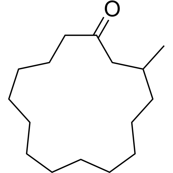
HY-N0633
MusconeTNF Receptor Inhibitor -

HY-158312
UH15-38RIPK3 Inhibitor -
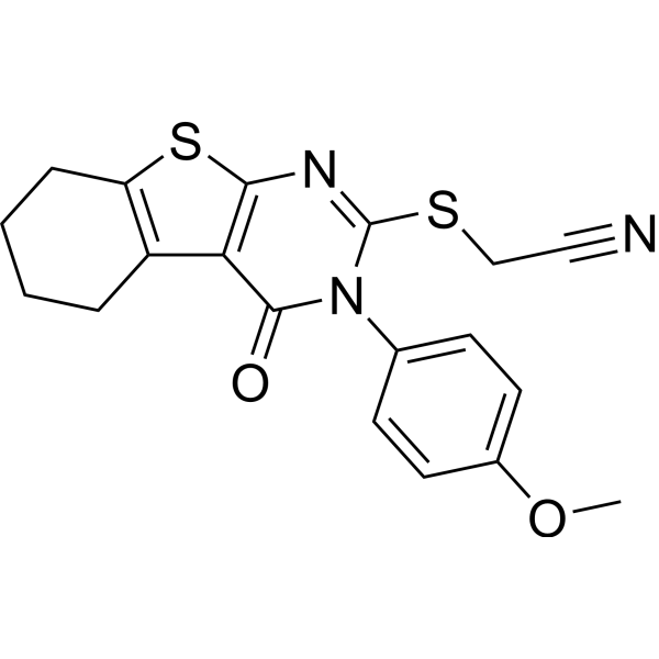
HY-124546
Necrostatin-5Necroptosis/RIP1 Inhibitor -
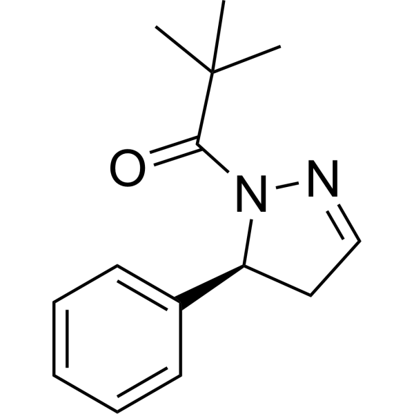
HY-103028A
GSK963RIP1 Kinase Inhibitor -
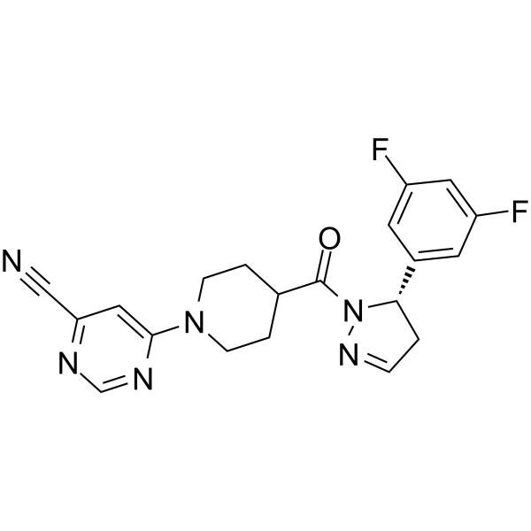
HY-114492
GSK547RIPK1 Inhibitor -
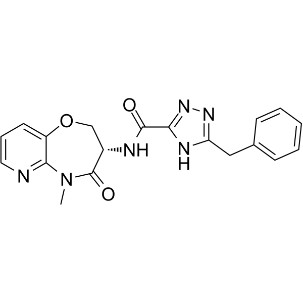
HY-114371
EclitasertibRIPK1 Inhibitor -
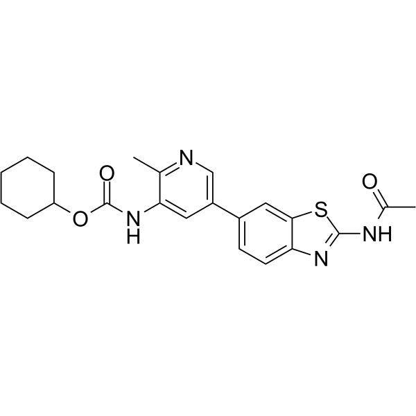
HY-128348
PK68RIPK1 Inhibitor -
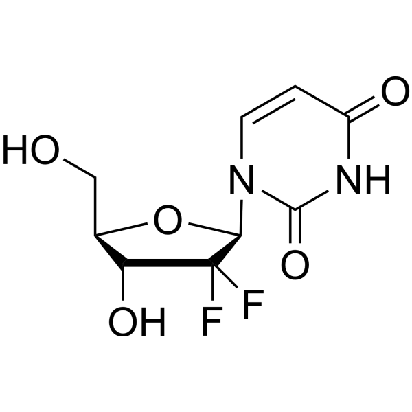
HY-138253
2′,2′-DifluorodeoxyuridineGemcitabine Metabolite -
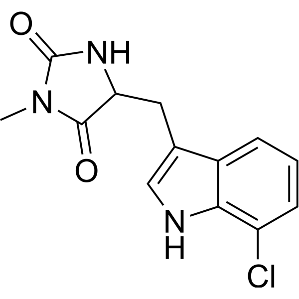
HY-14622A
Necrostatin 2 racemateRIPK1 Inhibitor -

HY-138253R
2′,2′-Difluorodeoxyuridine (Standard)Gemcitabine Metabolite -
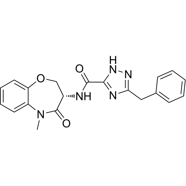
HY-101760
GSK2982772RIPK1 Inhibitor -
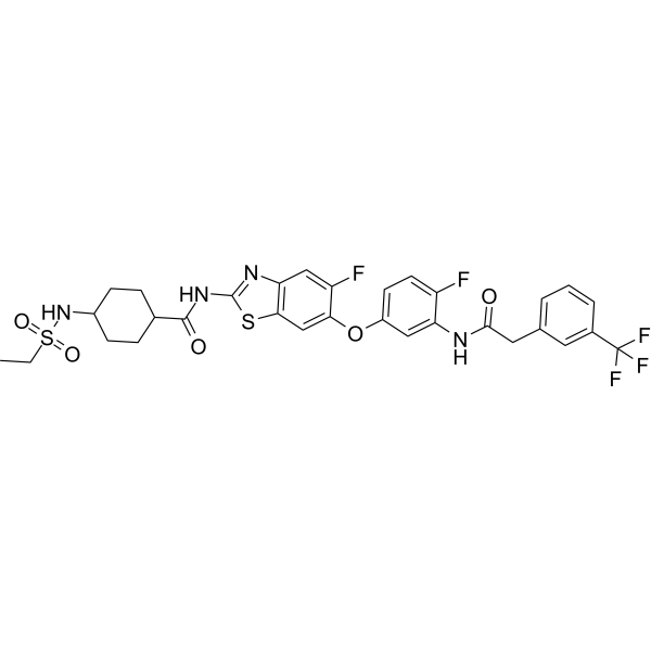
HY-149052
SZM-1209RIPK1 Inhibitor


Biological Activity
Description
Cellular Effect
|
Cell Line

|
Type | Value | Description | References |
|---|---|---|---|---|
| Jurkat | EC50 |
0.18 μM
Compound: 12
|
Effective concentration required for inhibition of necroptosis in FADD deficient Jurkat T cells treated with TNF-alpha
Effective concentration required for inhibition of necroptosis in FADD deficient Jurkat T cells treated with TNF-alpha
|
[PMID: 16153840] |
| Jurkat | EC50 |
180 nM
Compound: 10
|
Inhibition of cellular necroptosis in TNFalpha treated FADD deficient human Jurkat cells
Inhibition of cellular necroptosis in TNFalpha treated FADD deficient human Jurkat cells
|
[PMID: 18408713] |
| Jurkat | IC50 |
0.18 μM
Compound: N3
|
Antiproliferative activity against human Jurkat cells assessed as reduction in cell viability incubated for 24 hrs by MTT assay
Antiproliferative activity against human Jurkat cells assessed as reduction in cell viability incubated for 24 hrs by MTT assay
|
[PMID: 38199165] |
| Jurkat | IC50 |
182 nM
Compound: 3, 7-cl-Nec-1
|
Inhibition of death receptor signaling mediated necroptotic cell death in human Jurkat cell deficient in FADD assessed as cell viability after 30 hrs by ATP based viability assay in presence of TNFalpha
Inhibition of death receptor signaling mediated necroptotic cell death in human Jurkat cell deficient in FADD assessed as cell viability after 30 hrs by ATP based viability assay in presence of TNFalpha
|
[PMID: 16408008] |


Chemical Information
-
CAS No. 862377-51-3
-
Molecular Weight 293.77
-
Formula C13H12ClN3OS
-
SMILES
O=C1N(C)C(NC1CC2=CNC3=C2C=CC=C3Cl)=S
-
Shipping
Room temperature in continental US; may vary elsewhere.
-
Storage
Please store the product under the recommended conditions in the Certificate of Analysis.

Protocols
-
Apoptosis
Apoptosis, also called programmed cell death, is generally characterized by distinct morphological characteristics.
-
Apoptosis Solutions
Apoptosis is a regulated, generally non-lytic cell-death pathway that removes unwanted, damaged, infected, or abnormal cells through coordinated morphological changes, caspase activation, DNA fragmentation, and membrane remodeling. The intrinsic apoptosis pathway is controlled mainly by mitochondrial outer membrane permeabilization, BCL-2 family proteins, cytochrome c release, apoptosome formation, caspase-9 activation, and downstream executioner caspase-3/7 activation. The extrinsic apoptosis pathway is initiated by death receptors such as Fas, TNFR, and TRAIL receptors, which recruit adaptor proteins and activate caspase-8 before engaging executioner caspases or mitochondrial amplification through BID cleavage. Apoptosis is linked to many phenotypes, including cancer cell killing, tissue homeostasis, immune regulation, neurodegeneration, infection response, and treatment-induced cytotoxicity; unresolved questions include how apoptosis interacts with necroptosis, pyroptosis, ferroptos
-
How to Choose the Right Model Animal
Choosing the right model animal is a validity-driven decision in which the species, strain, sex, age, genetic background, disease-induction method, outcome measures, and welfare burden must match the scientific question rather than laboratory tradition or convenience. A model should be selected by judging face validity, construct validity, and predictive validity: whether it resembles the human phenotype, whether it reproduces relevant mechanisms, and whether results are likely to predict human biology or treatment response. Animal studies often fail to translate because of species differences, weak disease resemblance, poor experimental design, inadequate reporting, publication bias, and underuse of randomization, blinding, and sample-size justification. Unresolved questions include how to rank competing models objectively, how much human-disease complexity must be reproduced for a given objective, and when non-animal systems such as organoids, ex vivo tissue, or computational models


Purity & Documentation
References
[1]. Degterev A, et al. Identification of RIP1 kinase as a specific cellular target of necrostatins. Nat Chem Biol. 2008 May;4(5):313-21. [Content Brief]
[2]. Teng X, et al. Structure-activity relationship study of novel necroptosis inhibitors. Bioorg Med Chem Lett. 2005 Nov 15;15(22):5039-44. [Content Brief]
[3]. Degterev A, et al. Chemical inhibitor of nonapoptotic cell death with therapeutic potential for ischemic brain injury. Nat Chem Biol. 2005 Jul;1(2):112-9. [Content Brief]

Calculators
Concentration (start) × Volume (start) = Concentration (final) × Volume (final)


Cl-Necrostatin-1 Related Classifications
HY-168770 Related Classifications
 Powered by Bioz