



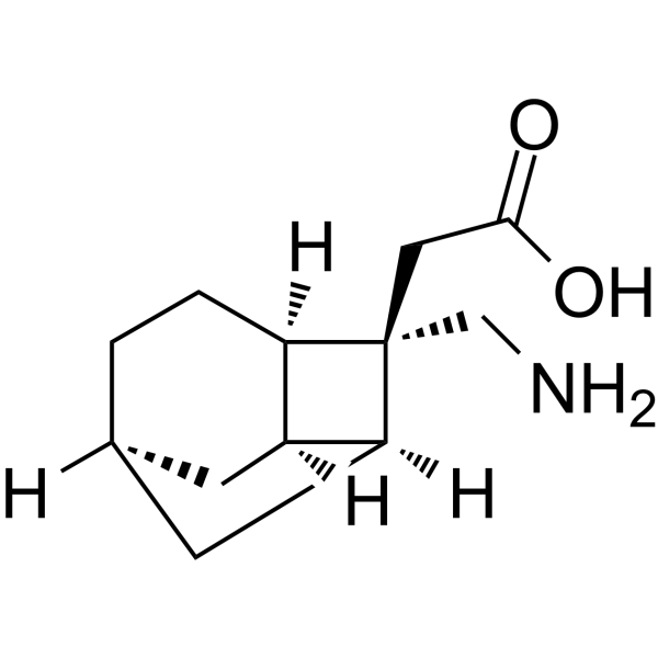
Crisugabalin

Based on 1 Customer Validation
Crisugabalin is an orally active, selective ligand for the α2δ subunit of voltage-gated calcium channels, with a target IC50 of 3.96 nM in rats. Crisugabalin inhibits the binding of [3H]gabapentin to the α2δ subunit, reduces calcium influx, decreases neuronal excitability, and impairs nociceptive transmission. Crisugabalin alleviates mechanical allodynia, neuropathic pain and inflammatory pain in rats, and reduces phase II pain behaviors. Crisugabalin can be used in research related to chronic pain, neuropathic pain, diabetic neuropathy, fibromyalgia, inflammatory pain, diabetic peripheral neuropathy and postherpetic neuralgia.
For research use only. We do not sell to patients.
- Purity : 99.88%
- CAS No.: 2209104-84-5
- Formula: C12H19NO2
- Molecular Weight:209.28
-
Storage:
Please store the product under the recommended conditions in the Certificate of Analysis.

To place orders, for customer services and technical support, please contact: MedChemExpress USA
Tel: 609-228-6898 E-mail: [email protected] [email protected]


-
 Biological Activity
-
Chemical Information
-
Solvent & Solubility
- Protocols
- Purity & Documentation
- References
-
 Help & FAQs


All Calcium Channel Isoforms
More

Biological Activity
Description
In Vitro
Crisugabalin (incubated at 25 °C for 30 min) potently binds to the VGCC α2δ subunit in the cerebral cortex cell membranes of Wistar rats, with an IC50 of 3.96 nM[1].
MedChemExpress (MCE) has not independently confirmed the accuracy of these methods. They are for reference only.
Parmacokinetics
In Vivo
Crisugabalin (1-30 mg/kg; p.o.; single administration) dose-dependently increases the mechanical pain threshold in streptozotocin (HY-13753)-induced diabetic neuropathy rats[1].
Crisugabalin (p.o.; single administration; 30 mg/kg) inhibits intermittent cold stress (ICS)-induced mechanical hyperalgesia in fibromyalgia mice[1].
Crisugabalin (10-30 mg/kg; p.o.; single administration) dose-dependently alleviates formalin-induced phase II inflammatory pain behaviors[1].
Crisugabalin (100 mg/kg; p.o.; single administration) induces sedative effects in rats[1].
MedChemExpress (MCE) has not independently confirmed the accuracy of these methods. They are for reference only.
-
Animal Model:Sprague-Dawley (male, 160-180 g, CCI-induced neuropathic pain)[1]
-
Dosage:1 mg/kg;3 mg/kg; 10 mg/kg; 30 mg/kg
-
Administration:p.o.; single dose
-
Result:Increased 50% PWT to 3.24-fold that of vehicle controls at 2 hours post-dosing (30 mg/kg, P < 0.001), with effects peaking at 6 hours post-dosing.
Produced PWT values of 12.93 g and 13.66 g at 2 and 4 hours post-dosing, respectively (10 mg/kg), similar to the 30 mg/kg group (11.93 g, 13.55 g).
Had a lower PWT than the 30 mg/kg group at 6 hours post-dosing (10 mg/kg) but efficacy comparable to 30 mg/kg pregabalin.
Significantly increased 50% PWT to 7.33 g vs.
3.11 g in vehicle controls at 4 hours post-dosing (3 mg/kg).
Had a minimum effective dose (MED) of 10 mg/kg for increased AUC of 50% PWT vs.
time.
-
Animal Model:Sprague-Dawley (male, 160-180 g, STZ-induced diabetic neuropathy)[1]
-
Dosage:1 mg/kg;3 mg/kg; 10 mg/kg; 30 mg/kg
-
Administration:p.o.; single dose
-
Result:Significantly increased 50% PWT at 2 hours post-dosing (30 mg/kg), with efficacy persisting up to 24 hours post-dosing.
Increased 50% PWT to 2.4-fold at 2 hours post-dosing (10 mg/kg), with significant increases at 4 hours (10.55 g vs.
4.35 g) and 6 hours (11.74 g vs.
4.24 g), and efficacy persisting up to 8 hours post-dosing.
Doubled PWT vs.
vehicle controls at 6 hours post-dosing (3 mg/kg).
Had a MED of 10 mg/kg for increased AUC of 50% PWT vs.
time, with efficacy at 10 mg/kg comparable to 30 mg/kg pregabalin.
-
Animal Model:C57BL/6 (male, 18-25 g, ICS-induced fibromyalgia)[1]
-
Dosage:30 mg/kg
-
Administration:p.o.; single dose
-
Result:Had a MED of 30 mg/kg for increased 50% PWT.
Induced a 2.6-fold increase in 50% PWT, with efficacy comparable to 30 mg/kg pregabalin.
-
Animal Model:ICR (male, 25-35 g, formalin-induced inflammatory pain)[1]
-
Dosage:10 mg/kg; 30 mg/kg
-
Administration:p.o.; single dose
-
Result:Decreased formalin-induced phase II nociceptive motion counts in a dose-dependent manner.
Produced a 1.6-fold and 2.2-fold decrease in phase II motion counts vs.
vehicle controls at 10 mg/kg and 30 mg/kg, respectively.
Had efficacy at 30 mg/kg comparable to 30 mg/kg pregabalin.
Had no significant effect on phase I motion counts at 30 mg/kg.
-
Animal Model:Sprague-Dawley (male, 160-180 g)[1]
-
Dosage:100 mg/kg
-
Administration:p.o.; single dose
-
Result:Produced a 3-fold decrease in total distance traveled vs.
vehicle controls.
Had a MED of 100 mg/kg for sedative effects, with efficacy comparable to 100 mg/kg pregabalin.


Chemical Information
-
CAS No. 2209104-84-5
-
Appearance Solid
-
Molecular Weight 209.28
-
Formula C12H19NO2
-
Color White to off-white
-
SMILES
O=C(O)C[C@@]1(CN)[C@](C2)([H])[C@@]3([H])C[C@]2([H])CC[C@@]13[H]
-
Synonyms
HSK16149
-
Shipping
Room temperature in continental US; may vary elsewhere.
-
Storage
Please store the product under the recommended conditions in the Certificate of Analysis.


Solvent & Solubility
In Vitro:
DMSO : ≥ 100 mg/mL (477.83 mM; Hygroscopic DMSO has a significant impact on the solubility of product, please use newly opened DMSO)
* "≥" means soluble, but saturation unknown.
Concentration (start) × Volume (start) = Concentration (final) × Volume (final)


Purity & Documentation
-
Data Sheet (282 KB)
-
SDS (252 KB)
- English - EN (252 KB)
- Français - FR (252 KB)
- Deutsch - DE (252 KB)
- Norwegian - NO (252 KB)
- Español - ES (252 KB)
- Swedish - SV (252 KB)
- Italian - IT (252 KB)
- Korean - KR (252 KB)
- Portuguese - PT (252 KB)
-
Handling Instructions (2659 KB)
References
[1]. Gou X, et al. Pharmacology and Mechanism of Action of HSK16149, a Selective Ligand of α2δ Subunit of Voltage-Gated Calcium Channel with Analgesic Activity in Animal Models of Chronic Pain. J Pharmacol Exp Ther. 2021 Mar;376(3):330-337. [Content Brief]
[2]. Wang Z, et al. Quantification of crisugabalin (HSK16149) in biological matrix by LC-MS/MS method: An application to rat pharmacokinetic and tissue distribution studies. J Chromatogr B Analyt Technol Biomed Life Sci. 2025;1251:124396. [Content Brief]


Complete Stock Solution Preparation Table
| Optional Solvent | Concentration Solvent Mass | 1 mg | 5 mg | 10 mg | 25 mg |
|---|---|---|---|---|---|
| DMSO | 1 mM | 4.7783 mL | 23.8914 mL | 47.7829 mL | 119.4572 mL |
| 5 mM | 0.9557 mL | 4.7783 mL | 9.5566 mL | 23.8914 mL | |
| 10 mM | 0.4778 mL | 2.3891 mL | 4.7783 mL | 11.9457 mL | |
| 15 mM | 0.3186 mL | 1.5928 mL | 3.1855 mL | 7.9638 mL | |
| 20 mM | 0.2389 mL | 1.1946 mL | 2.3891 mL | 5.9729 mL | |
| 25 mM | 0.1911 mL | 0.9557 mL | 1.9113 mL | 4.7783 mL | |
| 30 mM | 0.1593 mL | 0.7964 mL | 1.5928 mL | 3.9819 mL | |
| 40 mM | 0.1195 mL | 0.5973 mL | 1.1946 mL | 2.9864 mL | |
| 50 mM | 0.0956 mL | 0.4778 mL | 0.9557 mL | 2.3891 mL | |
| 60 mM | 0.0796 mL | 0.3982 mL | 0.7964 mL | 1.9910 mL | |
| 80 mM | 0.0597 mL | 0.2986 mL | 0.5973 mL | 1.4932 mL | |
| 100 mM | 0.0478 mL | 0.2389 mL | 0.4778 mL | 1.1946 mL |


Crisugabalin Related Classifications
HY-142240 Related Classifications
 Powered by Bioz
Keywords
- Crisugabalin
- 2209104-84-5
- HSK16149
- HSK 16149
- HSK-16149
- Calcium Channel
- ligand for the α2δ subunit of voltage-gated calcium channels
- Sprague-Dawley rat
- C57BL/6 mice
- ICR mice
- chronic pain
- neuropathic pain
- diabetic neuropathy
- fibromyalgia
- inflammatory pain
- diabetic peripheral neuropathic pain
- postherpetic neuralgia
- Inhibitor
- inhibitor
- inhibit