



[D-Ala2]-GIP (human)

Based on 1 Customer Validation
[D-Ala2]-GIP (human) is a GIP receptor agonist (EC50 = 630 pM). [D-Ala2]-GIP (human) improves glucose tolerance. [D-Ala2]-GIP (human) shows neuroprotective activity in MPTP (HY-W114750)-induced Parkinson's disease model. [D-Ala2]-GIP (human) also improves cognitive function and hippocampal synaptic plasticity in obese diabetic rats. [D-Ala2]-GIP (human) can be used for research of type 2 diabetes, Parkinson's disease, etc
For research use only. We do not sell to patients.
- Purity : 99.57%
- CAS No.: 444073-04-5
- Formula: C226H338N60O66S
- Molecular Weight:4983.60
-
Storage:
Sealed storage, away from moisture and light.
Powder -80°C, 2 years , -20°C, 1 year* In solvent : -80°C, 6 months; -20°C, 1 month (sealed storage, away from moisture and light)
![[D-Ala2]-GIP (human) Chemical Structure](https://file.medchemexpress.com/product_pic/hy-p3247.gif "Click to View [D-Ala2]-GIP (human) Chemical Structure")

To place orders, for customer services and technical support, please contact: MedChemExpress USA
Tel: 609-228-6898 E-mail: [email protected] [email protected]


-
 Biological Activity
-
Chemical Information
-
Solvent & Solubility
- Protocols
- Purity & Documentation
- References
-
 Help & FAQs


-
HY-P3462
CagrilintideAMYR/CTR agonist -
HY-P0276
GIP, humanInsulin Secretion Stimulator -

HY-114118
SemaglutideGLP-1R Agonist -

HY-P10138
human GIP(3-30), amideGIP Antagonist -
HY-P2542
GIP (3-42), humanGIP Receptor Agonist -

HY-103545
GIP (1-39)Insulinotropic Peptide -
HY-P3579
Gastric Inhibitory Peptide, porcineInsulinotropic Polypeptide -

HY-P2541
GIP (1-30) amide, porcineInsulinotropic Polypeptide -
HY-P3255
DA-JC4GLP-1/GIP Receptor Agonist


Biological Activity
Description
In Vivo
[D-Ala2]-GIP (25 nmol/kg, s.c.) can improve the cognitive function, hippocampal synaptic plasticity and glucose homeostasis of mice fed a high-fat diet[2].
[D-Ala2]-GIP (50-100 nmol/kg, i.p.) can attenuate the behavioral phenotype of Parkinson's disease caused by MPTP (HY-W114750) administration in mice[3].
MedChemExpress (MCE) has not independently confirmed the accuracy of these methods. They are for reference only.
-
Animal Model:Male C57BL/6 mice[3]
-
Dosage:50-100 nmol/kg
-
Administration:Intraperitoneal injection (i.p.)
-
Result:Reversed the decline in motor ability caused by MPTP.
Reverse the increase in muscle stiffness.
Reduce the increased tremor amplitude at all frequencies except 7 Hz, and observe maximum protection in the frequency range of 1-6 Hz.
Significantly inhibited the increase of MDA levels in the brain.
-
Animal Model:Male Wistar rats (~290 g) were fasted overnight and then given an oral glucose tolerance test (OGTT) (1 g glucose/kg body wt)[1]
-
Dosage:8 nmol/kg
-
Administration:Subcutaneous injection (s.c.)
-
Result:Resulted in a more pronounced reduction in the glycemic profile and an enhanced insulin time course than that of native GIP during an OGTT.
-
Animal Model:Young (8 week-old) male NIH Swiss mice[2]
-
Dosage:25 nmol/kg
-
Administration:Subcutaneous injection (s.c.)
-
Result:Significantly decreased (P < 0.001) blood glucose levels in high-fat fed mice.
Fully rescued the deleterious effects of high-fat feeding on long-term potentiation.


Chemical Information
-
CAS No. 444073-04-5
-
Appearance Solid
-
Molecular Weight 4983.60
-
Formula C226H338N60O66S
-
Color White to off-white
-
Sequence
Tyr-{d-Ala}-Glu-Gly-Thr-Phe-Ile-Ser-Asp-Tyr-Ser-Ile-Ala-Met-Asp-Lys-Ile-His-Gln-Gln-Asp-Phe-Val-Asn-Trp-Leu-Leu-Ala-Gln-Lys-Gly-Lys-Lys-Asn-Asp-Trp-Lys-His-Asn-Ile-Thr-Gln
-
Sequence Shortening
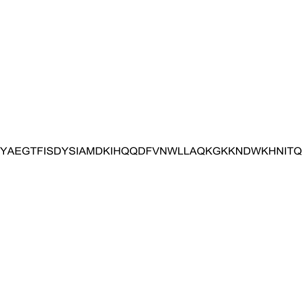
Y-{d-Ala}-EGTFISDYSIAMDKIHQQDFVNWLLAQKGKKNDWKHNITQ
-
Shipping
Room temperature in continental US; may vary elsewhere.
-
Storage
Sealed storage, away from moisture and light
Powder -80°C 2 years -20°C 1 year * In solvent : -80°C, 6 months; -20°C, 1 month (sealed storage, away from moisture and light)


Solvent & Solubility
In Vitro:
DMSO : 50 mg/mL (10.03 mM; Need ultrasonic; Hygroscopic DMSO has a significant impact on the solubility of product, please use newly opened DMSO)
Please refer to the solubility information to select the appropriate solvent. Once prepared, please aliquot and store the solution to prevent product inactivation from repeated freeze-thaw cycles.
Storage method and period of stock solution: -80°C, 6 months; -20°C, 1 month (sealed storage, away from moisture and light). When stored at -80°C, please use it within 6 months. When stored at -20°C, please use it within 1 month.
Please refer to the solubility information to select the appropriate solvent. Once prepared, please aliquot and store the solution to prevent product inactivation from repeated freeze-thaw cycles.
Storage method and period of stock solution: -80°C, 6 months; -20°C, 1 month (sealed storage, away from moisture and light). When stored at -80°C, please use it within 6 months. When stored at -20°C, please use it within 1 month.
Concentration (start) × Volume (start) = Concentration (final) × Volume (final)
In Vivo:
Select the appropriate dissolution method based on your experimental animal and administration route.
- For the following dissolution methods, please ensure to first prepare a clear stock solution using an In Vitro approach and then sequentially add co-solvents:
- To ensure reliable experimental results, the clarified stock solution can be appropriately stored based on storage conditions. As for the working solution for In Vivo experiments, it is recommended to prepare freshly and use it on the same day.
- The percentages shown for the solvents indicate their volumetric ratio in the final prepared solution. If precipitation or phase separation occurs during preparation, heat and/or sonication can be used to aid dissolution.
Add each solvent one by one: 10% DMSO 40% PEG300 5% Tween-80 45% Saline
Solubility: ≥ 2.5 mg/mL (0.50 mM); Clear solution
This protocol yields a clear solution of ≥ 2.5 mg/mL (saturation unknown).
Taking 1 mL working solution as an example, add 100 μL DMSO stock solution (25.0 mg/mL) to 400 μL PEG300, and mix evenly; then add 50 μL Tween-80 and mix evenly; then add 450 μL Saline to adjust the volume to 1 mL.
Preparation of Saline: Dissolve 0.9 g sodium chloride in ddH₂O and dilute to 100 mL to obtain a clear Saline solution.
Add each solvent one by one: 10% DMSO 90% (20% SBE-β-CD in Saline)
Solubility: ≥ 2.5 mg/mL (0.50 mM); Clear solution
This protocol yields a clear solution of ≥ 2.5 mg/mL (saturation unknown).
Taking 1 mL working solution as an example, add 100 μL DMSO stock solution (25.0 mg/mL) to 900 μL 20% SBE-β-CD in Saline, and mix evenly.
Preparation of 20% SBE-β-CD in Saline (4°C, storage for one week): 2 g SBE-β-CD powder is dissolved in 10 mL Saline, completely dissolve until clear.
In Vivo Dissolution Calculator
Please enter the basic information of animal experiments:
-
-
-
-
Recommended: Prepare an additional quantity of animals to account for potential losses during experiments.
Please enter your animal formula composition:
-
%DMSO +
Recommended: Keep the proportion of DMSO in working solution below 2% if your animal is weak.
-
%+
-
+%Tween-80 + +
-
%Saline +
The co-solvents required include: DMSO, . All of co-solvents are available by MedChemExpress (MCE). , Tween 80. All of co-solvents are available by MedChemExpress (MCE).
Working solution concentration: 0.22 mg/mL
Method for preparing stock solution: mg drug dissolved in μL DMSO. Stock solution concentration: mg/mL. * In solvent : -80°C, 6 months; -20°C, 1 month (sealed storage, away from moisture and light)
1. Take μL DMSO stock solution;
2. Add μL .
μL , mix evenly;
3. Then add μL Tween 80, mix evenly;
4. Then add μL
Please ensure that the stock solution in the first step is dissolved to a clear state, and add co-solvents in sequence. You can use ultrasonic heating (ultrasonic cleaner, recommended frequency 20-40 kHz), vortexing, etc. to assist dissolution.

Protocols
-
Human pluripotent stem cell midbrain dopaminergic neuron differentiation
Human pluripotent stem cells are directed toward midbrain dopaminergic neurons by first inducing a neural floor-plate-like progenitor state, then patterning cells with ventralizing SHH signaling and midbrain/WNT-FGF cues, and finally maturing progenitors into neurons expressing dopaminergic markers such as TH, NURR1/NR4A2, PITX3, DAT/SLC6A3, VMAT2/SLC18A2, GIRK2/KCNJ6, FOXA2, LMX1A, and EN1. The main readouts are loss of pluripotency, acquisition of FOXA2+/LMX1A+ midbrain floor-plate progenitors, emergence of βIII-tubulin+/MAP2+ neurons, and production of TH+ dopaminergic neurons with molecular, dopamine-release, and electrophysiological features of midbrain dopaminergic identity.
-
Protocol for Water Maze
The Morris Water Maze is a rodent spatial learning and memory assay in which a mouse or rat swims in opaque water to find an escape platform; in the hidden-platform version, the animal cannot see the platform and must use distal extra-maze cues to learn its fixed spatial location. The assay primarily measures hippocampus-dependent spatial learning during acquisition trials and spatial reference memory during probe trials after platform removal; readouts include escape latency, swim path length, swim speed, quadrant occupancy, platform-site crossings, and proximity to the former platform location.
-
Research Protocol for Metabolic Diseases
AMP-activated protein kinase, AMPK, is a conserved cellular energy sensor that responds to reduced cellular energy status and coordinates metabolism by increasing ATP-generating catabolic pathways while suppressing ATP-consuming anabolic processes. In metabolic disease research, the AMPK pathway is experimentally relevant because it regulates hepatic lipid synthesis, fatty acid oxidation, glucose production, skeletal-muscle glucose disposal, mTORC1-linked biosynthesis, autophagy, mitochondrial homeostasis, and whole-body energy balance. The central pathway logic is that energy stress, metformin, exercise-like stimulation, or direct AMPK activators increase AMPKα Thr172 phosphorylation and downstream substrate phosphorylation, including ACC and RAPTOR. Phosphorylation of ACC suppresses lipogenesis and supports fatty acid oxidation, whereas phosphorylation of RAPTOR suppresses mTORC1 signaling and links cellular energy status to growth and protein synthesis control. The pathway is linked
-
How to Choose the Right Model Animal
Choosing the right model animal is a validity-driven decision in which the species, strain, sex, age, genetic background, disease-induction method, outcome measures, and welfare burden must match the scientific question rather than laboratory tradition or convenience. A model should be selected by judging face validity, construct validity, and predictive validity: whether it resembles the human phenotype, whether it reproduces relevant mechanisms, and whether results are likely to predict human biology or treatment response. Animal studies often fail to translate because of species differences, weak disease resemblance, poor experimental design, inadequate reporting, publication bias, and underuse of randomization, blinding, and sample-size justification. Unresolved questions include how to rank competing models objectively, how much human-disease complexity must be reproduced for a given objective, and when non-animal systems such as organoids, ex vivo tissue, or computational models


Purity & Documentation
-
Data Sheet (291 KB)
-
SDS (254 KB)
- English - EN (254 KB)
- Français - FR (254 KB)
- Deutsch - DE (254 KB)
- Norwegian - NO (254 KB)
- Español - ES (254 KB)
- Swedish - SV (254 KB)
- Italian - IT (254 KB)
- Korean - KR (254 KB)
- Portuguese - PT (254 KB)
-
Handling Instructions (2659 KB)
References
[1]. Hinke SA, et al. Dipeptidyl peptidase IV-resistant [D-Ala(2)]glucose-dependent insulinotropic polypeptide (GIP) improves glucose tolerance in normal and obese diabetic rats. Diabetes. 2002 Mar;51(3):652-61. [Content Brief]
[2]. Porter DW,et al. Prolonged GIP receptor activation improves cognitive function, hippocampal synaptic plasticity and glucose homeostasis in high-fat fed mice. Eur J Pharmacol. 2011 Jan 15;650(2-3):688-93. [Content Brief]
[3]. Verma MK, et al. Effect of D-Ala2GIP, a stable GIP receptor agonist on MPTP-induced neuronal impairments in mice. Eur J Pharmacol. 2017 Jun 5;804:38-45. [Content Brief]


Complete Stock Solution Preparation Table
Please refer to the solubility information to select the appropriate solvent. Once prepared, please aliquot and store the solution to prevent product inactivation from repeated freeze-thaw cycles.
Storage method and period of stock solution: -80°C, 6 months; -20°C, 1 month (sealed storage, away from moisture and light). When stored at -80°C, please use it within 6 months. When stored at -20°C, please use it within 1 month.
| Optional Solvent | Concentration Solvent Mass | 1 mg | 5 mg | 10 mg | 25 mg |
|---|---|---|---|---|---|
| DMSO | 1 mM | 0.2007 mL | 1.0033 mL | 2.0066 mL | 5.0165 mL |
| 5 mM | 0.0401 mL | 0.2007 mL | 0.4013 mL | 1.0033 mL | |
| 10 mM | 0.0201 mL | 0.1003 mL | 0.2007 mL | 0.5016 mL |


[D-Ala2]-GIP (human) Related Classifications
HY-P3247 Related Classifications
 Powered by Bioz