



Delphinidin 3-rutinoside chloride
Based on 1 publication(s) in Google Scholar
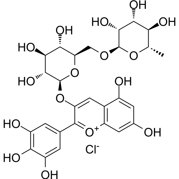
Delphinidin 3-rutinoside chloride is an anthocyanin component. Delphinidin 3-rutinoside chloride is isolable from the fruits of blackcurrant Ribes nigrum L. Delphinidin 3-rutinoside chloride activates the ETB receptor and stimulates the NO/cGMP pathway. Delphinidin 3-rutinoside chloride increases cyclic guanosine monophosphate production and reduces the phosphorylation level of Myosin regulatory light chain. Delphinidin 3-rutinoside chloride stimulates GLP-1 secretion. It significantly induces relaxation of bovine ciliary muscle strips contracted by ET-1 and inhibits ET-1-induced contraction of bovine ciliary muscle strips. Delphinidin 3-rutinoside chloride is applicable to research related to type 2 diabetes.
For research use only. We do not sell to patients.
- Purity : 98.83%
- CAS No.: 15674-58-5
- Formula: C27H31ClO16
- Molecular Weight:646.98
-
Storage:
-20°C, sealed storage, away from moisture
* In solvent : -80°C, 6 months; -20°C, 1 month (sealed storage, away from moisture)

To place orders, for customer services and technical support, please contact: MedChemExpress USA
Tel: 609-228-6898 E-mail: [email protected] [email protected]


-
 Biological Activity
-
Chemical Information
-
Solvent & Solubility
- Protocols
- Purity & Documentation
- References
-
 Help & FAQs


-
HY-105935
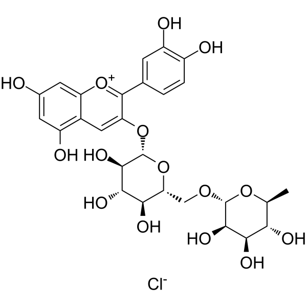
Keracyanin chlorideAntioxidant Agent -
HY-108052
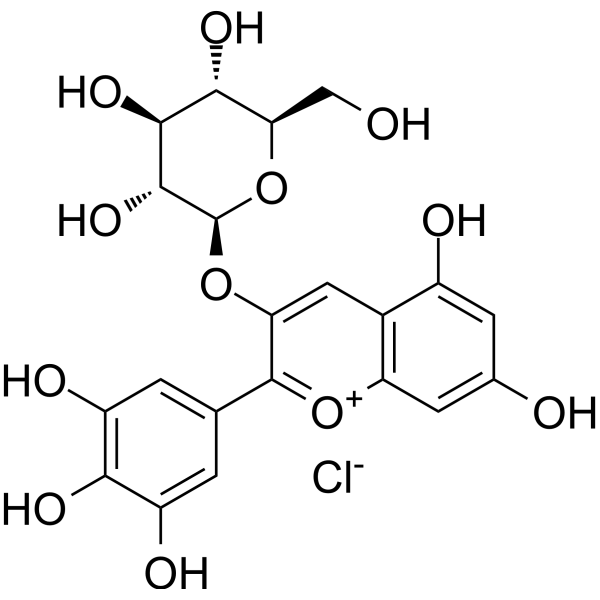
Delphinidin 3-glucoside chlorideEGFR Inhibitor -
HY-N0831
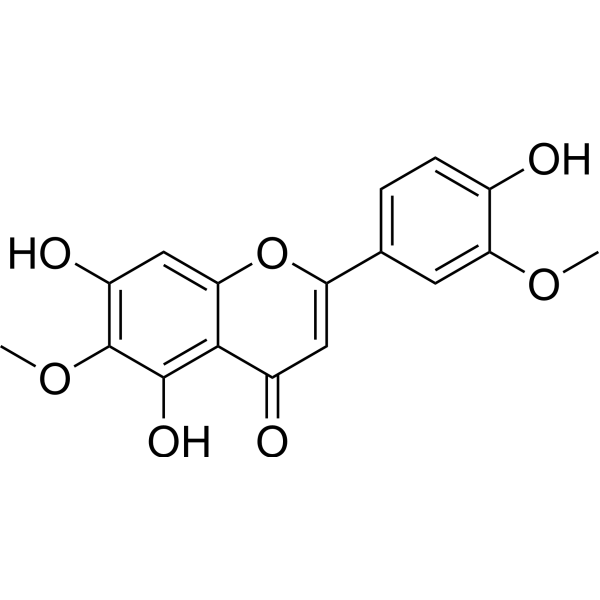
JaceosidinBax Activator -
HY-N2533
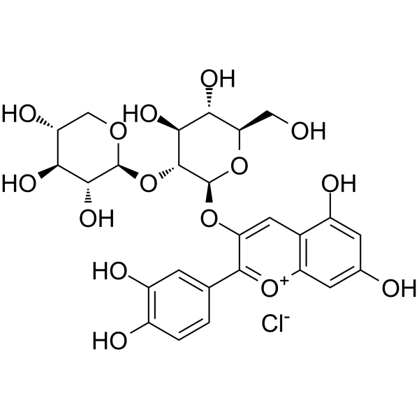
Cyanidin 3-sambubioside chlorideAnthocyanin Compound -
HY-N6606
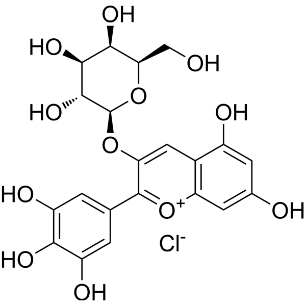
Delphinidin-3-O-galactoside chlorideAnthocyanin -
HY-N0796
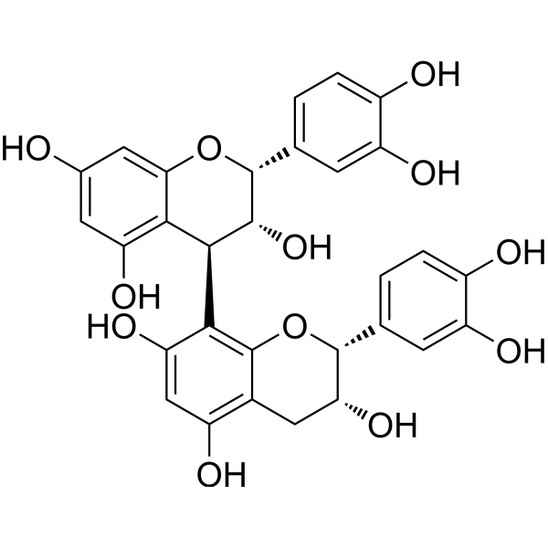
Procyanidin B2Anticancer Agent -
HY-N0796R
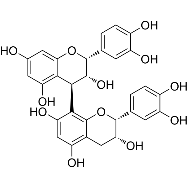
Procyanidin B2 (Standard) -
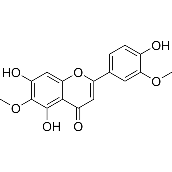
HY-N0831R
Jaceosidin (Standard) -

HY-105935R
Keracyanin chloride (Standard)Antioxidant Agent
Publications Citing Use of MedChemExpress (MCE) Delphinidin 3-rutinoside chloride
More

Biological Activity
Description
IC50 & Target
[1]|
ETB |
In Vitro
Delphinidin 3-rutinoside (3×10⁻⁵ M) chloride significantly induces relaxation of bovine ciliary muscle strips pre-contracted by ET-1[1].
Delphinidin 3-rutinoside (1×10⁻⁴ M) chloride inhibits ET-1-induced contraction of bovine ciliary muscle strips via an ETB receptor/NO/guanylate cyclase-dependent pathway, but exerts no effect on carbachol-induced contraction[1].
Delphinidin 3-rutinoside (100 μM; 2 h) chloride significantly increases GLP-1 secretion in mouse GLUTag enteroendocrine L cells[2].
MedChemExpress (MCE) has not independently confirmed the accuracy of these methods. They are for reference only. Further protocols information, click here.


Chemical Information
-
CAS No. 15674-58-5
-
Appearance Solid
-
Molecular Weight 646.98
-
Formula C27H31ClO16
-
Color Brown to reddish brown
-
SMILES
OC1=CC(O)=CC2=C1C=C(C(C3=CC(O)=C(C(O)=C3)O)=[O+]2)O[C@@H]4O[C@@H]([C@H]([C@@H]([C@H]4O)O)O)CO[C@H]5[C@@H]([C@@H]([C@H]([C@@H](O5)C)O)O)O.[Cl-]
-
Synonyms
Delphinidin 3-O-rutinoside chloride
-
Structure Classification
-
Initial Source
-
Shipping
Room temperature in continental US; may vary elsewhere.
-
Storage
-20°C, sealed storage, away from moisture
* In solvent : -80°C, 6 months; -20°C, 1 month (sealed storage, away from moisture)


Publications (1)
-
Journal Impact Factor
-
Most Recent
-
Food Chem
Flavonoid-mediated metabolic underpinning quality variation in red bud-sport pear mutants. [Abstract]2025 Oct 15:489:144992. PMID: 40466530


Solvent & Solubility
In Vitro:
DMSO : 50 mg/mL (77.28 mM; Need ultrasonic; Hygroscopic DMSO has a significant impact on the solubility of product, please use newly opened DMSO)
H2O : 25 mg/mL (38.64 mM; ultrasonic and warming and heat to 60°C)
Please refer to the solubility information to select the appropriate solvent. Once prepared, please aliquot and store the solution to prevent product inactivation from repeated freeze-thaw cycles.
Storage method and period of stock solution: -80°C, 6 months; -20°C, 1 month (sealed storage, away from moisture). When stored at -80°C, please use it within 6 months. When stored at -20°C, please use it within 1 month.
* Note: If you choose water as the stock solution, please dilute it to the working solution, then filter and sterilize it with a 0.22 μm filter before use.
Please refer to the solubility information to select the appropriate solvent. Once prepared, please aliquot and store the solution to prevent product inactivation from repeated freeze-thaw cycles.
Storage method and period of stock solution: -80°C, 6 months; -20°C, 1 month (sealed storage, away from moisture). When stored at -80°C, please use it within 6 months. When stored at -20°C, please use it within 1 month.
* Note: If you choose water as the stock solution, please dilute it to the working solution, then filter and sterilize it with a 0.22 μm filter before use.
Concentration (start) × Volume (start) = Concentration (final) × Volume (final)

Protocols
-
Kinase activity and phosphorylation assays
Kinase activity assays measure the ability of kinases to transfer phosphate groups from ATP to specific substrates, while phosphorylation assays detect the presence and levels of phosphorylated proteins. Common methods include radiolabeled ATP incorporation (e. g. ,), ADP release detection via bioluminescence (e. g. ,[3]), enzyme-linked immunosorbent assays (ELISA) for phospho-specific epitopes (e. g. ,[6]), and microtiter-based formats for high-throughput screening (e. g. ,[8]). The ADP-Glo assay quantifies kinase activity by measuring ADP produced during phosphorylation using a luciferase-based system. Radiometric assays involve autoradiography or scintillation counting after incorporation of 32P-labeled ATP into substrate proteins. ELISA-based approaches rely on phospho-specific antibodies to detect activated kinases in cell lysates or purified samples.
-
Western Blot
Western blotting (WB) is a commonly used experimental method in molecular biology, biochemistry, and immunogenetics for identifying and quantifying target proteins. It combines gel electrophoresis with immunoassay, enabling researchers to analyze protein expression, post-translational modifications, and molecular weight.
-
Protocol for Kinase activity and phosphorylation assays
Kinase activity assays measure transfer of phosphate from ATP to a protein or peptide substrate, generating phosphorylated substrate, ADP, or incorporated radiolabeled phosphate as the readout; phosphorylation assays measure site-specific phosphorylation in cells or tissues as a proxy for kinase-pathway activation, inhibition, or substrate regulation. Phosphorylation can be detected by phospho-specific Western blot, immunoprecipitation kinase assay, phospho-immunofluorescence, phospho-flow cytometry, luminescent ADP detection, radiolabeled ATP incorporation, or reporter-based pathway assays, and these readouts can be applied to cancer cells, primary neurons, mouse tumors, organoids, inflammatory macrophages, ferroptosis studies, and mitophagy studies when the kinase target is biologically relevant.
-
Research Protocol for Metabolic Diseases
AMP-activated protein kinase, AMPK, is a conserved cellular energy sensor that responds to reduced cellular energy status and coordinates metabolism by increasing ATP-generating catabolic pathways while suppressing ATP-consuming anabolic processes. In metabolic disease research, the AMPK pathway is experimentally relevant because it regulates hepatic lipid synthesis, fatty acid oxidation, glucose production, skeletal-muscle glucose disposal, mTORC1-linked biosynthesis, autophagy, mitochondrial homeostasis, and whole-body energy balance. The central pathway logic is that energy stress, metformin, exercise-like stimulation, or direct AMPK activators increase AMPKα Thr172 phosphorylation and downstream substrate phosphorylation, including ACC and RAPTOR. Phosphorylation of ACC suppresses lipogenesis and supports fatty acid oxidation, whereas phosphorylation of RAPTOR suppresses mTORC1 signaling and links cellular energy status to growth and protein synthesis control. The pathway is linked


Purity & Documentation
-
Data Sheet (284 KB)
-
SDS (252 KB)
- English - EN (252 KB)
- Français - FR (252 KB)
- Deutsch - DE (252 KB)
- Norwegian - NO (252 KB)
- Español - ES (252 KB)
- Swedish - SV (252 KB)
- Italian - IT (252 KB)
- Korean - KR (252 KB)
- Portuguese - PT (252 KB)
-
Handling Instructions (2659 KB)
References
[1]. Matsumoto H, et al. Delphinidin-3-rutinoside relaxes the bovine ciliary smooth muscle through activation of ETB receptor and NO/cGMP pathway. Exp Eye Res. 2005;80(3):313-322. [Content Brief]
[2]. Tani T, et al. Delphinidin 3-rutinoside-rich blackcurrant extract ameliorates glucose tolerance by increasing the release of glucagon-like peptide-1 secretion. Food Sci Nutr. 2017;5(4):929-933. Published 2017 Apr 7. [Content Brief]


Complete Stock Solution Preparation Table
Please refer to the solubility information to select the appropriate solvent. Once prepared, please aliquot and store the solution to prevent product inactivation from repeated freeze-thaw cycles.
Storage method and period of stock solution: -80°C, 6 months; -20°C, 1 month (sealed storage, away from moisture). When stored at -80°C, please use it within 6 months. When stored at -20°C, please use it within 1 month.
| Optional Solvent | Concentration Solvent Mass | 1 mg | 5 mg | 10 mg | 25 mg |
|---|---|---|---|---|---|
| H2O / DMSO | 1 mM | 1.5456 mL | 7.7282 mL | 15.4564 mL | 38.6411 mL |
| 5 mM | 0.3091 mL | 1.5456 mL | 3.0913 mL | 7.7282 mL | |
| 10 mM | 0.1546 mL | 0.7728 mL | 1.5456 mL | 3.8641 mL | |
| 15 mM | 0.1030 mL | 0.5152 mL | 1.0304 mL | 2.5761 mL | |
| 20 mM | 0.0773 mL | 0.3864 mL | 0.7728 mL | 1.9321 mL | |
| 25 mM | 0.0618 mL | 0.3091 mL | 0.6183 mL | 1.5456 mL | |
| 30 mM | 0.0515 mL | 0.2576 mL | 0.5152 mL | 1.2880 mL | |
| DMSO | 40 mM | 0.0386 mL | 0.1932 mL | 0.3864 mL | 0.9660 mL |
| 50 mM | 0.0309 mL | 0.1546 mL | 0.3091 mL | 0.7728 mL | |
| 60 mM | 0.0258 mL | 0.1288 mL | 0.2576 mL | 0.6440 mL |
* Note: If you choose water as the stock solution, please dilute it to the working solution, then filter and sterilize it with a 0.22 μm filter before use.


Delphinidin 3-rutinoside chloride Related Classifications
HY-114367 Related Classifications
 Powered by Bioz
Keywords