



Deoxypodophyllotoxin

Based on 1 Customer Validation
Deoxypodophyllotoxin (DPT), a derivative of podophyllotoxin, is a lignan with potent antimitotic, anti-inflammatory and antiviral properties isolated from Anthriscus sylvestris. Deoxypodophyllotoxin, targets the microtubule, has a major impact in oncology not only as anti-mitotics but also as potent inhibitors of angiogenesis. Deoxypodophyllotoxin induces cell autophagy and apoptosis. Deoxypodophyllotoxin evokes increase of intracellular Ca2+ concentrations in DRG neurons.
For research use only. We do not sell to patients.
- Purity : 99.52%
- CAS No.: 19186-35-7
- Formula: C22H22O7
- Molecular Weight:398.41
-
Storage:Powder -20°C, 3 years , 4°C, 2 years ; In solvent -80°C, 6 months , -20°C, 1 month

To place orders, for customer services and technical support, please contact: MedChemExpress USA
Tel: 609-228-6898 E-mail: [email protected] [email protected]


-
 Biological Activity
-
Chemical Information
-
Solvent & Solubility
- Protocols
- Purity & Documentation
- References
-
 Help & FAQs


-
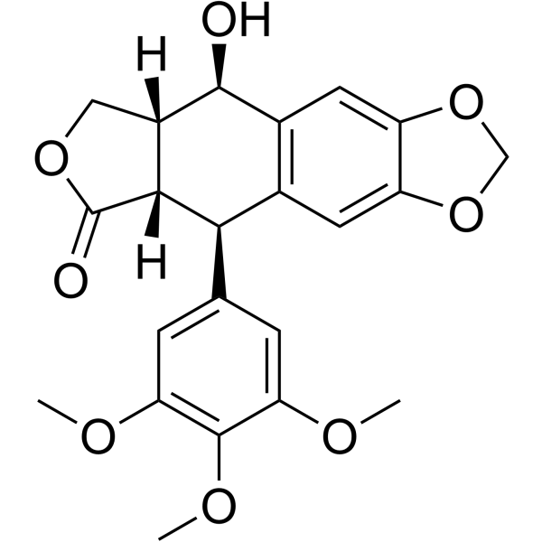
HY-15494
PicropodophyllinApoptosis Inducer -
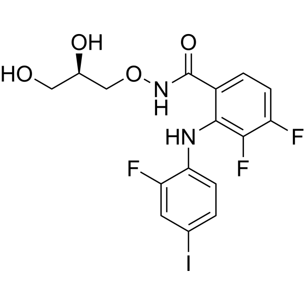
HY-10254
MirdametinibMEK Inhibitor -
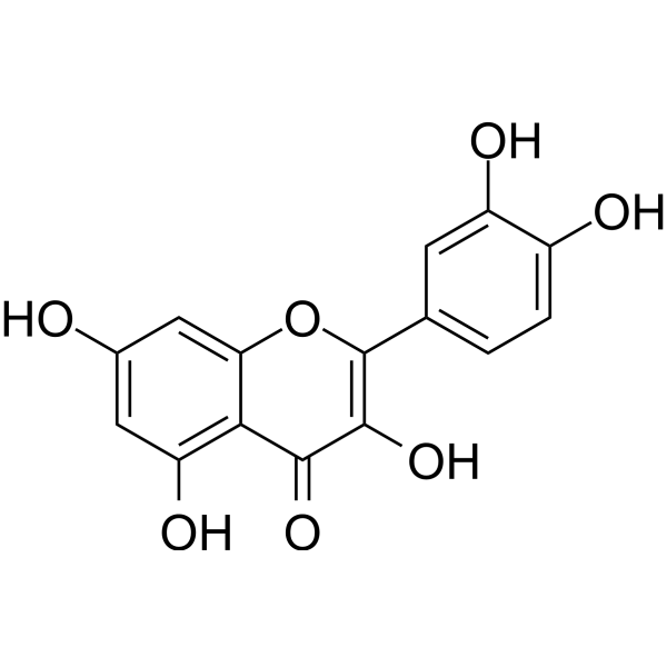
HY-18085
QuercetinFlavonoid Antioxidant -
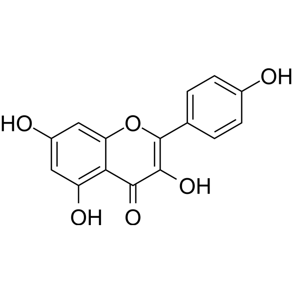
HY-14590
KaempferolEstrogen Receptor α Inhibitor -
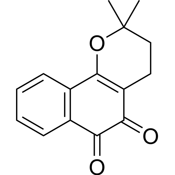
HY-13555
β-LapachoneAutophagy Inducer -
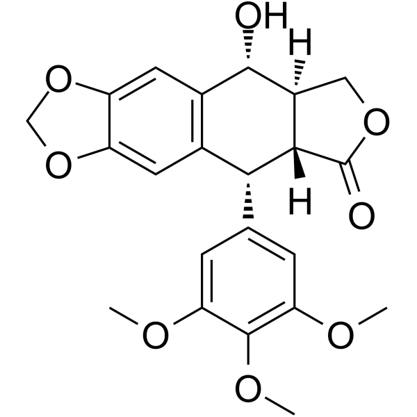
HY-15552
PodofiloxMicrotubule/Tubulin Inhibitor -
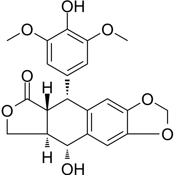
HY-N2323
4'-DemethylpodophyllotoxinAnticancer Agent -
HY-N1977
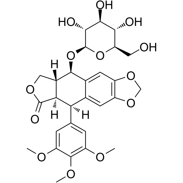
Podophyllotoxin glucosidePodophyllotoxin Derivative -
HY-N7654
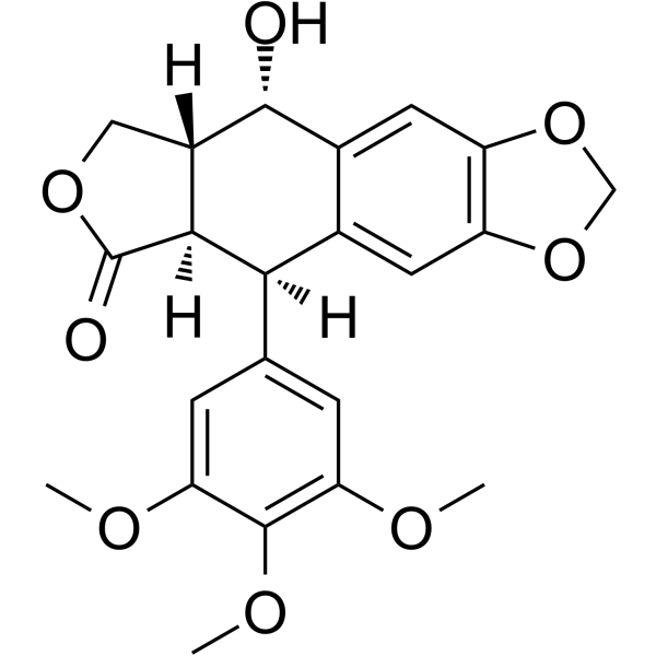
(-)-EpipodophyllotoxinApoptosis Inducer


Biological Activity
Description
Cellular Effect
|
Cell Line

|
Type | Value | Description | References |
|---|---|---|---|---|
| A-431 | ED50 |
4.3 μg/mL
Compound: 9
|
Cytotoxicity against human A431 cells
Cytotoxicity against human A431 cells
|
[PMID: 8277312] |
| A549 | ED50 |
15.5 nM
Compound: Deoxypodophylltoxin (DPT)
|
Cytotoxic activity against human lung cancer A549 cell line was determined
Cytotoxic activity against human lung cancer A549 cell line was determined
|
[PMID: 11844709] |
| A549 | ED50 |
23 nM
Compound: DPT (1)
|
In vitro cytotoxicity against A549 (human lung carcinoma) cell line.
In vitro cytotoxicity against A549 (human lung carcinoma) cell line.
|
[PMID: 12419378] |
| A549 | ED50 |
0.012 μg/mL
Compound: DPT
|
Cytotoxicity in A549 (human carcinoma) cell line.
Cytotoxicity in A549 (human carcinoma) cell line.
|
[PMID: 12873481] |
| A549 | IC50 |
0.47 μM
Compound: 2, DPT
|
Cytotoxicity against human A549 cells after 48 hrs by MTT assay
Cytotoxicity against human A549 cells after 48 hrs by MTT assay
|
[PMID: 21733601] |
| A549 | IC50 |
1.38 μM
Compound: 8, DPT
|
Cytotoxicity against human A549 cells assessed as growth inhibition after 48 hrs by MTT assay
Cytotoxicity against human A549 cells assessed as growth inhibition after 48 hrs by MTT assay
|
[PMID: 22041063] |
| A549 | IC50 |
1.38 μM
Compound: 9, DPT
|
Cytotoxicity against human A549 cells after 48 hrs by MTT assay
Cytotoxicity against human A549 cells after 48 hrs by MTT assay
|
[PMID: 22244588] |
| A549 | IC50 |
0.004 μg/mL
Compound: 10
|
Antiproliferative activity against human A549 cells assessed as inhibition of cell viability by MTT assay
Antiproliferative activity against human A549 cells assessed as inhibition of cell viability by MTT assay
|
[PMID: 32223924] |
| A549 | IC50 |
<0.006 μM
Compound: 2 (Deoxypodophyllotoxin)
|
Antineoplastic activity against A-549 (human lung carcinoma) cell line.
Antineoplastic activity against A-549 (human lung carcinoma) cell line.
|
10.1016/0960-894X(95)00432-S |
| BC1 cell line | ED50 |
0.001 μg/mL
Compound: 9
|
Cytotoxicity against human BC1 cells
Cytotoxicity against human BC1 cells
|
[PMID: 8277312] |
| BXPC-3 | ED50 |
0.0029 μg/mL
Compound: 2
|
Anticancer activity against human BxPC3 cells after 48 hrs by SRB assay
Anticancer activity against human BxPC3 cells after 48 hrs by SRB assay
|
[PMID: 14987061] |
| BXPC-3 | GI50 |
0.00044 μg/mL
Compound: 1
|
Growth inhibition of human BxPC3 cells measured after 48 hrs by sulforhodamine B assay
Growth inhibition of human BxPC3 cells measured after 48 hrs by sulforhodamine B assay
|
[PMID: 26938998] |
| BXPC-3 | GI50 |
0.44 ng/mL
Compound: 1
|
Growth inhibition of human BxPC3 cells measured after 48 hrs by sulforhodamine B assay
Growth inhibition of human BxPC3 cells measured after 48 hrs by sulforhodamine B assay
|
[PMID: 26938998] |
| Col2 | ED50 |
0.01 μg/mL
Compound: 9
|
Cytotoxicity against human Col2 cells
Cytotoxicity against human Col2 cells
|
[PMID: 8277312] |
| CV-1 | IC50 |
0.025 μM
Compound: 7
|
Antiviral activity against HSV1 infected in monkey CV-1 cells assessed as inhibition of plaque formation
Antiviral activity against HSV1 infected in monkey CV-1 cells assessed as inhibition of plaque formation
|
[PMID: 9834179] |
| CV-1 | IC50 |
0.006 μM
Compound: 2 (Deoxypodophyllotoxin)
|
Antineoplastic activity against CV-1 (monkey kidney fibroblast) cell line.
Antineoplastic activity against CV-1 (monkey kidney fibroblast) cell line.
|
10.1016/0960-894X(95)00432-S |
| DU-145 | ED50 |
0.0028 μg/mL
Compound: 2
|
Anticancer activity against human DU145 cells after 48 hrs by SRB assay
Anticancer activity against human DU145 cells after 48 hrs by SRB assay
|
[PMID: 14987061] |
| DU-145 | GI50 |
0.0017 μg/mL
Compound: 1
|
Growth inhibition of human DU145 cells measured after 48 hrs by sulforhodamine B assay
Growth inhibition of human DU145 cells measured after 48 hrs by sulforhodamine B assay
|
[PMID: 26938998] |
| GLC4 cell line | IC50 |
0.008 μM
Compound: 23
|
Cytotoxic effect against GLC4 (human small cell lung carcinoma cell line) using the microculture tetrazolium (MTT) assay based on continuous incubation
Cytotoxic effect against GLC4 (human small cell lung carcinoma cell line) using the microculture tetrazolium (MTT) assay based on continuous incubation
|
[PMID: 7783142] |
| GLC4 cell line | IC50 |
0.12 μM
Compound: 23
|
Cytotoxic effect against GLC4 (human small cell lung carcinoma cell line) using the microculture tetrazolium (MTT) assay based on 2 hr incubation
Cytotoxic effect against GLC4 (human small cell lung carcinoma cell line) using the microculture tetrazolium (MTT) assay based on 2 hr incubation
|
[PMID: 7783142] |
| HCT-116 | IC50 |
14.5 nM
Compound: DPT
|
Antiproliferative activity against human HCT-116 cells measured after 72 hrs by MTT assay
Antiproliferative activity against human HCT-116 cells measured after 72 hrs by MTT assay
|
[PMID: 36471625] |
| HeLa | IC50 |
0.0069 μM
Compound: 7
|
Cytotoxicity against human HeLa cells after 72 hrs by MTT assay
Cytotoxicity against human HeLa cells after 72 hrs by MTT assay
|
[PMID: 21570846] |
| HeLa | IC50 |
36 μM
Compound: 2, DPT
|
Cytotoxicity against human HeLa cells after 48 hrs by MTT assay
Cytotoxicity against human HeLa cells after 48 hrs by MTT assay
|
[PMID: 21733601] |
| HeLa | IC50 |
1.98 μM
Compound: 8, DPT
|
Cytotoxicity against human HeLa cells assessed as growth inhibition after 48 hrs by CCK-8 assay
Cytotoxicity against human HeLa cells assessed as growth inhibition after 48 hrs by CCK-8 assay
|
[PMID: 22041063] |
| HeLa | IC50 |
6.01 μM
Compound: 9, DPT
|
Cytotoxicity against human HeLa cells after 48 hrs by CCK-8 assay
Cytotoxicity against human HeLa cells after 48 hrs by CCK-8 assay
|
[PMID: 22244588] |
| HepG2 | IC50 |
14.5 μg/mL
Compound: 10
|
Antiproliferative activity against human HepG2 cells assessed as inhibition of cell viability by MTT assay
Antiproliferative activity against human HepG2 cells assessed as inhibition of cell viability by MTT assay
|
[PMID: 32223924] |
| HL-60 | IC50 |
0.47 μM
Compound: 8, DPT
|
Cytotoxicity against human HL60 cells assessed as growth inhibition after 48 hrs by CCK-8 assay
Cytotoxicity against human HL60 cells assessed as growth inhibition after 48 hrs by CCK-8 assay
|
[PMID: 22041063] |
| HL-60 | IC50 |
0.47 μM
Compound: 9, DPT
|
Cytotoxicity against human HL60 cells after 48 hrs by CCK-8 assay
Cytotoxicity against human HL60 cells after 48 hrs by CCK-8 assay
|
[PMID: 22244588] |
| HT | ED50 |
0.003 μg/mL
Compound: 9
|
Cytotoxicity against human HT cells
Cytotoxicity against human HT cells
|
[PMID: 8277312] |
| HT-29 | IC50 |
2.5 ng/mL
Compound: Deoxypodophyllotoxin
|
Growth inhibition of human HT29 cells
Growth inhibition of human HT29 cells
|
[PMID: 15332833] |
| HT-29 | IC50 |
16.6 nM
Compound: DPT
|
Antiproliferative activity against human HT-29 cells measured after 72 hrs by MTT assay
Antiproliferative activity against human HT-29 cells measured after 72 hrs by MTT assay
|
[PMID: 36471625] |
| HT-29 | IC50 |
0.006 μM
Compound: 2 (Deoxypodophyllotoxin)
|
Antineoplastic activity against HT-29 (human colon carcinoma) cell line.
Antineoplastic activity against HT-29 (human colon carcinoma) cell line.
|
10.1016/0960-894X(95)00432-S |
| K562 | IC50 |
16 nM
Compound: DPT
|
Antiproliferative activity against human K562 cells measured after 72 hrs by MTT assay
Antiproliferative activity against human K562 cells measured after 72 hrs by MTT assay
|
[PMID: 36471625] |
| KB | IC50 |
0.0089 μM
Compound: 7
|
Cytotoxicity against human KB cells after 72 hrs by MTT assay
Cytotoxicity against human KB cells after 72 hrs by MTT assay
|
[PMID: 21570846] |
| KB | IC50 |
0.003 μM
Compound: Dppt
|
Cytotoxicity against human KB cells assessed as inhibition of cell proliferation after 72 hrs by MTS assay
Cytotoxicity against human KB cells assessed as inhibition of cell proliferation after 72 hrs by MTS assay
|
[PMID: 22429510] |
| KB | ED50 |
0.032 μg/mL
Compound: 4
|
Cytotoxicity against human KB cells
Cytotoxicity against human KB cells
|
[PMID: 521819] |
| KB | ED50 |
0.05 μg/mL
Compound: 9
|
Cytotoxicity against human KB cells
Cytotoxicity against human KB cells
|
[PMID: 8277312] |
| KB-V1 | ED50 |
0.06 μg/mL
Compound: 9
|
Cytotoxicity against human KBV1 cells
Cytotoxicity against human KBV1 cells
|
[PMID: 8277312] |
| KM-20L2 | ED50 |
0.0028 μg/mL
Compound: 2
|
Anticancer activity against human KM20L2 cells after 48 hrs by SRB assay
Anticancer activity against human KM20L2 cells after 48 hrs by SRB assay
|
[PMID: 14987061] |
| KM-20L2 | GI50 |
0.0014 μg/mL
Compound: 1
|
Growth inhibition of human KM20L2 cells measured after 48 hrs by sulforhodamine B assay
Growth inhibition of human KM20L2 cells measured after 48 hrs by sulforhodamine B assay
|
[PMID: 26938998] |
| LNCaP | IC50 |
0.03 μM
Compound: 4
|
Cytotoxicity against androgen sensitive human LNCaP cells
Cytotoxicity against androgen sensitive human LNCaP cells
|
[PMID: 17256902] |
| LNCaP | ED50 |
0.01 μg/mL
Compound: 9
|
Cytotoxicity against human LNCAP cells
Cytotoxicity against human LNCAP cells
|
[PMID: 8277312] |
| Lu1 | ED50 |
0.002 μg/mL
Compound: 9
|
Cytotoxicity against human Lu1 cells
Cytotoxicity against human Lu1 cells
|
[PMID: 8277312] |
| MCF7 | ED50 |
11.7 nM
Compound: Deoxypodophylltoxin (DPT)
|
Cytotoxic activity against human breast cancer MCF-7 cell line was determined
Cytotoxic activity against human breast cancer MCF-7 cell line was determined
|
[PMID: 11844709] |
| MCF7 | ED50 |
11 nM
Compound: DPT (1)
|
In vitro cytotoxicity against MCF-7 (human breast carcinoma) cell line.
In vitro cytotoxicity against MCF-7 (human breast carcinoma) cell line.
|
[PMID: 12419378] |
| MCF7 | ED50 |
0.0021 μg/mL
Compound: 2
|
Anticancer activity against human MCF7 cells after 48 hrs by SRB assay
Anticancer activity against human MCF7 cells after 48 hrs by SRB assay
|
[PMID: 14987061] |
| MCF7 | GI50 |
0.0027 μg/mL
Compound: 1
|
Growth inhibition of human MCF7 cells measured after 48 hrs by sulforhodamine B assay
Growth inhibition of human MCF7 cells measured after 48 hrs by sulforhodamine B assay
|
[PMID: 26938998] |
| MCF7 | IC50 |
6 nM
Compound: DPT
|
Antiproliferative activity against human MCF7 cells measured after 72 hrs by MTT assay
Antiproliferative activity against human MCF7 cells measured after 72 hrs by MTT assay
|
[PMID: 36471625] |
| MDA-MB-231 | IC50 |
0.07 μg/mL
Compound: 10
|
Antiproliferative activity against human MDA-MB-231 cells assessed as inhibition of cell viability by MTT assay
Antiproliferative activity against human MDA-MB-231 cells assessed as inhibition of cell viability by MTT assay
|
[PMID: 32223924] |
| NCI-H460 | ED50 |
0.0027 μg/mL
Compound: 2
|
Anticancer activity against human NCI-H460 cells after 48 hrs by SRB assay
Anticancer activity against human NCI-H460 cells after 48 hrs by SRB assay
|
[PMID: 14987061] |
| NCI-H460 | GI50 |
0.0018 μg/mL
Compound: 1
|
Growth inhibition of human NCI-H460 cells measured after 48 hrs by sulforhodamine B assay
Growth inhibition of human NCI-H460 cells measured after 48 hrs by sulforhodamine B assay
|
[PMID: 26938998] |
| P388 | ED50 |
0.0023 μg/mL
Compound: 2
|
Anticancer activity against mouse P388 cells after 48 hrs
Anticancer activity against mouse P388 cells after 48 hrs
|
[PMID: 14987061] |
| P388 | IC50 |
2.5 ng/mL
Compound: Deoxypodophyllotoxin
|
Growth inhibition of mouse P388 cells
Growth inhibition of mouse P388 cells
|
[PMID: 15332833] |
| P388 | ED50 |
0.029 μg/mL
Compound: 1
|
Growth inhibition of mouse P388 cells measured after 48 hrs by sulforhodamine B assay
Growth inhibition of mouse P388 cells measured after 48 hrs by sulforhodamine B assay
|
[PMID: 26938998] |
| P388 | ED50 |
0.002 μg/mL
Compound: 9
|
Cytotoxicity in mouse P388 cells
Cytotoxicity in mouse P388 cells
|
[PMID: 8277312] |
| P388 | IC50 |
0.01 μM
Compound: 2 (Deoxypodophyllotoxin)
|
Antineoplastic activity against P-388 (murine leukemia) cell line.
Antineoplastic activity against P-388 (murine leukemia) cell line.
|
10.1016/0960-894X(95)00432-S |
| PC-3 | IC50 |
0.032 μM
Compound: 4
|
Cytotoxicity against androgen-independent human PC3 cells
Cytotoxicity against androgen-independent human PC3 cells
|
[PMID: 17256902] |
| PC-3 | IC50 |
0.018 μM
Compound: 8
|
Antiproliferative activity against human PC3 cells after 72 hrs by MTT assay
Antiproliferative activity against human PC3 cells after 72 hrs by MTT assay
|
[PMID: 27214307] |
| SF-268 | ED50 |
0.0033 μg/mL
Compound: 2
|
Anticancer activity against human SF268 cells after 48 hrs by SRB assay
Anticancer activity against human SF268 cells after 48 hrs by SRB assay
|
[PMID: 14987061] |
| SF-268 | GI50 |
0.00056 μg/mL
Compound: 1
|
Growth inhibition of human SF268 cells measured after 48 hrs by sulforhodamine B assay
Growth inhibition of human SF268 cells measured after 48 hrs by sulforhodamine B assay
|
[PMID: 26938998] |
| SF-268 | GI50 |
0.56 ng/mL
Compound: 1
|
Growth inhibition of human SF268 cells measured after 48 hrs by sulforhodamine B assay
Growth inhibition of human SF268 cells measured after 48 hrs by sulforhodamine B assay
|
[PMID: 26938998] |
| SiHa | IC50 |
10.2 μM
Compound: 2, DPT
|
Cytotoxicity against human SiHa cells after 48 hrs by MTT assay
Cytotoxicity against human SiHa cells after 48 hrs by MTT assay
|
[PMID: 21733601] |
| SiHa | IC50 |
6.01 μM
Compound: 8, DPT
|
Cytotoxicity against human SiHa cells assessed as growth inhibition after 48 hrs by MTT assay
Cytotoxicity against human SiHa cells assessed as growth inhibition after 48 hrs by MTT assay
|
[PMID: 22041063] |
| SiHa | IC50 |
1.98 μM
Compound: 9, DPT
|
Cytotoxicity against human SiHa cells after 48 hrs by MTT assay
Cytotoxicity against human SiHa cells after 48 hrs by MTT assay
|
[PMID: 22244588] |
| SK-MEL-2 | ED50 |
8.4 nM
Compound: Deoxypodophylltoxin (DPT)
|
Cytotoxic activity against human melanoma SK-MEL-2 was determined
Cytotoxic activity against human melanoma SK-MEL-2 was determined
|
[PMID: 11844709] |
| SK-MEL-2 | ED50 |
14 nM
Compound: DPT (1)
|
In vitro cytotoxicity against SK-MEL-2 (human melanoma) cell line.
In vitro cytotoxicity against SK-MEL-2 (human melanoma) cell line.
|
[PMID: 12419378] |
| SK-MEL-2 | ED50 |
0.009 μg/mL
Compound: DPT
|
Cytotoxicity in SK-MEL-2 (human melanoma) cell line.
Cytotoxicity in SK-MEL-2 (human melanoma) cell line.
|
[PMID: 12873481] |
| U-373MG ATCC | ED50 |
0.001 μg/mL
Compound: 9
|
Cytotoxicity against human U373 cells
Cytotoxicity against human U373 cells
|
[PMID: 8277312] |
| ZR-75-1 | ED50 |
2 μg/mL
Compound: 9
|
Cytotoxicity against human ZR-75-1 cells
Cytotoxicity against human ZR-75-1 cells
|
[PMID: 8277312] |
In Vitro
Deoxypodophyllotoxin (25-75 nM; 6-48 hours) increases the percentage of early apoptotic cell population from 2.05 to 5.62 and 18.49% for 24 h and 48 h, respectively[1]. Deoxypodophyllotoxin (25-75 nM; 6-48 hours) treats SGC-7901 cells arrested in G2/M phase in time- and dose- dependent manners[1]. Deoxypodophyllotoxin (25-75 nM; 6-48 hours) results in a remarkably time- and dose-dependent decrease in Cdc2 and Cdc25C expression levels and increases cyclin B1 within 6h, decreases PARP, Bcl-2 and caspase-3 activity[1].
MedChemExpress (MCE) has not independently confirmed the accuracy of these methods. They are for reference only. Further protocols information, click here.
-
Cell Line:SGC-7901 cells
-
Concentration:25, 50, 75 nM
-
Incubation Time:6, 12, 24, 48 hours
-
Result:Induced apoptosis in SGC-7901 Cells.
-
Cell Line:SGC-7901 cells
-
Concentration:25, 50, 75 nM
-
Incubation Time:6, 12, 24, 48 hours
-
Result:Induced G2/M cell cycle arrest in SGC-7901 Cells
-
Cell Line:SGC-7901 cells
-
Concentration:25, 50, 75 nM
-
Incubation Time:6, 12, 24, 48 hours
-
Result:Altered the expression of cyclin B1, Cdc2,Cdc25C, p-PARP, Bcl-2 and p-caspase-3 proteins.
In Vivo
MedChemExpress (MCE) has not independently confirmed the accuracy of these methods. They are for reference only.
-
Animal Model:Xenograft model of gastric cancer in nude mice with SGC-7901 cells[1]
-
Dosage:5, 10, and 20 mg/kg
-
Administration:Intravenously injected; 5, 10, and 20 mg/kg; 3 times a week; 28 days
-
Result:Inhibited the growth of gastric cancer tumors.


Chemical Information
-
CAS No. 19186-35-7
-
Appearance Solid
-
Molecular Weight 398.41
-
Formula C22H22O7
-
Color White to off-white
-
SMILES
O=C1[C@]2([H])[C@H](C3=CC(OC)=C(OC)C(OC)=C3)C4=CC(OCO5)=C5C=C4C[C@@]2([H])CO1
-
Structure Classification
-
Shipping
Room temperature in continental US; may vary elsewhere.
-
Storage
Powder -20°C 3 years 4°C 2 years In solvent -80°C 6 months -20°C 1 month


Solvent & Solubility
In Vitro:
DMSO : 100 mg/mL (251.00 mM; Need ultrasonic; Hygroscopic DMSO has a significant impact on the solubility of product, please use newly opened DMSO)
Please refer to the solubility information to select the appropriate solvent. Once prepared, please aliquot and store the solution to prevent product inactivation from repeated freeze-thaw cycles.
Storage method and period of stock solution: -80°C, 6 months; -20°C, 1 month. When stored at -80°C, please use it within 6 months. When stored at -20°C, please use it within 1 month.
Please refer to the solubility information to select the appropriate solvent. Once prepared, please aliquot and store the solution to prevent product inactivation from repeated freeze-thaw cycles.
Storage method and period of stock solution: -80°C, 6 months; -20°C, 1 month. When stored at -80°C, please use it within 6 months. When stored at -20°C, please use it within 1 month.
Concentration (start) × Volume (start) = Concentration (final) × Volume (final)
In Vivo:
Select the appropriate dissolution method based on your experimental animal and administration route.
- For the following dissolution methods, please ensure to first prepare a clear stock solution using an In Vitro approach and then sequentially add co-solvents:
- To ensure reliable experimental results, the clarified stock solution can be appropriately stored based on storage conditions. As for the working solution for In Vivo experiments, it is recommended to prepare freshly and use it on the same day.
- The percentages shown for the solvents indicate their volumetric ratio in the final prepared solution. If precipitation or phase separation occurs during preparation, heat and/or sonication can be used to aid dissolution.
Add each solvent one by one: 10% DMSO 40% PEG300 5% Tween-80 45% Saline
Solubility: ≥ 2.5 mg/mL (6.27 mM); Clear solution
This protocol yields a clear solution of ≥ 2.5 mg/mL (saturation unknown).
Taking 1 mL working solution as an example, add 100 μL DMSO stock solution (25.0 mg/mL) to 400 μL PEG300, and mix evenly; then add 50 μL Tween-80 and mix evenly; then add 450 μL Saline to adjust the volume to 1 mL.
Preparation of Saline: Dissolve 0.9 g sodium chloride in ddH₂O and dilute to 100 mL to obtain a clear Saline solution.
Add each solvent one by one: 10% DMSO 90% (20% SBE-β-CD in Saline)
Solubility: ≥ 2.5 mg/mL (6.27 mM); Clear solution
This protocol yields a clear solution of ≥ 2.5 mg/mL (saturation unknown).
Taking 1 mL working solution as an example, add 100 μL DMSO stock solution (25.0 mg/mL) to 900 μL 20% SBE-β-CD in Saline, and mix evenly.
Preparation of 20% SBE-β-CD in Saline (4°C, storage for one week): 2 g SBE-β-CD powder is dissolved in 10 mL Saline, completely dissolve until clear.
In Vivo Dissolution Calculator
Please enter the basic information of animal experiments:
-
-
-
-
Recommended: Prepare an additional quantity of animals to account for potential losses during experiments.
Please enter your animal formula composition:
-
%DMSO +
Recommended: Keep the proportion of DMSO in working solution below 2% if your animal is weak.
-
%+
-
+%Tween-80 + +
-
%Saline +
The co-solvents required include: DMSO, . All of co-solvents are available by MedChemExpress (MCE). , Tween 80. All of co-solvents are available by MedChemExpress (MCE).
Working solution concentration: 0.22 mg/mL
Method for preparing stock solution: mg drug dissolved in μL DMSO. Stock solution concentration: mg/mL.
1. Take μL DMSO stock solution;
2. Add μL .
μL , mix evenly;
3. Then add μL Tween 80, mix evenly;
4. Then add μL
Please ensure that the stock solution in the first step is dissolved to a clear state, and add co-solvents in sequence. You can use ultrasonic heating (ultrasonic cleaner, recommended frequency 20-40 kHz), vortexing, etc. to assist dissolution.

Protocols
-
Research Protocol for Infectious Diseases
Infectious-disease experiments test how pathogens interact with host barriers, innate immune receptors, inflammatory signaling, pathogen replication, and tissue injury; pattern-recognition receptors such as TLRs, RIG-I-like receptors, NOD-like receptors, and inflammasomes detect microbial molecules and activate NF-κB, interferon, and cytokine responses. The central hypothesis is that infection severity reflects the balance between pathogen burden and host response: protective inflammation restricts pathogen growth, whereas excessive or mislocalized inflammation contributes to tissue damage and disease phenotype. Unresolved questions include which host pathways are protective versus pathogenic, why some infection models fail to translate to human disease, and which combined readouts best predict clinically relevant infection outcomes.
-
Human pluripotent stem cell endothelial-cell differentiation
Human pluripotent stem cell endothelial differentiation is based on stepwise developmental patterning: early activation of WNT/GSK3β inhibition promotes mesodermal or vascular progenitor entry, followed by endothelial specification using VEGF-related signaling, BMP4, FGF2, Notch modulation, or cAMP depending on the published protocol. Endothelial differentiation is read out by acquisition of CD31, CD34, VE-cadherin/CD144, KDR/VEGFR2, vWF, Tie2, NOS3, acetylated LDL uptake, tube/network formation, barrier function, and in vivo vessel-forming capacity where tested.
-
Vascular/Branching Fractal Analysis
Vascular/branching fractal analysis quantifies the geometric complexity of vessel trees or vascular networks from segmented 2D images, commonly by converting vessels into binary and/or skeletonized maps and estimating fractal dimension using box-counting or related approaches. Fractal dimension is interpreted as an image-derived readout of vascular branching complexity, space filling, or density, and has been applied to retinal photographs, fluorescein angiography, OCT angiography, capillary perfusion maps, and in vitro Matrigel angiogenesis networks. The assay readout is generated from vessel-positive pixels after image preprocessing, vessel segmentation, binarization, and optional skeletonization; reported outputs include fractal dimension, vessel density, branchpoint density, endpoint density, vessel length density, tortuosity, and generation-based branching metrics when VESGEN-style analysis is used. The biological interpretation is limited to quantitative vascular patterning and s
-
Annexin V plus membrane-impermeant dye apoptosis staining
Annexin V-based apoptosis assays rely on the detection of phosphatidylserine (PS) externalization from the inner leaflet of the plasma membrane to the outer leaflet, an early biochemical hallmark of apoptosis. Fluorescently labeled Annexin V binds PS in a calcium-dependent manner, enabling identification of early apoptotic cells by flow cytometry or fluorescence microscopy. When combined with a membrane-impermeant DNA-binding dye (e. g. , propidium iodide), this approach allows discrimination between viable (Annexin V−/dye−), early apoptotic (Annexin V+/dye−), and late apoptotic or necrotic (Annexin V+/dye+) cell populations by assessing membrane integrity and PS exposure.
-
Research Protocol for Inflammation-related Diseases
The NLRP3 inflammasome is a cytosolic innate immune signaling platform that integrates priming signals and danger-signal activation to promote caspase-1 activation, maturation of IL-1β and IL-18, and gasdermin D-mediated pyroptotic cell death. The core experimental logic is to determine whether inflammatory disease phenotypes are driven by increased NLRP3 expression, ASC-containing inflammasome assembly, caspase-1 cleavage, GSDMD cleavage, and extracellular release of IL-1β/IL-18 rather than by nonspecific cell injury alone. The pathway is strongly linked to inflammation-related disease phenotypes because monosodium urate crystals activate NALP3/NLRP3 inflammasome signaling in gout-like crystal inflammation, cholesterol crystals activate NLRP3 inflammasomes in atherogenesis models, and DSS-induced intestinal inflammation has been reported to involve NLRP3 inflammasome activity. However, experimental colitis studies also show context-dependent protective effects of NLRP3 inflammasome co
-
Ca2+ Staining Technique
Ca2+ staining is an experimental technique that utilizes specific fluorescent probes (such as Fluo-4 AM, Fura-2, etc.) to qualitatively or quantitatively detect dynamic changes in intracellular Ca2+ concentrations; this is achieved by monitoring the changes in fluorescent signals generated when these probes bind to free intracellular calcium ions. The underlying principle relies primarily on the presence of chelating groups within the probe's molecular structure that possess high affinity for calcium ions.
-
Apoptosis
Apoptosis, also called programmed cell death, is generally characterized by distinct morphological characteristics.
-
TUNEL staining for apoptotic DNA fragmentation
TUNEL staining detects DNA strand breaks by using terminal deoxynucleotidyl transferase to add labeled nucleotides to exposed 3′-OH DNA termini, generating either microscopic staining in fixed cells or tissue sections, or fluorescence/cytometric signal in cell suspensions. TUNEL positivity reflects DNA fragmentation but should not be interpreted alone as definitive apoptosis, because TUNEL can also label necrotic, autolytic, mechanically damaged, or DNA-repair-associated DNA breaks.
-
Endothelial Tube Formation Assay
Endothelial tube formation assay evaluates the ability of endothelial cells to attach, migrate, align, and organize into capillary-like networks when cultured on gelled basement membrane extract or Matrigel; the readout is the morphology and quantity of tube-like networks, which reflects an in vitro endothelial morphogenesis step related to angiogenesis. Basement membrane extract/Matrigel provides laminin-rich extracellular matrix cues that support endothelial differentiation into capillary-like structures, but it can contain biologically active growth factors, so growth-factor-reduced matrix is preferred when testing defined angiogenic stimulators or inhibitors.
-
Autophagy
Autophagy is a process in which eukaryotic cells use lysosomes to degrade their own cytoplasmic proteins and damaged organelles under the regulation of autophagy related gene (Atg). Microtubule-associated proteins light chain 3 (LC3) is recognized as autophagy marker, which transfers from cytoplasmic LC3 (LC3-I) to membrane type (LC3-II). LC3-II/I ratio could be detected by Western Blot and fluorescence microscopy.
-
Apoptosis Solutions
Apoptosis is a regulated, generally non-lytic cell-death pathway that removes unwanted, damaged, infected, or abnormal cells through coordinated morphological changes, caspase activation, DNA fragmentation, and membrane remodeling. The intrinsic apoptosis pathway is controlled mainly by mitochondrial outer membrane permeabilization, BCL-2 family proteins, cytochrome c release, apoptosome formation, caspase-9 activation, and downstream executioner caspase-3/7 activation. The extrinsic apoptosis pathway is initiated by death receptors such as Fas, TNFR, and TRAIL receptors, which recruit adaptor proteins and activate caspase-8 before engaging executioner caspases or mitochondrial amplification through BID cleavage. Apoptosis is linked to many phenotypes, including cancer cell killing, tissue homeostasis, immune regulation, neurodegeneration, infection response, and treatment-induced cytotoxicity; unresolved questions include how apoptosis interacts with necroptosis, pyroptosis, ferroptos
-
Lysosome and acidic-vesicle live-cell staining
Lysosome and acidic-vesicle live-cell staining detects acidic intracellular compartments by using membrane-permeant acidotropic probes that accumulate in low-pH vesicles, including lysosomes, late endosomes, autolysosomes, and acidic phagosomes. LysoTracker staining is commonly used as an intensity-based readout of acidic lysosomal compartment abundance or enlargement, while acridine orange produces green fluorescence in less concentrated compartments and red fluorescence after concentration-dependent accumulation in acidic vesicular organelles. Loss or reduction of acridine-orange red signal can be used as a readout of lysosomal membrane permeabilization or reduced acidic-vesicle integrity. This protocol is designed for live cultured cells and can be adapted for fluorescence microscopy, high-content imaging, plate-reader readout, or flow cytometry when the selected literature supports the readout. Because these dyes report acidotropic accumulation rather than lysosome identity alone,
-
Macroautophagy Solutions
Macroautophagy is a conserved lysosome-dependent degradation pathway in which cytoplasmic material is sequestered into double-membrane autophagosomes and delivered to lysosomes for degradation and recycling. The pathway supports cellular homeostasis during nutrient limitation, organelle stress, protein-aggregate accumulation, infection, differentiation, and tissue remodeling by coupling cargo sequestration, autophagosome maturation, lysosomal fusion, and degradation of cargo-derived macromolecules. The core molecular sequence includes initiation by nutrient- and stress-regulated autophagy machinery, autophagosome nucleation, LC3/ATG8-family conjugation to autophagosomal membranes, cargo selection through receptors such as SQSTM1/p62, autophagosome-lysosome fusion, and lysosomal degradation. LC3 was identified as a mammalian homolog of yeast Atg8 that localizes to autophagosomal membranes after processing, and p62/SQSTM1 was shown to connect ubiquitinated cargo with autophagic degradati
-
Endothelial Cell Migration/Angiogenic Sprouting Assay
Endothelial cell migration and angiogenic sprouting assays are in vitro (and partially ex vivo-adapted) functional models that quantify the ability of endothelial cells to undergo coordinated migration, extracellular matrix invasion, and multicellular organization into capillary-like sprouts in response to pro-angiogenic stimuli such as VEGF, bFGF, or conditioned microenvironments. These assays are used to model early angiogenic events including tip-cell formation, directional migration, and lumen-like sprout extension, which collectively reflect angiogenic activation and vascular morphogenesis processes observed in vivo.


Purity & Documentation
-
Data Sheet (284 KB)
-
SDS (480 KB)
- English - EN (480 KB)
- Français - FR (480 KB)
- Deutsch - DE (480 KB)
- Norwegian - NO (480 KB)
- Español - ES (480 KB)
- Swedish - SV (480 KB)
- Italian - IT (480 KB)
- Korean - KR (480 KB)
- Portuguese - PT (480 KB)
-
Handling Instructions (2659 KB)
References
[1]. Wang YR, et al. Deoxypodophyllotoxin induces G2/M cell cycle arrest and apoptosis in SGC-7901 cells and inhibits tumor growth in vivo. Molecules. 2015 Jan 20;20(1):1661-75. [Content Brief]
[2]. Kim SH, et al. Deoxypodophyllotoxin induces cytoprotective autophagy against apoptosis via inhibition of PI3K/AKT/mTOR pathway in osteosarcoma U2OS cells. Pharmacol Rep. 2017 Oct;69(5):878-884. [Content Brief]
[3]. Xu P, et al. Pharmacological effect of deoxypodophyllotoxin: a medicinal agent of plant origin, on mammalian neurons. Neurotoxicology. 2010 Dec;31(6):680-6. [Content Brief]


Complete Stock Solution Preparation Table
Please refer to the solubility information to select the appropriate solvent. Once prepared, please aliquot and store the solution to prevent product inactivation from repeated freeze-thaw cycles.
Storage method and period of stock solution: -80°C, 6 months; -20°C, 1 month. When stored at -80°C, please use it within 6 months. When stored at -20°C, please use it within 1 month.
| Optional Solvent | Concentration Solvent Mass | 1 mg | 5 mg | 10 mg | 25 mg |
|---|---|---|---|---|---|
| DMSO | 1 mM | 2.5100 mL | 12.5499 mL | 25.0998 mL | 62.7494 mL |
| 5 mM | 0.5020 mL | 2.5100 mL | 5.0200 mL | 12.5499 mL | |
| 10 mM | 0.2510 mL | 1.2550 mL | 2.5100 mL | 6.2749 mL | |
| 15 mM | 0.1673 mL | 0.8367 mL | 1.6733 mL | 4.1833 mL | |
| 20 mM | 0.1255 mL | 0.6275 mL | 1.2550 mL | 3.1375 mL | |
| 25 mM | 0.1004 mL | 0.5020 mL | 1.0040 mL | 2.5100 mL | |
| 30 mM | 0.0837 mL | 0.4183 mL | 0.8367 mL | 2.0916 mL | |
| 40 mM | 0.0627 mL | 0.3137 mL | 0.6275 mL | 1.5687 mL | |
| 50 mM | 0.0502 mL | 0.2510 mL | 0.5020 mL | 1.2550 mL | |
| 60 mM | 0.0418 mL | 0.2092 mL | 0.4183 mL | 1.0458 mL | |
| 80 mM | 0.0314 mL | 0.1569 mL | 0.3137 mL | 0.7844 mL | |
| 100 mM | 0.0251 mL | 0.1255 mL | 0.2510 mL | 0.6275 mL |


Deoxypodophyllotoxin Related Classifications
HY-N2500 Related Classifications
 Powered by Bioz