



DG013A
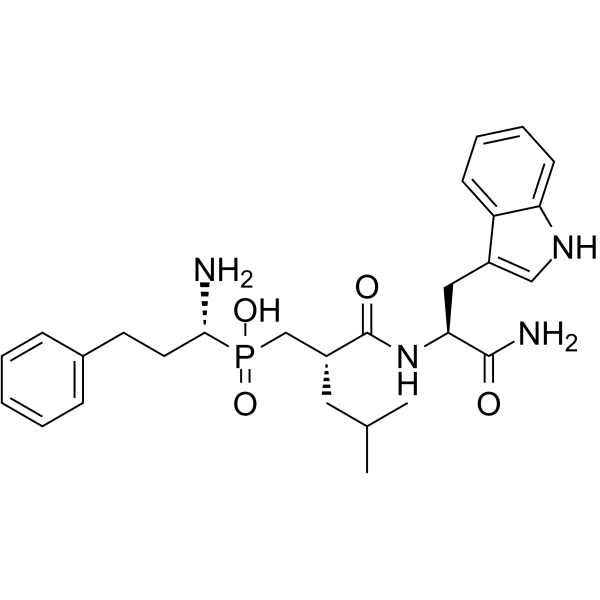
DG013A is a zinc-dependent M1-aminopeptidase inhibitor that exhibits inhibitory activity against ERAP1 (IC50 = 33 nM) and ERAP2 (IC50 = 11 nM). DG013A enhances SRHFLAFSFR epitope presentation, rescues GSW11 peptide neoantigen presentation, reduces SIINFEKL presentation, modulates the immunopeptidome, decreases HLA-B27 free heavy chain expression, and reduces IL-17A secretion. DG013A exhibits antiproliferative activity. DG013A can be used for research on autoimmune diseases and melanoma.
Nur für Forschungszwecke. Wir verkaufen nicht an Patienten.
- CAS. Nr.: 2007955-07-7
- Formel: C27H37N4O4P
- Molecular Weight:512.58
-
Speicherung:
Please store the product under the recommended conditions in the Certificate of Analysis.

To place orders, for customer services and technical support, please contact: MedChemExpress USA
Tel: 609-228-6898 E-mail: [email protected] [email protected]


-
 Biologische Aktivität
-
Chemical Information
- Protokoll
- Reinheit & Dokumentation
- Verweise
-
 Help & FAQs




Biologische Aktivität
Beschreibung
IC50 & Target
[2]|
IL-17A |
Cellular Effect
|
Cell Line

|
Type | Value | Description | References |
|---|---|---|---|---|
| HeLa | ED50 |
350 nM
|
Inhibition of ERAP1-dependent antigen presentation of the SIINFEKL epitope on the cell surface of HeLa cells stably expressing H-2 Kb infected with recombinant vaccinia virus, measured by flow cytometry after 18-22 hrs.
Inhibition of ERAP1-dependent antigen presentation of the SIINFEKL epitope on the cell surface of HeLa cells stably expressing H-2 Kb infected with recombinant vaccinia virus, measured by flow cytometry after 18-22 hrs.
|
31222486 |
In Vitro
DG013A is a moderate inhibitor of ERAP1 and ERAP2, but acts as a highly potent inhibitor of the off-target M1-aminopeptidase APN (IC50 = 0.0037 μM)[1].
DG013A exhibits negligible passive permeability in Caco-2 cells, which is inconsistent with its use as a convertible intracellular chemical probe[1].
DG013A is a potent nanomolar ERAP1 inhibitor (IC50 = 33 nM) and ERAP2 inhibitor (IC50 = 11 nM), and it also inhibits IRAP and APN, with its binding to ERAP2 involving coordination of its phosphonic acid group with the active-site zinc ion[2].
DG013A is a potent inhibitor of recombinant ERAP1 with an IC50 of 43.5 nM[3].
DG013A shows weak antiproliferative activity in HCT116 cells, possibly due to off-target effects[1].
DG013A (0-30 μM; 48 h) shows no toxicity toward A375 malignant melanoma cells at concentrations up to 30 μM for 48 h[3].
DG013A modulates antigen presentation and the immunopeptidome in HeLa-B27, CT26, and melanoma cells[2].
DG013A (1 μM; 6 days) does not significantly alter cell surface MHC-I levels in A375 malignant melanoma cells within 6 days[3].
DG013A (18-22 h) inhibits ERAP1-dependent SIINFEKL epitope antigen presentation in H-2 Kb-expressing HeLa cells with an apparent ED50 of 350 nM[3].
MedChemExpress (MCE) has not independently confirmed the accuracy of these methods. They are for reference only.
-
Cell Line:A375 human malignant melanoma cells
-
Concentration:0-30 μM
-
Incubation Time:48 h
-
Result:Did not appear to be toxic to A375 cells up to a concentration of 30 μM.


Chemical Information
-
CAS. Nr. 2007955-07-7
-
Molecular Weight 512.58
-
Formel C27H37N4O4P
-
SMILES
O=P([C@@H](N)CCC1=CC=CC=C1)(O)C[C@@H](CC(C)C)C(N[C@H](C(N)=O)CC2=CNC3=CC=CC=C23)=O
-
Versand
Room temperature in continental US; may vary elsewhere.
-
Speicherung
Please store the product under the recommended conditions in the Certificate of Analysis.


Reinheit & Dokumentation
Verweise

Calculators
Konzentration (Stammlösung) × Volumen (Stammlösung) = Konzentration (Ziellösung) × Volumen (Ziellösung)


DG013A Related Classifications
HY-139907 Related Classifications
 Powered by Bioz