



Echitamine chloride

Based on 1 Customer Validation
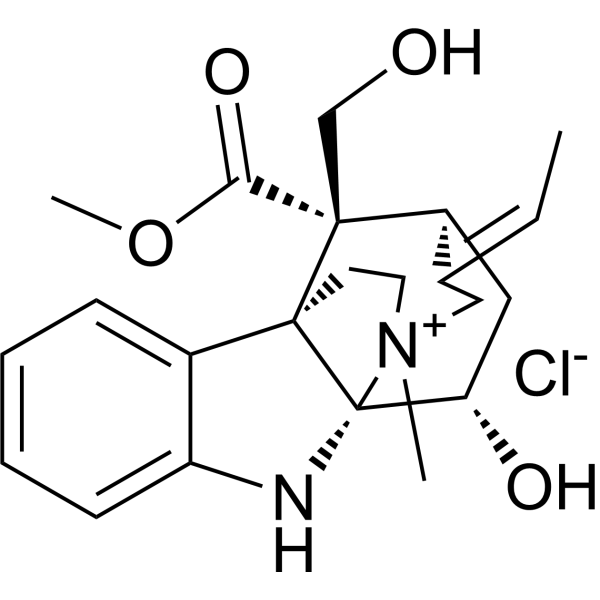
Echitamine (Ditaine) chloride is the major monoterpene indole alkaloid present in Alstonia scholaris with potent anti-tumour activity. Echitamine chloride induces DNA fragmentation and cells apoptosis. Echitamine chloride inhibits pancreatic lipase with an IC50 of 10.92 μM.
For research use only. We do not sell to patients.
- Purity : 98.0%
- CAS No.: 6878-36-0
- Formula: C22H29ClN2O4
- Molecular Weight:420.93
-
Storage:
4°C, protect from light
* In solvent : -80°C, 6 months; -20°C, 1 month (protect from light)

To place orders, for customer services and technical support, please contact: MedChemExpress USA
Tel: 609-228-6898 E-mail: [email protected] [email protected]


-
 Biological Activity
-
Chemical Information
- Protocols
- Purity & Documentation
- References
-
 Help & FAQs




Biological Activity
Description
IC50 & Target
IC50: 10.92 µM (Pancreatic lipase)[2]


Chemical Information
-
CAS No. 6878-36-0
-
Appearance Solid
-
Molecular Weight 420.93
-
Formula C22H29ClN2O4
-
Color White to off-white
-
SMILES
O=C([C@@]([C@@](C1=CC=CC=C1N2)(CC3)[C@]2([C@H]4O)[N+]3(C/C5=C/C)C)([C@H]5C4)CO)OC.[Cl-]
-
Synonyms
Ditaine chloride
-
Structure Classification
-
Initial Source
-
Shipping
Room temperature in continental US; may vary elsewhere.
-
Storage
4°C, protect from light
* In solvent : -80°C, 6 months; -20°C, 1 month (protect from light)

Protocols
-
Apoptosis
Apoptosis, also called programmed cell death, is generally characterized by distinct morphological characteristics.
-
TUNEL staining for apoptotic DNA fragmentation
TUNEL staining detects DNA strand breaks by using terminal deoxynucleotidyl transferase to add labeled nucleotides to exposed 3′-OH DNA termini, generating either microscopic staining in fixed cells or tissue sections, or fluorescence/cytometric signal in cell suspensions. TUNEL positivity reflects DNA fragmentation but should not be interpreted alone as definitive apoptosis, because TUNEL can also label necrotic, autolytic, mechanically damaged, or DNA-repair-associated DNA breaks.
-
Cell Cytotoxicity Assay
Cytotoxicity assays are usually based on the assessment of cell membrane damage, which can also be indirectly detected by measuring cell viability. Detection methods include MTT assay, CKK-8 assay, LDH assay and ATP assay, etc.
-
Annexin V plus membrane-impermeant dye apoptosis staining
Annexin V-based apoptosis assays rely on the detection of phosphatidylserine (PS) externalization from the inner leaflet of the plasma membrane to the outer leaflet, an early biochemical hallmark of apoptosis. Fluorescently labeled Annexin V binds PS in a calcium-dependent manner, enabling identification of early apoptotic cells by flow cytometry or fluorescence microscopy. When combined with a membrane-impermeant DNA-binding dye (e. g. , propidium iodide), this approach allows discrimination between viable (Annexin V−/dye−), early apoptotic (Annexin V+/dye−), and late apoptotic or necrotic (Annexin V+/dye+) cell populations by assessing membrane integrity and PS exposure.
-
Apoptosis Solutions
Apoptosis is a regulated, generally non-lytic cell-death pathway that removes unwanted, damaged, infected, or abnormal cells through coordinated morphological changes, caspase activation, DNA fragmentation, and membrane remodeling. The intrinsic apoptosis pathway is controlled mainly by mitochondrial outer membrane permeabilization, BCL-2 family proteins, cytochrome c release, apoptosome formation, caspase-9 activation, and downstream executioner caspase-3/7 activation. The extrinsic apoptosis pathway is initiated by death receptors such as Fas, TNFR, and TRAIL receptors, which recruit adaptor proteins and activate caspase-8 before engaging executioner caspases or mitochondrial amplification through BID cleavage. Apoptosis is linked to many phenotypes, including cancer cell killing, tissue homeostasis, immune regulation, neurodegeneration, infection response, and treatment-induced cytotoxicity; unresolved questions include how apoptosis interacts with necroptosis, pyroptosis, ferroptos


Purity & Documentation
-
Data Sheet (281 KB)
-
SDS (252 KB)
- English - EN (252 KB)
- Français - FR (252 KB)
- Deutsch - DE (252 KB)
- Norwegian - NO (252 KB)
- Español - ES (252 KB)
- Swedish - SV (252 KB)
- Italian - IT (252 KB)
- Korean - KR (252 KB)
- Portuguese - PT (252 KB)
-
Handling Instructions (2659 KB)
References
[1]. Ganesh Chandra Jagetia, et al. Evaluation of the Cytotoxic Effect of the Monoterpene Indole Alkaloid Echitamine In-Vitro and in Tumour-Bearing Mice. J Pharm Pharmacol. 2005 Sep;57(9):1213-9. [Content Brief]
[2]. Ginson George, et al. Development and Validation of a New HPTLC-HRMS Method for the Quantification of a Potent Pancreatic Lipase Inhibitory Lead Echitamine From Alstonia scholaris. Nat Prod Res. 2019 Dec 24;1-5. [Content Brief]

Calculators
Concentration (start) × Volume (start) = Concentration (final) × Volume (final)


Echitamine chloride Related Classifications
HY-N3797A Related Classifications
 Powered by Bioz