



Epiblastin A
Based on 1 publication(s) in Google Scholar
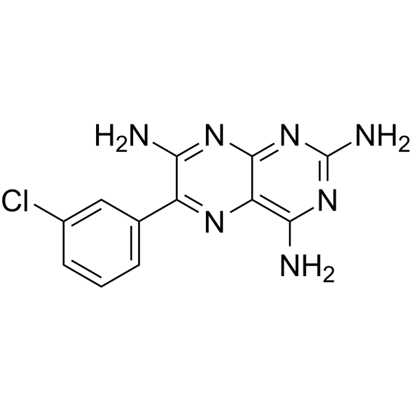
Epiblastin A is an ATP-competitive casein kinase 1 (CK1) inhibitor that inhibits CK1α, CK1δ, and CK1ε. Epiblastin A inhibits the kinase activity of STK10, BRSK1, EEF2K, EGFR, MKNK2, and RIPK2. Epiblastin A inhibits Ki-67 expression, induces Oct4-GFP expression, and enhances the conversion of mouse epiblast stem cells to embryonic stem cells. Epiblastin A selectively reduces the cell viability of colorectal cancer cell lines HCT116, HT29, and DLD1. Epiblastin A can be used in colorectal cancer-related research.
For research use only. We do not sell to patients.
- Purity : 99.44%
- CAS No.: 16470-02-3
- Formula: C12H10ClN7
- Molecular Weight:287.71
-
Storage:Powder -20°C, 3 years ; In solvent -80°C, 6 months , -20°C, 1 month

To place orders, for customer services and technical support, please contact: MedChemExpress USA
Tel: 609-228-6898 E-mail: [email protected] [email protected]


-
 Biological Activity
-
Chemical Information
-
Solvent & Solubility
- Protocols
- Purity & Documentation
- References
-
 Help & FAQs


-
HY-15490
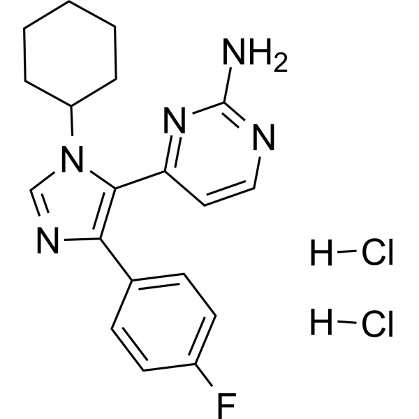
PF-670462 dihydrochlorideCasein Kinase Inhibitor -
HY-111820
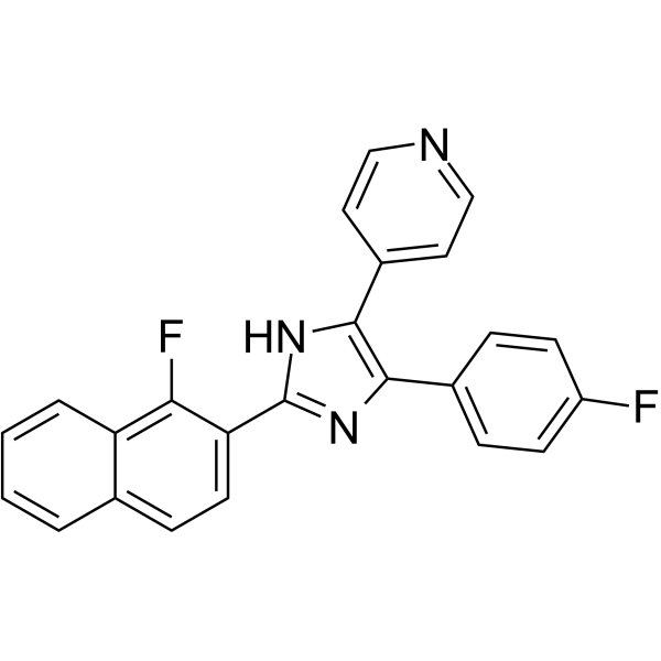
CK1-IN-1Casein Kinase 1 Inhibitor -
HY-12443
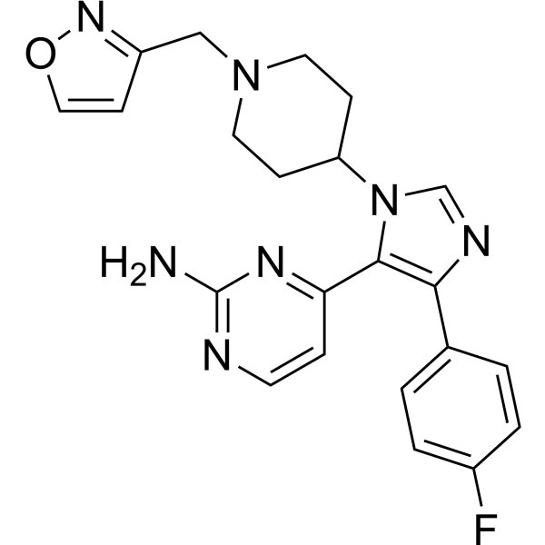
PF-5006739CK1δ/ε Inhibitor -
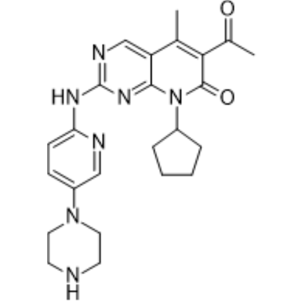
HY-50767
PalbociclibCDK4/6 Inhibitor -
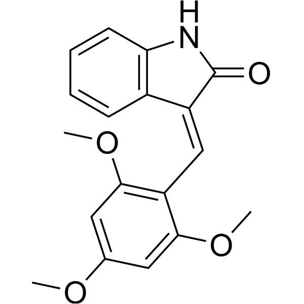
HY-12774
IC261CK1 Inhibitor -
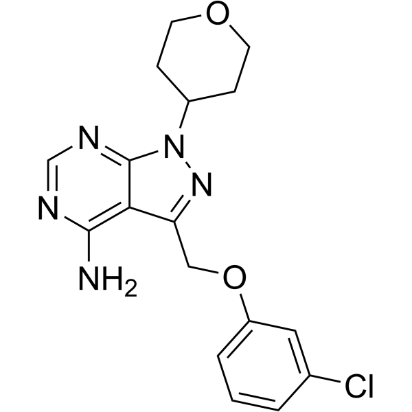
HY-12470
PF-4800567CK1 Inhibitor -
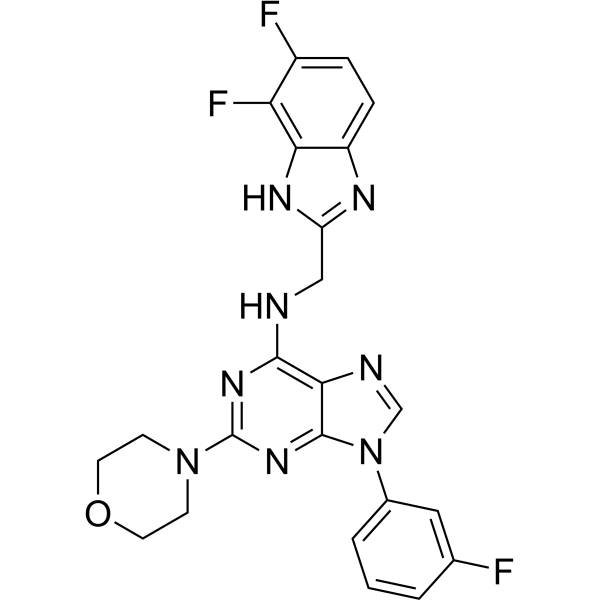
HY-100011
SR-3029CK1δ/ε Inhibitor -
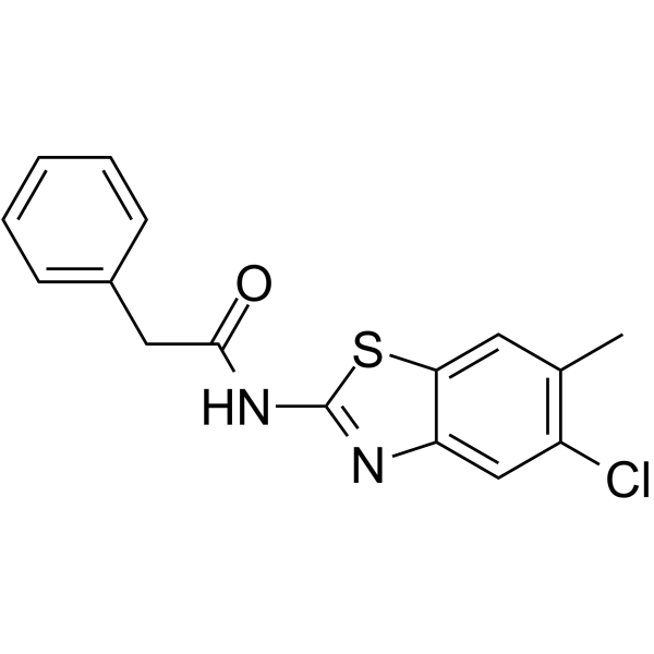
HY-15704
LH846Casein Kinase Inhibitor -

HY-100011R
SR-3029 (Standard)CK1δ/ε Inhibitor -
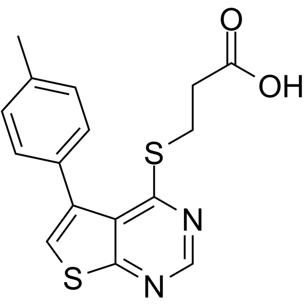
HY-15479
TTP 22Casein Kinase Inhibitor -
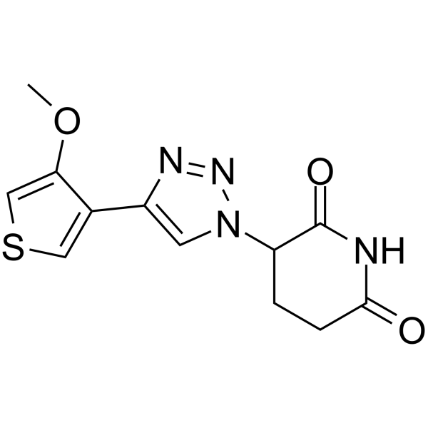
HY-145319
FPFT-2216IKZF1 IKZF3 CK-1α PDE6D CRBN Degrader -
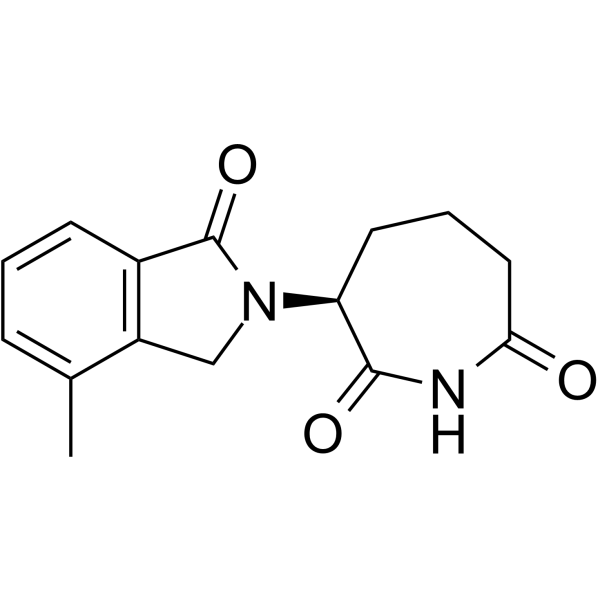
HY-120084
BTX161CK1α Molecular Glue Degrader
Publications Citing Use of MedChemExpress (MCE) Epiblastin A
MoreAll EGFR Isoforms
More

Biological Activity
Description
|
CK1δ |
CK1α |
CK1ε |
EGFR 8.3 μM (IC50) |
RIPK2 38 μM (IC50) |
Cellular Effect
|
Cell Line

|
Type | Value | Description | References |
|---|---|---|---|---|
| HT-29 | IC50 |
6.8 μM
|
Inhibition of viability of human colorectal carcinoma HT29 cells assessed after 48 hrs treatment by sulforhodamine B (SRB) assay.
Inhibition of viability of human colorectal carcinoma HT29 cells assessed after 48 hrs treatment by sulforhodamine B (SRB) assay.
|
PMC8798578 |
| HCT-116 | IC50 |
5.0 μM
|
Inhibition of viability of human colorectal carcinoma HCT116 cells assessed after 48 hrs treatment by sulforhodamine B (SRB) assay.
Inhibition of viability of human colorectal carcinoma HCT116 cells assessed after 48 hrs treatment by sulforhodamine B (SRB) assay.
|
PMC8798578 |
| DLD-1 | IC50 |
3.2 μM
|
Inhibition of viability of human colorectal carcinoma DLD1 cells assessed after 48 hrs treatment by sulforhodamine B (SRB) assay.
Inhibition of viability of human colorectal carcinoma DLD1 cells assessed after 48 hrs treatment by sulforhodamine B (SRB) assay.
|
PMC8798578 |
In Vitro
Epiblastin A (0.5-25 μM; 48 h) dose-dependently inhibits the viability of HT29, HCT116, and DLD1 colorectal cancer cell lines with IC50 values of 6.8, 5.0, and 3.2 μM, respectively, and exhibits no cytotoxicity against normal FHC colon cells[1].
Epiblastin A promotes the late-stage reprogramming of mouse epiblast stem cells (mEpiSCs) to mouse embryonic stem cells (mESCs), and it inhibits the CK1 family during this late-stage reprogramming process[2].
Epiblastin A is a potent inhibitor of CK1α, CK1δ, and CK1ε, with the strongest inhibitory potency against CK1δ; it also inhibits STK10 kinase activity[3].
MedChemExpress (MCE) has not independently confirmed the accuracy of these methods. They are for reference only.
-
Cell Line:HT29, HCT116, DLD1, FHC
-
Concentration:0.5, 1, 2.5, 5, 10, 25 μM
-
Incubation Time:48 h
-
Result:Suppressed the viability of cells.
Exhibited no apparent effect on the viability of FHC normal colonic cells.
Achieved 48 h IC50 values of 6.8 μM for HT29, 5.0 μM for HCT116, and 3.2 μM for DLD1 cells.
In Vivo
MedChemExpress (MCE) has not independently confirmed the accuracy of these methods. They are for reference only.
-
Animal Model:NOD.CB17-Prkdcscid/NcrCrl (NOD/SCID) (5-6 weeks old, Forty thousand DLD1 cells)[1]
-
Dosage:5 mg/kg
-
Administration:i.p.; every 72 h; 45 days
-
Result:Resulted in tumor sizes comparable to those in mice treated with 5 mg/kg Cetuximab (HY-P9905), with no apparently significant effect on mouse body weights.
Resulted in significantly smaller tumors than in vehicle-treated mice at the same time-point. Markedly reduced tumor growth compared to the vehicle-treated control group, with no apparent effect on mouse body weights.
Caused a 2.18-fold reduction in median tumor weight over the time-course of the animal study.
Profoundly suppressed the enhanced expression of CK1α (1.42-fold).
- Kinase activity and phosphorylation assays
- RNA extraction experimental
- Ki-67 Immunostaining Proliferation Assay
- Cell Cytotoxicity Assay
- Mammalian live/dead viability and cytotoxicity staining
- CCK-8/WST-8 Cell Proliferation Assay
- Protocol for Kinase activity and phosphorylation assays
- MTT Cell Proliferation Assay
- Cell Viability Determination by MTT Colorimetric Assay


Chemical Information
-
CAS No. 16470-02-3
-
Appearance Solid
-
Molecular Weight 287.71
-
Formula C12H10ClN7
-
Color Light yellow to green yellow
-
SMILES
NC1=NC(N)=C2N=C(C(N)=NC2=N1)C3=CC=CC(Cl)=C3
-
Shipping
Room temperature in continental US; may vary elsewhere.
-
Storage
Powder -20°C 3 years In solvent -80°C 6 months -20°C 1 month


Publications (1)
-
Journal Impact Factor
-
Most Recent
-
J Clin Invest
Molecular glue degrader function of SPOP enhances STING-dependent immunotherapy efficacy in melanoma models. [Abstract]2025 Oct 28:e191772. PMID: 41148213


Solvent & Solubility
In Vitro:
DMSO : 4.55 mg/mL (15.81 mM; ultrasonic and warming and heat to 60°C; Hygroscopic DMSO has a significant impact on the solubility of product, please use newly opened DMSO)
Please refer to the solubility information to select the appropriate solvent. Once prepared, please aliquot and store the solution to prevent product inactivation from repeated freeze-thaw cycles.
Storage method and period of stock solution: -80°C, 6 months; -20°C, 1 month. When stored at -80°C, please use it within 6 months. When stored at -20°C, please use it within 1 month.
Please refer to the solubility information to select the appropriate solvent. Once prepared, please aliquot and store the solution to prevent product inactivation from repeated freeze-thaw cycles.
Storage method and period of stock solution: -80°C, 6 months; -20°C, 1 month. When stored at -80°C, please use it within 6 months. When stored at -20°C, please use it within 1 month.
Concentration (start) × Volume (start) = Concentration (final) × Volume (final)


Purity & Documentation
-
Data Sheet (287 KB)
-
SDS (251 KB)
- English - EN (251 KB)
- Français - FR (251 KB)
- Deutsch - DE (251 KB)
- Norwegian - NO (251 KB)
- Español - ES (251 KB)
- Swedish - SV (251 KB)
- Italian - IT (251 KB)
- Korean - KR (251 KB)
- Portuguese - PT (251 KB)
-
Handling Instructions (2659 KB)
References


Complete Stock Solution Preparation Table
Please refer to the solubility information to select the appropriate solvent. Once prepared, please aliquot and store the solution to prevent product inactivation from repeated freeze-thaw cycles.
Storage method and period of stock solution: -80°C, 6 months; -20°C, 1 month. When stored at -80°C, please use it within 6 months. When stored at -20°C, please use it within 1 month.
| Optional Solvent | Concentration Solvent Mass | 1 mg | 5 mg | 10 mg | 25 mg |
|---|---|---|---|---|---|
| DMSO | 1 mM | 3.4757 mL | 17.3786 mL | 34.7572 mL | 86.8931 mL |
| 5 mM | 0.6951 mL | 3.4757 mL | 6.9514 mL | 17.3786 mL | |
| 10 mM | 0.3476 mL | 1.7379 mL | 3.4757 mL | 8.6893 mL | |
| 15 mM | 0.2317 mL | 1.1586 mL | 2.3171 mL | 5.7929 mL |


Epiblastin A Related Classifications
HY-114858 Related Classifications
 Powered by Bioz