
HDAC6-IN-36
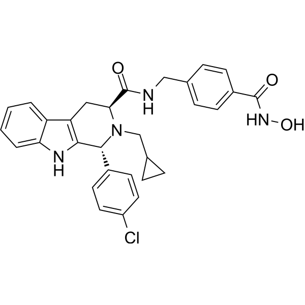
HDAC6-IN-36 (compound 11d) is an inhibitor of HDAC6 with IC50 value of 8.64 nM. HDAC6-IN-36 induces neurite outgrowth of PC12 cells without producing toxic effects.
Nos produits utilisent uniquement pour la recherche. Nous ne vendons pas aux patients.
- Formule: C30H29ClN4O3
- Masse moléculaire:529.03
-
Stockage:
Please store the product under the recommended conditions in the Certificate of Analysis.
Activité biologique
Description
IC50 & Target
|
HDAC1 1284 nM (IC50) |
HDAC6 8.64 nM (IC50) |
In Vitro
HDAC6-IN-36 (1 μM, 24 h) increases the acetylation level of histone H3 and ɑ-tubulin in the PC12 cell line.
HDAC6-IN-36 (1-10 μM, 48 h) stimulates neurite outgrowth induced by NGF (20 ng/mL) in PC12 cells.
MedChemExpress (MCE) has not independently confirmed the accuracy of these methods. They are for reference only. Further protocols information, click here.
-
Cell Line:PC12 cell
-
Concentration:0.1, 0.5 and 1 μM
-
Incubation Time:24 h
-
Result:Increased the acetylation level of α-tubulin in a dose-dependent manner.
Chemical Information
-
Masse moléculaire 529.03
-
Formule C30H29ClN4O3
-
SMILES
O=C(NCC1=CC=C(C(NO)=O)C=C1)[C@H](C2)N(CC3CC3)[C@H](C4=CC=C(Cl)C=C4)C5=C2C6=CC=CC=C6N5
-
Livraison
Room temperature in continental US; may vary elsewhere.
-
Stockage
Please store the product under the recommended conditions in the Certificate of Analysis.
Protocole
-
PC12 NGF-Induced Neuronal Differentiation Culture
PC12 cells are a rat adrenal pheochromocytoma clonal line that responds to NGF by stopping proliferation and extending branching neurite-like processes; after longer NGF exposure, cells develop long processes and neuronal-like ultrastructural and functional features. NGF-induced differentiation is read out mainly by neurite outgrowth, reduced proliferation, microtubule assembly, and neuronal differentiation-associated proteins such as MAPs, tau, GAP-43, and synapsin-1.
-
Cell Cytotoxicity Assay
Cytotoxicity assays are usually based on the assessment of cell membrane damage, which can also be indirectly detected by measuring cell viability. Detection methods include MTT assay, CKK-8 assay, LDH assay and ATP assay, etc.
-
SH-SY5Y Neuronal Differentiation Culture
SH-SY5Y neuronal differentiation culture uses sequential exposure to retinoic acid and neurotrophic factors to reduce proliferative neuroblastoma-like behavior and induce neuron-like morphology, including neurite extension, neuronal marker expression, and, in RA/BDNF protocols, greater synaptic-marker expression than undifferentiated culture. Retinoic acid is commonly used as the initiating differentiation cue, while BDNF in serum-reduced or serum-free medium supports later maturation and neurotrophic-factor-dependent neuron-like survival.
-
SH-SY5Y neuronal-like differentiation
SH-SY5Y neuronal-like differentiation uses defined culture conditions to shift proliferative human neuroblastoma cells toward a neuron-like state, mainly assessed by reduced proliferation, neurite extension, neuronal-marker expression, and, in some protocols, increased dependence on neurotrophic support. Retinoic acid (RA) is commonly used for the first differentiation phase, and sequential RA followed by brain-derived neurotrophic factor (BDNF) in serum-free medium is a well-characterized approach for generating neuron-like SH-SY5Y cultures with extensive neurite outgrowth. The primary readouts are morphology-based neurite outgrowth and marker-based confirmation using proteins such as βIII-tubulin, MAP2, GAP43, synaptophysin, NeuN, NSE, TH, or related neuronal/synaptic markers, depending on the study endpoint.
-
Cell differentiation
Cell differentiation refers to the process in which cells of the same origin gradually produce cell groups with different morphological structure and functional characteristics.
-
PC12 NGF-induced neuronal-like differentiation
PC12 cells are a rat adrenal pheochromocytoma-derived clonal cell line that responds to nerve growth factor by stopping proliferation and extending neurites, producing a sympathetic neuron-like phenotype used to study neuronal differentiation and neurite outgrowth. NGF acts through TrkA-dependent signaling, and neurite outgrowth is associated with ERK/Akt signaling, microtubule organization, neuronal-marker expression, and increased electrophysiological neuronal features such as sodium-channel density. The main assay readout is morphological differentiation, usually measured as the percentage of neurite-bearing cells, neurite length, neurite number, or total neurite length per cell. Additional readouts include GAP-43, tyrosine hydroxylase, βIII-tubulin, neurofilament, synapsin I, synaptophysin, ERK phosphorylation, Akt phosphorylation, and sodium-channel current density.
Pureté et documentation
Références
Calculators
Concentration (start) × Volume (start) = Concentration (final) × Volume (final)

