



IHMT-EZH2-426
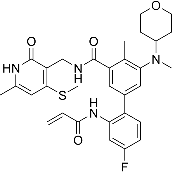
IHMT-EZH2-426 is an orally active and selective EZH2 degrader. IHMT-EZH2-426 forms a covalent bond with Cys663 of EZH2, inhibits its catalytic activity, and reduces the protein levels of EZH2 as well as the level of H3K27me3. IHMT-EZH2-426 acts as an antiproliferative agent against B-cell lymphoma and triple-negative breast cancer cells, and also exhibits anti-tumor related activity in Pfeiffer and MDA‑MB‑231 xenograft models. IHMT-EZH2-426 can be used in studies related to B-cell lymphoma and triple-negative breast cancer.
Nur für Forschungszwecke. Wir verkaufen nicht an Patienten.
- CAS. Nr.: 3018914-66-1
- Formel: C31H35FN4O4S
- Molecular Weight:578.70
-
Speicherung:
Please store the product under the recommended conditions in the Certificate of Analysis.

To place orders, for customer services and technical support, please contact: MedChemExpress USA
Tel: 609-228-6898 E-mail: [email protected] [email protected]


-
 Biologische Aktivität
-
Chemical Information
- Protokoll
- Reinheit & Dokumentation
- Verweise
-
 Help & FAQs


Alle Histone Methyltransferase Isoform-spezifische Produkte anzeigen
More

Biologische Aktivität
Beschreibung
IC50 & Target
[1]|
EZH2 WT 1.3 nM (IC50) |
Cellular Effect
|
Cell Line

|
Type | Value | Description | References |
|---|---|---|---|---|
| Pfeiffer | GI50 |
0.02 μM
|
Antiproliferative activity against EZH2-mutant (A677G) human Pfeiffer B-cell lymphoma cells assessed as reduction in cell viability incubated for 5 days by CellTiter-Glo assay.
Antiproliferative activity against EZH2-mutant (A677G) human Pfeiffer B-cell lymphoma cells assessed as reduction in cell viability incubated for 5 days by CellTiter-Glo assay.
|
37826933 |
| KARPAS-422 | GI50 |
0.085 μM
|
Antiproliferative activity against EZH2-mutant (Y641N) human Karpas-422 B-cell lymphoma cells assessed as reduction in cell viability incubated for 5 days by CellTiter-Glo assay.
Antiproliferative activity against EZH2-mutant (Y641N) human Karpas-422 B-cell lymphoma cells assessed as reduction in cell viability incubated for 5 days by CellTiter-Glo assay.
|
37826933 |
| SU-DHL-4 | GI50 |
1.42 μM
|
Antiproliferative activity against EZH2-mutant (Y641S) human SU-DHL-4 B-cell lymphoma cells assessed as reduction in cell viability incubated for 5 days by CellTiter-Glo assay.
Antiproliferative activity against EZH2-mutant (Y641S) human SU-DHL-4 B-cell lymphoma cells assessed as reduction in cell viability incubated for 5 days by CellTiter-Glo assay.
|
37826933 |
| SU-DHL-6 | GI50 |
1.95 μM
|
Antiproliferative activity against EZH2-mutant (Y641N) human SU-DHL-6 B-cell lymphoma cells assessed as reduction in cell viability incubated for 5 days by CellTiter-Glo assay.
Antiproliferative activity against EZH2-mutant (Y641N) human SU-DHL-6 B-cell lymphoma cells assessed as reduction in cell viability incubated for 5 days by CellTiter-Glo assay.
|
37826933 |
| WSUDLCL2 | GI50 |
3.85 μM
|
Antiproliferative activity against EZH2-mutant (Y641F) human WSU-DLCL2 B-cell lymphoma cells assessed as reduction in cell viability incubated for 5 days by CellTiter-Glo assay.
Antiproliferative activity against EZH2-mutant (Y641F) human WSU-DLCL2 B-cell lymphoma cells assessed as reduction in cell viability incubated for 5 days by CellTiter-Glo assay.
|
37826933 |
| Daudi | GI50 |
7.01 μM
|
Antiproliferative activity against EZH2 WT human Daudi B-cell lymphoma cells assessed as reduction in cell viability incubated for 5 days by CellTiter-Glo assay.
Antiproliferative activity against EZH2 WT human Daudi B-cell lymphoma cells assessed as reduction in cell viability incubated for 5 days by CellTiter-Glo assay.
|
37826933 |
| MDA-MB-231 | GI50 |
6.78 μM
|
Antiproliferative activity against human MDA-MB-231 TNBC cells assessed as reduction in cell viability incubated for 5 days by CellTiter-Glo assay.
Antiproliferative activity against human MDA-MB-231 TNBC cells assessed as reduction in cell viability incubated for 5 days by CellTiter-Glo assay.
|
37826933 |
| MDA-MB-468 | GI50 |
1.93 μM
|
Antiproliferative activity against human MDA-MB-468 TNBC cells assessed as reduction in cell viability incubated for 5 days by CellTiter-Glo assay.
Antiproliferative activity against human MDA-MB-468 TNBC cells assessed as reduction in cell viability incubated for 5 days by CellTiter-Glo assay.
|
37826933 |
In Vitro
IHMT-EZH2-426 (0.03-10 μM; 72 h) inhibits H3K27 trimethylation at a concentration of 0.03 μM and reduces EZH2 protein levels at 1 μM after 72 h of treatment in Karpas-422, Pfeiffer, MDA-MB-231 and MDA-MB-468 cell lines[1].
IHMT-EZH2-426 (10 μg/mL; 72 h treatment, up to 48 h washout) achieves sustained inhibition of H3K27me3 for at least 48 h after washout in MDA-MB-231 cells, confirming its persistent binding to the target[1].
IHMT-EZH2-426 (1-50 μM; 4 h compound treatment, 4 h probe incubation) binds to EZH2 in MDA-MB-231 cell lysates, and labels approximately 50% of available target proteins at a concentration of 1 μM[1].
IHMT-EZH2-426 forms a covalent adduct with purified EZH2 (SET domain) protein, confirming its covalent binding mechanism[1].
IHMT-EZH2-426 (for 14 days) inhibits colony formation of MDA-MB-231 and MDA-MB-468 triple-negative breast cancer (TNBC) cells in a dose-dependent manner[1].
MedChemExpress (MCE) has not independently confirmed the accuracy of these methods. They are for reference only.
-
Cell Line:B-cell lymphoma cell lines (Karpas-422, Pfeiffer), TNBC cell lines (MDA-MB-231, MDA-MB-468)
-
Concentration:0.03, 0.1, 0.3, 1, 3, 10 μM
-
Incubation Time:72 h
-
Result:Potently inhibited H3K27 trimethylation at 0.03 μM in Karpas-422, Pfeiffer, MDA-MB-231, and MDA-MB-468 cell lines.
Reduced EZH2 protein levels at 1 μM in Karpas-422, Pfeiffer, MDA-MB-231, and MDA-MB-468 cell lines.
-
Cell Line:MDA-MB-231 TNBC cells
-
Concentration:10 μg/mL
-
Incubation Time:72 h (treatment)
0, 2, 4, 6, 8, 12, 24, 48 h (washout) -
Result:Sustained inhibition of H3K27me3 for up to 48 h after washout.
-
Cell Line:Pfeiffer, Karpas‑422, SU‑DHL‑4, SU‑DHL‑6, WSU‑DHL2, Daudi, Farage, Raji
-
Concentration:0.1, 0.3, 1, 3, 10 μM
-
Incubation Time:5 days
-
Result:Inhibited proliferation of EZH2-mutant B-cell lymphoma cell lines (GI50 0.02-3.85 μM)
Exhibited moderate antiproliferative activity against EZH2 WT B-cell lymphoma cell lines and TNBC cells, including 3D patient-derived TNBC cells (GI50 1.93-7.01 μM).
Parmacokinetics
In Vivo
IHMT-EZH2-426 (100 mg/kg; p.o.; daily; 20 days) exhibits potent in vivo anti-tumor activity against Pfeiffer diffuse large B-cell lymphoma (DLBCL) xenografts[1].
MedChemExpress (MCE) has not independently confirmed the accuracy of these methods. They are for reference only.
-
Animal Model:BALB/c nude mice (female, 5-week old, 5 × 10⁶ MDA-MB-231 cells)[1]
-
Dosage:100 mg/kg
-
Administration:p.o.; daily; 14 days
-
Result:Suppressed H3K27 trimethylation in tumor tissues.
Reduced EZH2 levels in tumor tissues.
Showed no apparent body weight loss during treatment.
-
Animal Model:NCG mice (female, 5-week old, 1 × 10⁷ Pfeiffer cells)[1]
-
Dosage:100 mg/kg
-
Administration:p.o.; daily; 20 days
-
Result:Demonstrated potent antitumor activity with efficacy comparable to the reference compound.
Showed no apparent body weight loss during treatment.


Chemical Information
-
CAS. Nr. 3018914-66-1
-
Molecular Weight 578.70
-
Formel C31H35FN4O4S
-
SMILES
O=C(C1=CC(C(C=CC(F)=C2)=C2NC(C=C)=O)=CC(N(C3CCOCC3)C)=C1C)NCC4=C(SC)C=C(C)NC4=O
-
Versand
Room temperature in continental US; may vary elsewhere.
-
Speicherung
Please store the product under the recommended conditions in the Certificate of Analysis.


Reinheit & Dokumentation
Verweise

Calculators
Konzentration (Stammlösung) × Volumen (Stammlösung) = Konzentration (Ziellösung) × Volumen (Ziellösung)


IHMT-EZH2-426 Related Classifications
HY-156292 Related Classifications
 Powered by Bioz