



INDO 1
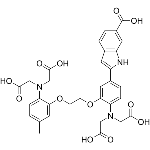
INDO 1 is a cell-impermeable, dual-emission ratiometric fluorescent calcium indicator with a KD·β value of 0.44 μM. INDO 1 binds to intracellular free calcium ions and undergoes wavelength-dependent fluorescence changes. In the unbound state (free form) without Ca2+, its fluorescence emission peak is at 485 nm upon excitation with ultraviolet light (350 nm). When INDO 1 pentasodium binds to Ca2+ (bound state), the emission peak shifts to 405 nm (Ex = 350 nm; dual emission at Em= 405/485 nm).
For research use only. We do not sell to patients.
- CAS No.: 96314-96-4
- Formula: C32H31N3O12
- Molecular Weight:649.60
-
Storage:
Please store the product under the recommended conditions in the Certificate of Analysis.

To place orders, for customer services and technical support, please contact: MedChemExpress USA
Tel: 609-228-6898 E-mail: [email protected] [email protected]


-
 Biological Activity
-
Chemical Information
- Protocols
- Purity & Documentation
- References
-
 Help & FAQs




Biological Activity
Description
In Vitro
INDO 1 pentapotassium solution is a calcium-sensitive fluorescent dye used in glass capillaries, with an apparent KD·β value of 0.44 μM; as calcium concentration increases, its emission at 405 nm enhances while emission at 470 nm diminishes[3].
MedChemExpress (MCE) has not independently confirmed the accuracy of these methods. They are for reference only.
Emission (Em)
475
Excitation (Ex)
350


Chemical Information
-
CAS No. 96314-96-4
-
Molecular Weight 649.60
-
Formula C32H31N3O12
-
SMILES
O=C(C1=CC2=C(C=C1)C=C(C3=CC=C(N(CC(O)=O)CC(O)=O)C(OCCOC4=CC(C)=CC=C4N(CC(O)=O)CC(O)=O)=C3)N2)O
-
Shipping
Room temperature in continental US; may vary elsewhere.
-
Storage
Please store the product under the recommended conditions in the Certificate of Analysis.


Purity & Documentation
References

Calculators
Concentration (start) × Volume (start) = Concentration (final) × Volume (final)


INDO 1 Related Classifications
HY-D0121 Related Classifications
 Powered by Bioz