



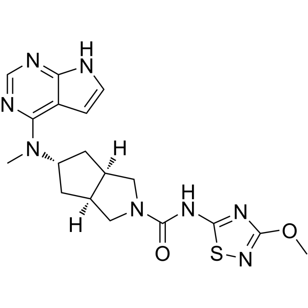
Ivarmacitinib

Based on 1 Customer Validation
Ivarmacitinib (SHR0302) is a potent and orally active all members of the JAK family inhibitor, particularly JAK1. The selectivity of Ivarmacitinib for JAK1 is >10-fold for JAK2, 77-fold for JAK3, 420-fold for Tyk2. Ivarmacitinib inhibits JAK1-STAT3 phosphorylation and induces the apoptosis of hepatic stellate cells. Ivarmacitinib has anti-proliferative and anti-inflammatory effects.
For research use only. We do not sell to patients.
- Purity : 99.62%
- CAS No.: 1445987-21-2
- Formula: C18H22N8O2S
- Molecular Weight:414.48
-
Storage:Powder -20°C, 3 years , 4°C, 2 years ; In solvent -80°C, 2 years , -20°C, 1 year

To place orders, for customer services and technical support, please contact: MedChemExpress USA
Tel: 609-228-6898 E-mail: [email protected] [email protected]


-
 Biological Activity
-
Chemical Information
-
Solvent & Solubility
- Protocols
- Purity & Documentation
- References
-
 Help & FAQs


-
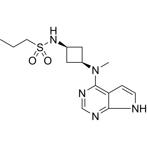
HY-107429
AbrocitinibJAK1 Inhibitor -
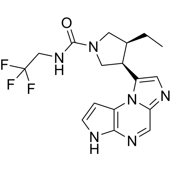
HY-19569
UpadacitinibJAK1 Inhibitor -
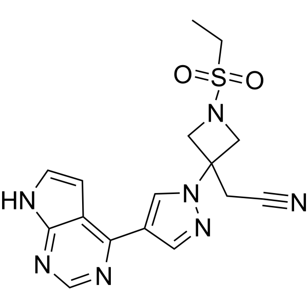
HY-15315
BaricitinibJAK1/2 Inhibitor -
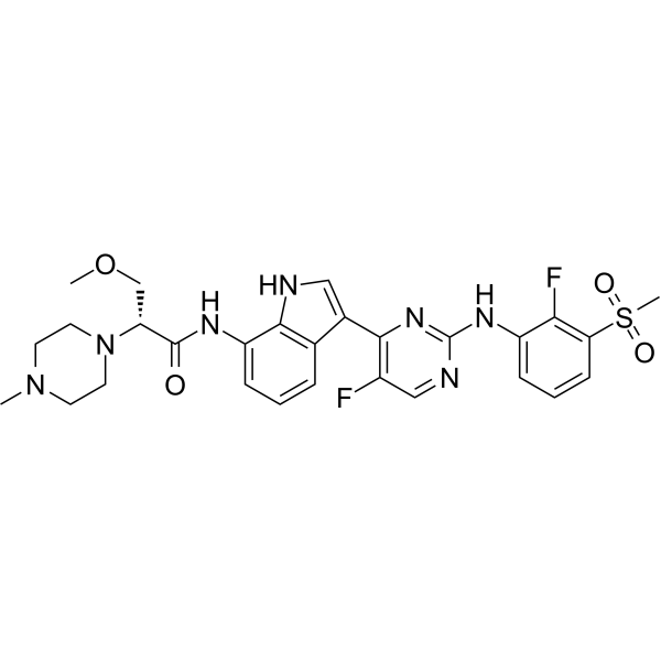
HY-126294
LondamocitinibJAK1 Inhibitor -
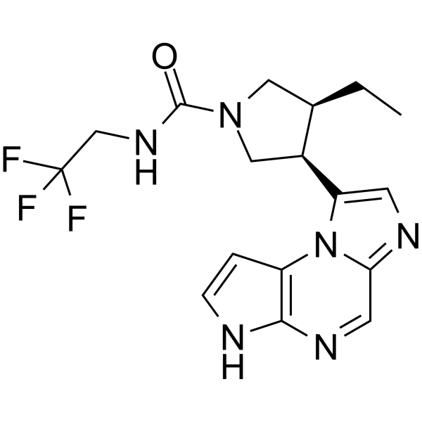
HY-19569R
Upadacitinib (Standard)JAK1 Inhibitor -
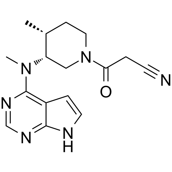
HY-40354
TofacitinibJAK1/JAK3 Inhibitor -
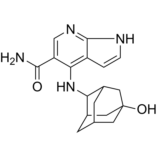
HY-19568
PeficitinibJAK Inhibitor -
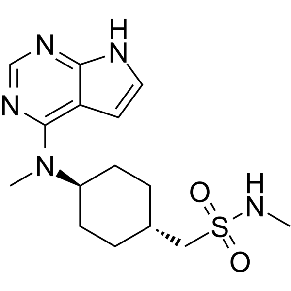
HY-13577
OclacitinibJAK Inhibitor -

HY-19568R
Peficitinib (Standard)JAK Inhibitor -
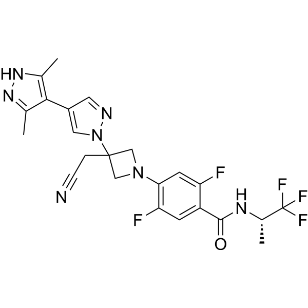
HY-145588
PovorcitinibJAK1 Inhibitor -
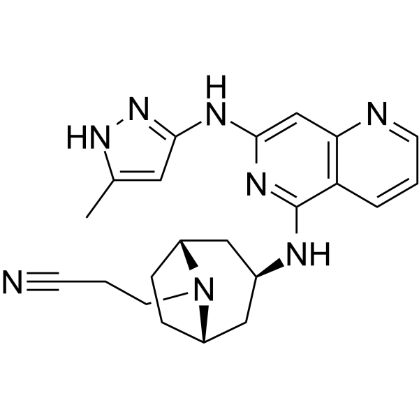
HY-109148
Izencitinibpan JAK Inhibitor -
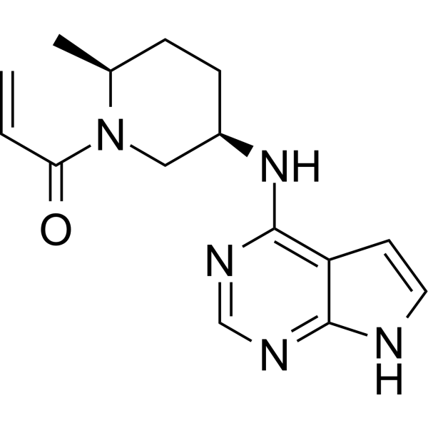
HY-100754
RitlecitinibJAK3 Inhibitor


Biological Activity
Description
IC50 & Target
[1]|
JAK1 |
JAK2 |
JAK3 |
Tyk2 |
In Vitro
Ivarmacitinib (SHR0302; 1 nM-10 µM; 48 hours; HSCs) treatment displays an inhibitory effect on the proliferation of HSCs in a concentration-dependent manner[2].
Ivarmacitinib (1 nM-10 µM) exerts an inhibitory effect on the activation, proliferation and migration of HSCs[2].
Ivarmacitinib (1 nM-10 µM; 48 hours; HSCs) treatment induces the apoptosis of HSCs[2].
Ivarmacitinib (1 nM-10 µM; 48 hours; HSCs) treatment significantly increases the activation of caspase-3 and Bax in HSCs, and decreases the expression of Bcl-2. SHR0302 also inhibits the activation of Akt signaling pathway[2].
MedChemExpress (MCE) has not independently confirmed the accuracy of these methods. They are for reference only.
-
Cell Line:Hepatic stellate cells (HSCs)
-
Concentration:1 nM, 10 nM, 100 nM, 1µM, 10 µM
-
Incubation Time:48 hours
-
Result:Displayed an inhibitory effect on the proliferation of HSCs, and that inhibition ocurred in a concentration-dependent manner.
-
Cell Line:Hepatic stellate cells (HSCs)
-
Concentration:1 nM, 10 nM, 100 nM, 1µM, 10 µM
-
Incubation Time:48 hours
-
Result:Induced the apoptosis of HSCs.
-
Cell Line:Hepatic stellate cells (HSCs)
-
Concentration:1 nM, 10 nM, 100 nM, 1µM, 10 µM
-
Incubation Time:48 hours
-
Result:Significantly increased the activation of caspase-3 and Bax in HSCs, and decreased the expression of Bcl-2. Also inhibited the activation of Akt signaling pathway.
In Vivo
Ivarmacitinib can inhibit the proliferation of T, B and fibroblast-like synoviocytes (FLS), and down-regulates cytokines TNF-α, IL-1β, IL-17 and antibody IgG1, IgG2a levels, and suppresses the proportion of Th17 and total B, and inhibits JAK1-STAT3 phosphorylation[1].
MedChemExpress (MCE) has not independently confirmed the accuracy of these methods. They are for reference only.
-
Animal Model:Male Sprague-Dawley (SD) rats (150-180 g) injected with complete Freund’s adjuvant (CFA)[1]
-
Dosage:0.3 mg/kg,1.0 mg/kg, 3.0 mg/kg
-
Administration:Intragastric administration; twice a day; for 14 days
-
Result:Suppressed the severity of adjuvant-induced arthritis (AA) rats by attenuating the arthritis index, arthritis global assessment and paw swelling degree, and alleviated histopathology of spleen and joint of AA rats.
- Kinase activity and phosphorylation assays
- Western Blot
- Detection of Apoptosis
- TUNEL staining for apoptotic DNA fragmentation
- Annexin V plus membrane-impermeant dye apoptosis staining
- Apoptosis Solutions
- Protocol for Kinase activity and phosphorylation assays
- Research Protocol for Inflammation-related Diseases
- Cell Viability Determination by MTT Colorimetric Assay
Clinical Trial
| NCT Number | Sponsor | Condition | Start Date |
Phase

|
|---|---|---|---|---|
| NCT01329991 | 2011-05 | PHASE1 |


Chemical Information
-
CAS No. 1445987-21-2
-
Appearance Solid
-
Molecular Weight 414.48
-
Formula C18H22N8O2S
-
Color White to light yellow
-
SMILES
O=C(N1C[C@@]2([H])[C@@](C[C@H](N(C)C3=C4C(NC=C4)=NC=N3)C2)([H])C1)NC5=NC(OC)=NS5
-
Synonyms
SHR0302
-
Shipping
Room temperature in continental US; may vary elsewhere.
-
Storage
Powder -20°C 3 years 4°C 2 years In solvent -80°C 2 years -20°C 1 year


Solvent & Solubility
In Vitro:
DMSO : 31.25 mg/mL (75.40 mM; Need ultrasonic; Hygroscopic DMSO has a significant impact on the solubility of product, please use newly opened DMSO)
Please refer to the solubility information to select the appropriate solvent. Once prepared, please aliquot and store the solution to prevent product inactivation from repeated freeze-thaw cycles.
Storage method and period of stock solution: -80°C, 2 years; -20°C, 1 year. When stored at -80°C, please use it within 2 years. When stored at -20°C, please use it within 1 year.
Please refer to the solubility information to select the appropriate solvent. Once prepared, please aliquot and store the solution to prevent product inactivation from repeated freeze-thaw cycles.
Storage method and period of stock solution: -80°C, 2 years; -20°C, 1 year. When stored at -80°C, please use it within 2 years. When stored at -20°C, please use it within 1 year.
Concentration (start) × Volume (start) = Concentration (final) × Volume (final)
In Vivo:
Select the appropriate dissolution method based on your experimental animal and administration route.
- For the following dissolution methods, please ensure to first prepare a clear stock solution using an In Vitro approach and then sequentially add co-solvents:
- To ensure reliable experimental results, the clarified stock solution can be appropriately stored based on storage conditions. As for the working solution for In Vivo experiments, it is recommended to prepare freshly and use it on the same day.
- The percentages shown for the solvents indicate their volumetric ratio in the final prepared solution. If precipitation or phase separation occurs during preparation, heat and/or sonication can be used to aid dissolution.
Add each solvent one by one: 10% DMSO 40% PEG300 5% Tween-80 45% Saline
Solubility: ≥ 2.08 mg/mL (5.02 mM); Clear solution
This protocol yields a clear solution of ≥ 2.08 mg/mL (saturation unknown).
Taking 1 mL working solution as an example, add 100 μL DMSO stock solution (20.8 mg/mL) to 400 μL PEG300, and mix evenly; then add 50 μL Tween-80 and mix evenly; then add 450 μL Saline to adjust the volume to 1 mL.
Preparation of Saline: Dissolve 0.9 g sodium chloride in ddH₂O and dilute to 100 mL to obtain a clear Saline solution.
Add each solvent one by one: 10% DMSO 90% (20% SBE-β-CD in Saline)
Solubility: ≥ 2.08 mg/mL (5.02 mM); Clear solution
This protocol yields a clear solution of ≥ 2.08 mg/mL (saturation unknown).
Taking 1 mL working solution as an example, add 100 μL DMSO stock solution (20.8 mg/mL) to 900 μL 20% SBE-β-CD in Saline, and mix evenly.
Preparation of 20% SBE-β-CD in Saline (4°C, storage for one week): 2 g SBE-β-CD powder is dissolved in 10 mL Saline, completely dissolve until clear.
In Vivo Dissolution Calculator
Please enter the basic information of animal experiments:
-
-
-
-
Recommended: Prepare an additional quantity of animals to account for potential losses during experiments.
Please enter your animal formula composition:
-
%DMSO +
Recommended: Keep the proportion of DMSO in working solution below 2% if your animal is weak.
-
%+
-
+%Tween-80 + +
-
%Saline +
The co-solvents required include: DMSO, . All of co-solvents are available by MedChemExpress (MCE). , Tween 80. All of co-solvents are available by MedChemExpress (MCE).
Working solution concentration: 0.22 mg/mL
Method for preparing stock solution: mg drug dissolved in μL DMSO. Stock solution concentration: mg/mL.
1. Take μL DMSO stock solution;
2. Add μL .
μL , mix evenly;
3. Then add μL Tween 80, mix evenly;
4. Then add μL
Please ensure that the stock solution in the first step is dissolved to a clear state, and add co-solvents in sequence. You can use ultrasonic heating (ultrasonic cleaner, recommended frequency 20-40 kHz), vortexing, etc. to assist dissolution.


Purity & Documentation
-
Data Sheet (281 KB)
-
SDS (396 KB)
- English - EN (396 KB)
- Français - FR (396 KB)
- Deutsch - DE (396 KB)
- Norwegian - NO (396 KB)
- Español - ES (396 KB)
- Swedish - SV (396 KB)
- Italian - IT (396 KB)
- Korean - KR (396 KB)
- Portuguese - PT (396 KB)
-
Handling Instructions (2659 KB)
References
[1]. Huaxun Wu, et al. JAK1-STAT3 Blockade by JAK Inhibitor SHR0302 Attenuates Inflammatory Responses of Adjuvant-Induced Arthritis Rats and Decreases Th17 and Total B Cells. Joint Bone Spine. 2016 Oct;83(5):525-32. [Content Brief]
[2]. Yuan-Jing Gu, et al. Targeted Blockade of JAK/STAT3 Signaling Inhibits Proliferation, Migration and Collagen Production as Well as Inducing the Apoptosis of Hepatic Stellate Cells. Int J Mol Med. 2016 Sep;38(3):903-11. [Content Brief]


Complete Stock Solution Preparation Table
Please refer to the solubility information to select the appropriate solvent. Once prepared, please aliquot and store the solution to prevent product inactivation from repeated freeze-thaw cycles.
Storage method and period of stock solution: -80°C, 2 years; -20°C, 1 year. When stored at -80°C, please use it within 2 years. When stored at -20°C, please use it within 1 year.
| Optional Solvent | Concentration Solvent Mass | 1 mg | 5 mg | 10 mg | 25 mg |
|---|---|---|---|---|---|
| DMSO | 1 mM | 2.4127 mL | 12.0633 mL | 24.1266 mL | 60.3165 mL |
| 5 mM | 0.4825 mL | 2.4127 mL | 4.8253 mL | 12.0633 mL | |
| 10 mM | 0.2413 mL | 1.2063 mL | 2.4127 mL | 6.0317 mL | |
| 15 mM | 0.1608 mL | 0.8042 mL | 1.6084 mL | 4.0211 mL | |
| 20 mM | 0.1206 mL | 0.6032 mL | 1.2063 mL | 3.0158 mL | |
| 25 mM | 0.0965 mL | 0.4825 mL | 0.9651 mL | 2.4127 mL | |
| 30 mM | 0.0804 mL | 0.4021 mL | 0.8042 mL | 2.0106 mL | |
| 40 mM | 0.0603 mL | 0.3016 mL | 0.6032 mL | 1.5079 mL | |
| 50 mM | 0.0483 mL | 0.2413 mL | 0.4825 mL | 1.2063 mL | |
| 60 mM | 0.0402 mL | 0.2011 mL | 0.4021 mL | 1.0053 mL |


Ivarmacitinib Related Classifications
HY-112724 Related Classifications
 Powered by Bioz