



Cytochalasin B
Based on 48 publication(s) in Google Scholar
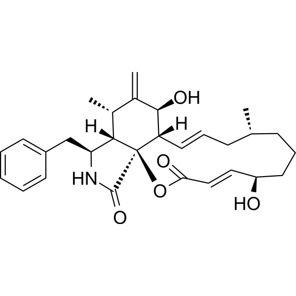
Cytochalasin B is a cell-permeable mycotoxin binding to the barbed end of actin filaments, disrupting the formation of actin polymers, with Kd value of 1.4-2.2 nM for F-actin. Cytochalasin B blocks cell migration.
For research use only. We do not sell to patients.
- Purity : 99.41%
- CAS No.: 14930-96-2
- Formula: C29H37NO5
- Molecular Weight:479.61
-
Storage:Powder -20°C, 3 years ; In solvent -80°C, 6 months , -20°C, 1 month

To place orders, for customer services and technical support, please contact: MedChemExpress USA
Tel: 609-228-6898 E-mail: [email protected] [email protected]


-
 Biological Activity
-
Chemical Information
-
Solvent & Solubility
- Protocols
- Purity & Documentation
- References
-
 Help & FAQs


-

HY-10071
Y-27632ROCK Inhibitor -
HY-19363
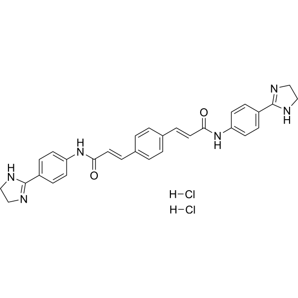
GW4869N-SMase Inhibitor -
HY-N6682
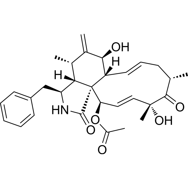
Cytochalasin DActin Polymerization Inhibitor -
HY-13520
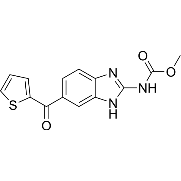
NocodazoleMicrotubule Inhibitor -
HY-16926
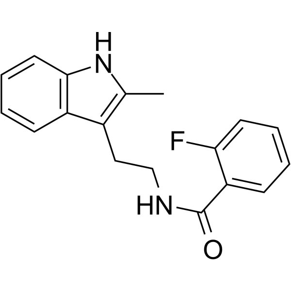
CK-666Arp2/3 Complex Inhibitor -
HY-16929
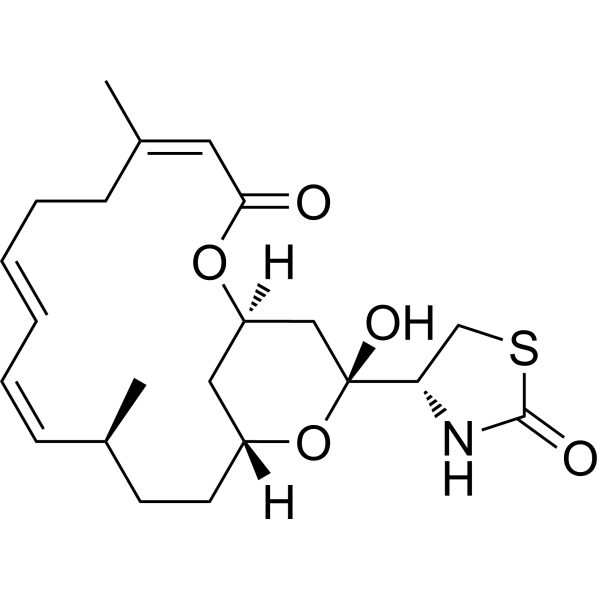
Latrunculin AG-actin Polymerization Inhibitor -

HY-18739
Phorbol 12-myristate 13-acetatePKC Activator -

HY-100558
Bafilomycin A1V-ATPase Inhibitor -
HY-10108
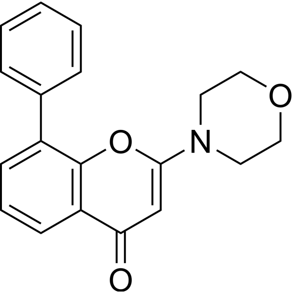
LY294002PI3K Inhibitor -
HY-15304
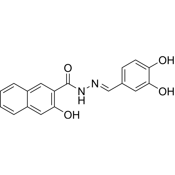
DynasoreAutophagy Inducer -
HY-16569
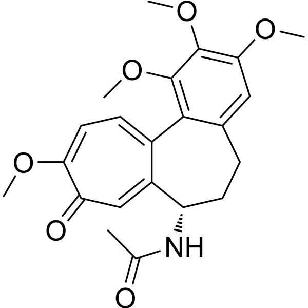
ColchicineTubulin Inhibitor -
HY-101840
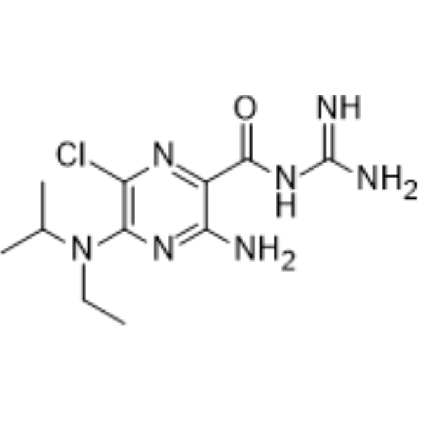
EIPATRPP3 Channel Inhibitor
Publications Citing Use of MedChemExpress (MCE) Cytochalasin B
More- Signal Transduct Target Ther. 2023 Dec 25;8(1):457. [Abstract]
- Nat Commun. 2026 Aug 22;17(1):10061.
- Nat Commun. 2024 Jan 5;15(1):296. [Abstract]
- Nat Commun. 2019 Sep 4;10(1):3981. [Abstract]
- Cell Discov. 2026 Mar 31;12(1):25. [Abstract]
- J Nanobiotechnology. 2025 Aug 11;23(1):559. [Abstract]
- Theranostics. 2024 Sep 9;14(15):5762-5777. [Abstract]
- Adv Sci (Weinh). 2025 Sep 15:e06294. [Abstract]
- Adv Sci (Weinh). 2025 Sep 17:e00358. [Abstract]
- Adv Sci (Weinh). 2025 Jun;12(23):e2416489. [Abstract]
- Adv Sci (Weinh). 2020 Jun 17;7(15):1903583. [Abstract]
- Cell Rep Med. 2026 May 19;7(5):102788. [Abstract]
- Chem Eng J. 2025 Sep 4;522:168081.
- Cell Commun Signal. 2021 Nov 16;19(1):114. [Abstract]
- Cell Commun Signal. 2020 Nov 30;18(1):187. [Abstract]
- Mater Today Bio. 2024 Aug 2:28:101175. [Abstract]
- Acta Pharmacol Sin. 2023 Aug;44(8):1665-1675. [Abstract]
- Dev Cell. 2025 Jun 23;60(12):1751-1767.e9. [Abstract]
- Free Radic Biol Med. 2022 Sep:190:1-14. [Abstract]
- Cell Rep. 2023 Oct 5;42(10):113213. [Abstract]
- Cell Prolif. 2024 Jan;57(1):e13530. [Abstract]
- Cell Prolif. 2023 Sep;56(9):e13442. [Abstract]
- Environ Sci Technol Lett. 2023 Sep 27.
- Environ Pollut. 2025 May 15:373:126119. [Abstract]
- NPJ Regen Med. 2021 Sep 6;6(1):51. [Abstract]
- J Cell Biol. 2026 Jun 1;225(6):e202502001. [Abstract]
- RSC Adv. 2024 Sep 6;14(39):28569-28584. [Abstract]
- Cell Biol Toxicol. 2024 Jul 12;40(1):54. [Abstract]
- Reprod Biol Endocrinol. 2022 Apr 18;20(1):67. [Abstract]
- Int J Mol Sci. 2025 Mar 26;26(7):3029. [Abstract]
- Mol Neurobiol. 2021 Oct;58(10):4906-4920. [Abstract]
- Front Cell Dev Biol. 2022 Apr 25;10:850052. [Abstract]
- Sci Rep. 2025 Mar 15;15(1):8989. [Abstract]
- J Mol Cell Cardiol. 2023 Sep:182:57-72. [Abstract]
- Antimicrob Agents Chemother. 2020 Sep 21;64(10):e00608-20. [Abstract]
- J Biol Chem. 2022 Nov;298(11):102511. [Abstract]
- J Cell Physiol. 2024 Nov;239(11):e31359. [Abstract]
- Microb Pathog. 2026 Apr:213:108348. [Abstract]
- Pathogens. 2023 Sep 9;12(9):1148. [Abstract]
- Cytokine. 2023 Sep:169:156276. [Abstract]
- Biochem Biophys Rep. 2022 Jul 2:31:101299. [Abstract]
- Tissue Cell. 2024 Aug:89:102431. [Abstract]
- Platelets. 2022 Apr 3;33(3):381-389. [Abstract]
- Asian J Androl. 2025 Jul 1;27(4):537-542. [Abstract]
- Mutat Res Genet Toxicol Environ Mutagen. 2023 Jul:889:503653. [Abstract]
- Res Sq. 2025 jun 19.
- bioRxiv. 2025 Nov 13:2025.11.11.687895. [Abstract]
- Journal of Dermatologic Science and Cosmetic Technology. 2025 Jun.
 Customer Validation & Images

-
Cell Imaging/Staining
-
IF
-
WB
-
IF
-
WB


Biological Activity
Description
IC50 & Target
Kd: 2.2 nM (F-actin, with Mg2+), 1.4 nM (F-actin, with Mg2+/K+)[1]
Cellular Effect
|
Cell Line

|
Type | Value | Description | References |
|---|---|---|---|---|
| A549 | IC50 |
2.13 μM
Compound: 5
|
Cytotoxicity against human A549 cells after 48 hrs by SRB assay
Cytotoxicity against human A549 cells after 48 hrs by SRB assay
|
[PMID: 22483395] |
| CHO | IC50 |
0.26 μg/mL
Compound: Cytochalasin B
|
Cytotoxicity against CHO cells transfected with ICAM1 by MTT assay
Cytotoxicity against CHO cells transfected with ICAM1 by MTT assay
|
[PMID: 15165136] |
| HCT-116 | IC50 |
12 μM
Compound: 8
|
Cytotoxicity against human HCT-116 cells assessed as reduction in cell viability after 72 hrs by MTT assay
Cytotoxicity against human HCT-116 cells assessed as reduction in cell viability after 72 hrs by MTT assay
|
[PMID: 34846891] |
| HCT-15 | IC50 |
1.94 μM
Compound: 5
|
Cytotoxicity against human HCT15 cells after 48 hrs by SRB assay
Cytotoxicity against human HCT15 cells after 48 hrs by SRB assay
|
[PMID: 22483395] |
| HEK293 | IC50 |
>10 μM
Compound: 5311281
|
Inhibition of the Glucose Transporter (GLUT2, SLC2A2) as assessed by a FRET based flow cytometry assay using a genetically-encoded biosensor for measuring free glucose (FLII12Pglu-700uDelta6) in HEK293 JumpIN TRex SLC2A2 WT-OE cells
Inhibition of the Glucose Transporter (GLUT2, SLC2A2) as assessed by a FRET based flow cytometry assay using a genetically-encoded biosensor for measuring free glucose (FLII12Pglu-700uDelta6) in HEK293 JumpIN TRex SLC2A2 WT-OE cells
|
10.5281/zenodo.7360664 |
| HEK293 | IC50 |
>10 μM
Compound: 5311281
|
Inhibition of the Glucose Transporter (GLUT2, SLC2A2) as assessed by a FRET based flow cytometry assay using a genetically-encoded biosensor for measuring free glucose (FLII12Pglu-700uDelta6) in HEK293 JumpIN TRex SLC2A2 WT-OE cells (PubChem AID: 1794829)
Inhibition of the Glucose Transporter (GLUT2, SLC2A2) as assessed by a FRET based flow cytometry assay using a genetically-encoded biosensor for measuring free glucose (FLII12Pglu-700uDelta6) in HEK293 JumpIN TRex SLC2A2 WT-OE cells (PubChem AID: 1794829)
|
10.5281/zenodo.7360664 |
| HEK293 | IC50 |
3.9 μM
Compound: 5311281
|
Inhibition of the Glucose Transporter (GLUT4, SLC2A4) as assessed by a FRET based flow cytometry assay using a genetically-encoded biosensor for measuring free glucose (FLII12Pglu-700uDelta6) in HEK293 JumpIN TRex SLC2A4 WT-OE cells
Inhibition of the Glucose Transporter (GLUT4, SLC2A4) as assessed by a FRET based flow cytometry assay using a genetically-encoded biosensor for measuring free glucose (FLII12Pglu-700uDelta6) in HEK293 JumpIN TRex SLC2A4 WT-OE cells
|
10.5281/zenodo.7360638 |
| HEK293 | IC50 |
3.9 μM
Compound: 5311281
|
Inhibition of the Glucose Transporter (GLUT3, SLC2A3) as assessed by a FRET based flow cytometry assay using a genetically-encoded biosensor for measuring free glucose (FLII12Pglu-700uDelta6) in HEK293 JumpIN TRex SLC2A3 WT-OE cells
Inhibition of the Glucose Transporter (GLUT3, SLC2A3) as assessed by a FRET based flow cytometry assay using a genetically-encoded biosensor for measuring free glucose (FLII12Pglu-700uDelta6) in HEK293 JumpIN TRex SLC2A3 WT-OE cells
|
10.5281/zenodo.7360656 |
| HEK293 | IC50 |
3.9 μM
Compound: 5311281
|
Inhibition of the Glucose Transporter (GLUT4, SLC2A4) as assessed by a FRET based flow cytometry assay using a genetically-encoded biosensor for measuring free glucose (FLII12Pglu-700uDelta6) in HEK293 JumpIN TRex SLC2A4 WT-OE cells (PubChem AID: 1794828)
Inhibition of the Glucose Transporter (GLUT4, SLC2A4) as assessed by a FRET based flow cytometry assay using a genetically-encoded biosensor for measuring free glucose (FLII12Pglu-700uDelta6) in HEK293 JumpIN TRex SLC2A4 WT-OE cells (PubChem AID: 1794828)
|
10.5281/zenodo.7360638 |
| HEK293 | IC50 |
3.9 μM
Compound: 5311281
|
Inhibition of the Glucose Transporter (GLUT3, SLC2A3) as assessed by a FRET based flow cytometry assay using a genetically-encoded biosensor for measuring free glucose (FLII12Pglu-700uDelta6) in HEK293 JumpIN TRex SLC2A3 WT-OE cells (PubChem AID: 1794827)
Inhibition of the Glucose Transporter (GLUT3, SLC2A3) as assessed by a FRET based flow cytometry assay using a genetically-encoded biosensor for measuring free glucose (FLII12Pglu-700uDelta6) in HEK293 JumpIN TRex SLC2A3 WT-OE cells (PubChem AID: 1794827)
|
10.5281/zenodo.7360656 |
| HEK293 | IC50 |
4.1 μM
Compound: 5311281
|
Inhibition of the Glucose Transporter (GLUT1, SLC2A1) as assessed by a FRET based flow cytometry assay using a genetically-encoded biosensor for measuring free glucose (FLII12Pglu-700uDelta6) in HEK293 JumpIN TRex SLC2A1 WT-OE cells
Inhibition of the Glucose Transporter (GLUT1, SLC2A1) as assessed by a FRET based flow cytometry assay using a genetically-encoded biosensor for measuring free glucose (FLII12Pglu-700uDelta6) in HEK293 JumpIN TRex SLC2A1 WT-OE cells
|
10.5281/zenodo.7360676 |
| HEK293 | IC50 |
4.1 μM
Compound: 5311281
|
Inhibition of the Glucose Transporter (GLUT1, SLC2A1) as assessed by a FRET based flow cytometry assay using a genetically-encoded biosensor for measuring free glucose (FLII12Pglu-700uDelta6) in HEK293 JumpIN TRex SLC2A1 WT-OE cells (PubChem AID: 1794830
Inhibition of the Glucose Transporter (GLUT1, SLC2A1) as assessed by a FRET based flow cytometry assay using a genetically-encoded biosensor for measuring free glucose (FLII12Pglu-700uDelta6) in HEK293 JumpIN TRex SLC2A1 WT-OE cells (PubChem AID: 1794830
|
10.5281/zenodo.7360676 |
| HeLa | IC50 |
0.25 μg/mL
Compound: Cytochalasin B
|
Inhibition of LFA1/ICAM1-mediated CFSE-labeled TPA-stimulated human HL60 cell adhesion to ICAM1 expressing human HeLa cells after 45 mins by fluorescence analysis
Inhibition of LFA1/ICAM1-mediated CFSE-labeled TPA-stimulated human HL60 cell adhesion to ICAM1 expressing human HeLa cells after 45 mins by fluorescence analysis
|
[PMID: 11374943] |
| HeLa | IC50 |
5.5 μg/mL
Compound: cytochalasin B
|
Inhibition of LFA1:CD11a/CD18/ICAM1-mediated human HL60 cell adhesion to human HeLa cells expressing ICAM1 by fluorescence analysis
Inhibition of LFA1:CD11a/CD18/ICAM1-mediated human HL60 cell adhesion to human HeLa cells expressing ICAM1 by fluorescence analysis
|
[PMID: 12880315] |
| HeLa | IC50 |
7.9 μM
Compound: 5
|
Cytotoxicity against human HeLa cells after 48 hrs by SRB assay
Cytotoxicity against human HeLa cells after 48 hrs by SRB assay
|
[PMID: 22483395] |
| HepG2 | IC50 |
9.5 μM
Compound: 8
|
Cytotoxicity against human HepG2 cells assessed as reduction in cell viability after 72 hrs by MTT assay
Cytotoxicity against human HepG2 cells assessed as reduction in cell viability after 72 hrs by MTT assay
|
[PMID: 34846891] |
| HL-60 | IC50 |
>30 μg/mL
Compound: Cytochalasin B
|
Cytotoxicity against human HL60 cells after 7 days by XTT/PMS assay
Cytotoxicity against human HL60 cells after 7 days by XTT/PMS assay
|
[PMID: 11374943] |
| HL-60 | IC50 |
>30 μg/mL
Compound: cytochalasin B
|
Cytotoxicity against human HL60 cells by XTT assay
Cytotoxicity against human HL60 cells by XTT assay
|
[PMID: 12880315] |
| HL-60 | IC50 |
40 μg/mL
Compound: Cytochalasin B
|
Cytotoxicity against human HL60 cells by XTT assay
Cytotoxicity against human HL60 cells by XTT assay
|
[PMID: 15165136] |
| HL-60 | IC50 |
7.1 μg/mL
Compound: Cytochalasin B
|
Inhibition of LFA1/ICAM1-mediated adhesion of HL60 cells to CHO-ICAM1 cells
Inhibition of LFA1/ICAM1-mediated adhesion of HL60 cells to CHO-ICAM1 cells
|
[PMID: 15165136] |
| MCF7 | IC50 |
5.2 μM
Compound: 8
|
Cytotoxicity against human MCF7 cells assessed as reduction in cell viability after 72 hrs by MTT assay
Cytotoxicity against human MCF7 cells assessed as reduction in cell viability after 72 hrs by MTT assay
|
[PMID: 34846891] |
| MRC5 | IC50 |
0.27 μM
Compound: 1; CytB
|
Cytotoxicity against human MRC5 cells assessed as inhibition of cell growth incubated for 72 hrs by WST-1 assay
Cytotoxicity against human MRC5 cells assessed as inhibition of cell growth incubated for 72 hrs by WST-1 assay
|
[PMID: 38283219] |
| RAW264.7 | IC50 |
5.6 μg/mL
Compound: Cytochalasin B
|
Inhibition of iNOS production in LPS-activated mouse RAW264.7 cells after 20 hrs
Inhibition of iNOS production in LPS-activated mouse RAW264.7 cells after 20 hrs
|
[PMID: 15165136] |
| RAW264.7 | IC50 |
6.5 μg/mL
Compound: Cytochalasin B
|
Cytotoxicity against mouse RAW264.7 cells after 2 days by MTT assay
Cytotoxicity against mouse RAW264.7 cells after 2 days by MTT assay
|
[PMID: 15165136] |
| SK-MEL-2 | IC50 |
0.67 μM
Compound: 5
|
Cytotoxicity against human SK-MEL-2 cells after 48 hrs by SRB assay
Cytotoxicity against human SK-MEL-2 cells after 48 hrs by SRB assay
|
[PMID: 22483395] |
| SK-OV-3 | IC50 |
0.46 μM
Compound: 5
|
Cytotoxicity against human SKOV3 cells after 48 hrs by SRB assay
Cytotoxicity against human SKOV3 cells after 48 hrs by SRB assay
|
[PMID: 22483395] |
| XF498 | IC50 |
2.36 μM
Compound: 5
|
Cytotoxicity against human XF498 cells after 48 hrs by SRB assay
Cytotoxicity against human XF498 cells after 48 hrs by SRB assay
|
[PMID: 22483395] |
In Vitro
Cytochalasin B is a cell-permeable mycotoxin binding to the barbed end of actin filaments, inhibits the enlongation and shortening of actin filaments, with Kds of 2.2 nM and 1.4 nM for F-actin in the presence of MgCl2 (2 mM) or MgCl2 (2 mM) plus KCl, respectively[1]. Cytochalasin B (0.1-10 μM) shows inhibitory effect on multiple murine cancer cell lines, with IC50s of 2.56 μM (M109c), 10.46 μM (B16BL6), 105.5 μM (P388/ADR), 51.9 μM (P388/S) and IC80s of 12.23 μM (M109c), 44.86 μM (B16BL6), 188.4 μM (P388/ADR), 84.1 μM (P388/S) after treatment for 3 h, with IC50s of 0.25 μM (M109c), 0.37 μM (B16F10), 0.87 μM (B16BL6), and IC80s of 0.75 μM (M109c), 1.21 μM (B16F10), 10.41 μM (B16BL6) after treatment for 4 days[2]. Cytochalasin B (6 μM) increases the myofibrillar fragmentation index (MFI), which is attributed to the intensely breaking of myofibrillar proteins into short segments. Cytochalasin B also accelerates the disruption of actin filaments. In addition, Cytochalasin B accelerates the transformation from F-actin to G-actin, lowering the content of F-actin and significantly increasing G-actin bands during postmortem conditioning[3].
MedChemExpress (MCE) has not independently confirmed the accuracy of these methods. They are for reference only.
In Vivo
MedChemExpress (MCE) has not independently confirmed the accuracy of these methods. They are for reference only.
- Scratch/Wound-Healing Migration Assay
- Cell migration
- Zigmond Chamber Chemotaxis Assay
- Under-Agarose Cell Migration Assay
- Neural Crest/Neuronal Cell Migration Explant Assay
- Transwell/Boyden Chamber Migration Assay
- 3D Collagen/Hydrogel Matrix Migration Assay
- Cell-Exclusion Zone Migration Assay
- Dunn/Insall Chamber Chemotaxis Assay
- Phalloidin F-actin cytoskeleton staining
Clinical Trial
| NCT Number | Sponsor | Condition | Start Date |
Phase
|
|---|---|---|---|---|
| NCT01329991 | Plexxikon| | 2011-05 | PHASE1 |


Chemical Information
-
CAS No. 14930-96-2
-
Appearance Solid
-
Molecular Weight 479.61
-
Formula C29H37NO5
-
Color White to off-white
-
SMILES
O=C1N[C@@H](CC2=CC=CC=C2)[C@@]3([H])[C@]14[C@](/C=C/C[C@H](C)CCC[C@@H](O)/C=C/C(O4)=O)([H])[C@H](O)C([C@H]3C)=C
-
Synonyms
Phomin
-
Structure Classification
-
Initial Source
Mould
-
Shipping
Room temperature in continental US; may vary elsewhere.
-
Storage
Powder -20°C 3 years In solvent -80°C 6 months -20°C 1 month


Publications (48)
-
Journal Impact Factor
-
Most Recent
-
Signal Transduct Target Ther
2023 Dec 25;8(1):457. PMID: 38143263
Cytochalasin B purchased from MedChemExpress. Usage Cited in: Signal Transduct Target Ther. 2023 Dec 25;8(1):457. [Abstract]
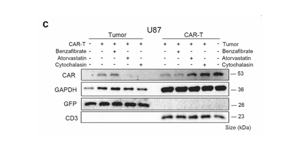
U87 cells with high EGFRvIII density were cocultured with EGFRvIII-targeted CAR-T cells, supplemented with indicated drugs. Cells were magnetically separated for CAR molecule detection. Representative immunoblotting images from three independent duplications showed that Atorvastatin (5μM, 3 days) and Cytochalasin (10μg/mL, 1 day) alleviated CAR molecule transfer.
-
-
Nat Commun
Nuclear membrane protein SUN2 promotes replication of flaviviruses through modulating cytoskeleton reorganization mediated by NS1. [Abstract]2024 Jan 5;15(1):296. PMID: 38177122 -
Nat Commun
CD36 inhibits β-catenin/c-myc-mediated glycolysis through ubiquitination of GPC4 to repress colorectal tumorigenesis. [Abstract]2019 Sep 4;10(1):3981. PMID: 31484922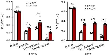
Cytochalasin B purchased from MedChemExpress. Usage Cited in: Nat Commun. 2019 Sep 4;10(1):3981. [Abstract]
Cell viability of SW480 and LoVo cells was measured by MTT assays with Cytochalasin B (20 μM, 48 h).
-
Cell Discov
Loss-of-function variants in ODAD1 disrupt ODA docking and induce actin cytoskeletal remodeling in primary ciliary dyskinesia. [Abstract]2026 Mar 31;12(1):25. PMID: 41916967 -
J Nanobiotechnology
Dynamic three-dimensional culture enhances tunneling nanotubes-mediated mitochondrial transfer in mesenchymal stromal cells to accelerate wound healing. [Abstract]2025 Aug 11;23(1):559. PMID: 40790215 -
Theranostics
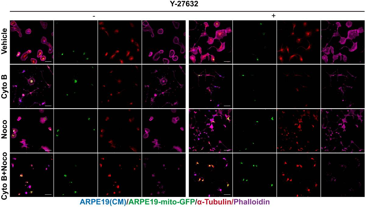
ROCK inhibitor enhances mitochondrial transfer via tunneling nanotubes in retinal pigment epithelium. [Abstract]2024 Sep 9;14(15):5762-5777. PMID: 39346535
Cytochalasin B purchased from MedChemExpress. Usage Cited in: Theranostics. 2024 Sep 9;14(15):5762-5777. [Abstract]
ARPE19-mito-GFP cells (green) co-cultured with ARPE19 cells (blue) in control and Y-27632 groups were treated with Cytochalasin B (10µM, 24h).
-
Adv Sci (Weinh)
2025 Sep 15:e06294. PMID: 40953331 -
Adv Sci (Weinh)
Microfilament-Myosin II Regulates the Differentiation of Multinucleated Cysts into Oocytes and Influences Oocyte Developmental Potential in Mice. [Abstract]2025 Sep 17:e00358. PMID: 40959902 -
Adv Sci (Weinh)
2025 Jun;12(23):e2416489. PMID: 40390546 -
Adv Sci (Weinh)
Heterogeneous Responses to Mechanical Force of Prostate Cancer Cells Inducing Different Metastasis Patterns. [Abstract]2020 Jun 17;7(15):1903583. PMID: 32775149
Cytochalasin B purchased from MedChemExpress. Usage Cited in: Adv Sci (Weinh). 2020 Jun 17;7(15):1903583. [Abstract]
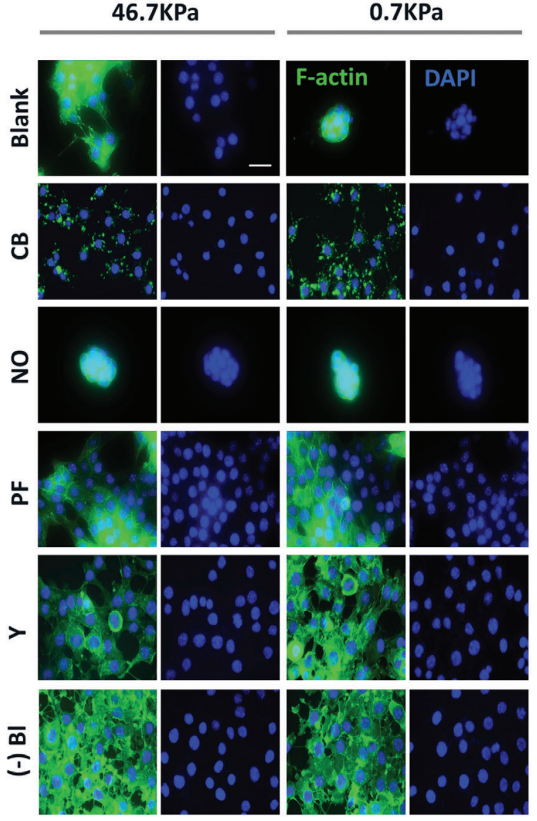
Fluorescence imaging of LNCaP cells treated with the F-actin inhibitor cytoskeleton B (CB), microtubule inhibitor nocodazole (NO), FAK inhibitor PF573288 (PF), ROCK inhibitor Y-27632 (Y), and myosin II (Myo-II) inhibitor (-)-blebbistatin ((-)Bl). After the cells were cultured on different substrates for 3 days, inhibitors were added and incubated for 24 h. (green, F-actin; scale bar, 25 µm).
Cytochalasin B purchased from MedChemExpress. Usage Cited in: Adv Sci (Weinh). 2020 Jun 17;7(15):1903583. [Abstract]
Expression of CTC cluster-related (plakoglobin and CD44) and EMT-related (vimentin and E-cadherin, 4 days) proteins in LNCaP cells grown on different substrates after treatment with different inhibitors (Cytoskeleton B (CB), Nocodazole (NO), PF-573288 (PF), Y-27632 (Y), and (-)-Blebbistatin ((-) Bl)).
-
Cell Rep Med
Taxane chemotherapy promotes response to TIM-3 checkpoint blockade via STING-mediated ER stress and HMGB1 secretion. [Abstract]2026 May 19;7(5):102788. PMID: 42097146 -
Cytochalasin B purchased from MedChemExpress. Usage Cited in: Chem Eng J. 2025 Sep 4;522:168081.
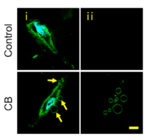
Fluorescence image of MSCCXCR4/PSGL-1 showing the production of MVs induced by Cytochalasin B (10 μg/mL, 30 min).
-
Cell Commun Signal
Estradiol increases risk of topoisomerase IIβ-mediated DNA strand breaks to initiate Xp11.2 translocation renal cell carcinoma. [Abstract]2021 Nov 16;19(1):114. PMID: 34784933 -
Cell Commun Signal
Pathogenic effects of inhibition of mTORC1/STAT3 axis facilitates Staphylococcus aureus-induced pyroptosis in human macrophages. [Abstract]2020 Nov 30;18(1):187. PMID: 33256738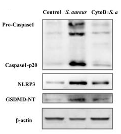
Cytochalasin B purchased from MedChemExpress. Usage Cited in: Cell Commun Signal. 2020 Nov 30;18(1):187. [Abstract]
Expression of pyroptosis-related proteins in response to S. aureus invasion and Cytochalasin B (10 μg/mL, 30 min) was assessed by western blot in THP-1.
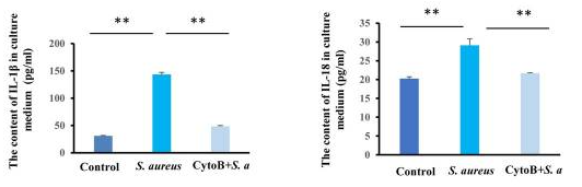
Cytochalasin B purchased from MedChemExpress. Usage Cited in: Cell Commun Signal. 2020 Nov 30;18(1):187. [Abstract]
The levels of IL-18 and IL-1β were quantified by an ELISA in THP-1, and the levels of IL-18 and IL-1β were less prominent than S. aureus invasion after Cytochalasin B (10 μg/mL, 30 min) treated.
-
Mater Today Bio
Melatonin-loaded bioactive microspheres accelerate aged bone regeneration by formation of tunneling nanotubes to enhance mitochondrial transfer. [Abstract]2024 Aug 2:28:101175. PMID: 39171100 -
Acta Pharmacol Sin
2023 Aug;44(8):1665-1675. PMID: 37016043 -
Dev Cell
STAT3-controlled CHI3L1/SPP1 positive feedback loop demonstrates the spatial heterogeneity and immune characteristics of glioblastoma. [Abstract]2025 Jun 23;60(12):1751-1767.e9. PMID: 39933531 -
Free Radic Biol Med
2022 Sep:190:1-14. PMID: 35933052 -
Cell Rep
Mechanotransduction in response to ECM stiffening impairs cGAS immune signaling in tumor cells. [Abstract]2023 Oct 5;42(10):113213. PMID: 37804510 -
Cell Prolif
The mitochondrial-endoplasmic reticulum co-transfer in dental pulp stromal cell promotes pulp injury repair. [Abstract]2024 Jan;57(1):e13530. PMID: 37493094 -
Cell Prolif
2023 Sep;56(9):e13442. PMID: 37086012 -
-
Environ Pollut
Associations of welding-related metals and hypertension in male welders: Roles of cytokinesis-block micronucleus cytome assay parameters. [Abstract]2025 May 15:373:126119. PMID: 40139295 -
NPJ Regen Med
Cyclic pulsation stress promotes bone formation of tissue engineered laminae through the F-actin/YAP-1/β-Catenin signaling axis. [Abstract]2021 Sep 6;6(1):51. PMID: 34489466 -
J Cell Biol
ER-derived caveolin-coated vesicles transport newly synthesized cholesterol to the plasma membrane. [Abstract]2026 Jun 1;225(6):e202502001. PMID: 41973050 -
RSC Adv
Dual inhibitory potential of ganoderic acid A on GLUT1/3: computational and in vitro insights into targeting glucose metabolism in human lung cancer. [Abstract]2024 Sep 6;14(39):28569-28584. PMID: 39247503 -
Cell Biol Toxicol
TRAF6 promotes spinal microglial M1 polarization to aggravate neuropathic pain by activating the c-JUN/NF-kB signaling pathway. [Abstract]2024 Jul 12;40(1):54. PMID: 38995476 -
Reprod Biol Endocrinol
Rapamycin improves the quality and developmental competence of mice oocytes by promoting DNA damage repair during in vitro maturation. [Abstract]2022 Apr 18;20(1):67. PMID: 35436937 -
Int J Mol Sci
Cytochalasin B Mitigates the Inflammatory Response in Lipopolysaccharide-Induced Mastitis by Suppressing Both the ARPC3/ARPC4-Dependent Cytoskeletal Changes and the Association Between HSP70 and the NLRP3 Inflammasome. [Abstract]2025 Mar 26;26(7):3029. PMID: 40243637 -
Mol Neurobiol
SETD3 Downregulation Mediates PTEN Upregulation-Induced Ischemic Neuronal Death Through Suppression of Actin Polymerization and Mitochondrial Function. [Abstract]2021 Oct;58(10):4906-4920. PMID: 34218417 -
Front Cell Dev Biol
Homozygous Loss of Septin12, but not its Haploinsufficiency, Leads to Male Infertility and Fertilization Failure. [Abstract]2022 Apr 25;10:850052. PMID: 35547809 -
Sci Rep
Coal dust particles can upregulate the expression of NLRP3 inflammasome components in rat alveolar macrophages through phagocytosis. [Abstract]2025 Mar 15;15(1):8989. PMID: 40089559 -
J Mol Cell Cardiol
Novel signaling axis of FHOD1-RNF213-Col1α/Col3α in the pathogenesis of hypertension-induced tunica media thickening. [Abstract]2023 Sep:182:57-72. PMID: 37482037 -
Antimicrob Agents Chemother
Inhibitory Effect of PIK-24 on Respiratory Syncytial Virus Entry by Blocking Phosphatidylinositol-3 Kinase Signaling. [Abstract]2020 Sep 21;64(10):e00608-20. PMID: 32718963 -
J Biol Chem
SARS-CoV-2 hijacks macropinocytosis to facilitate its entry and promote viral spike-mediated cell-to-cell fusion. [Abstract]2022 Nov;298(11):102511. PMID: 36259516 -
J Cell Physiol
Ankrd1 inhibits the FAK/Rho-GTPase/F-actin pathway by downregulating ITGA6 transcriptional to regulate myoblast functions. [Abstract]2024 Nov;239(11):e31359. PMID: 38988048 -
Microb Pathog
Phosphorylation of silkworm thymosin promotes the proliferation of Bombyx mori nucleopolyhedrovirus by facilitating the assembly of microfilaments. [Abstract]2026 Apr:213:108348. PMID: 41628838 -
Pathogens
Sputum from People with Cystic Fibrosis Reduces the Killing of Methicillin-Resistant Staphylococcus aureus by Neutrophils and Diminishes Phagosomal Production of Reactive Oxygen Species. [Abstract]2023 Sep 9;12(9):1148. PMID: 37764956 -
Cytokine
Phagocyte extracellular traps formation contributes to host defense against Clostridium perfringens infection. [Abstract]2023 Sep:169:156276. PMID: 37339556 -
Biochem Biophys Rep
Exosome-mediated transduction of mechanical force regulates prostate cancer migration via microRNA. [Abstract]2022 Jul 2:31:101299. PMID: 35812347 -
Tissue Cell
Characteristics of tunneling nanotube-like structures formed by human dermal microvascular pericytes in vitro. [Abstract]2024 Aug:89:102431. PMID: 38870572 -
Platelets
2022 Apr 3;33(3):381-389. PMID: 33979555 -
Asian J Androl
Micronucleus counts correlating with male infertility: a clinical analysis of chromosomal abnormalities and reproductive parameters. [Abstract]2025 Jul 1;27(4):537-542. PMID: 39789711 -
Mutat Res Genet Toxicol Environ Mutagen
Assessment of radiation doses and DNA damage in pediatric patients undergoing interventional procedures for vascular anomalies. [Abstract]2023 Jul:889:503653. PMID: 37491112 -
-
bioRxiv
2025 Nov 13:2025.11.11.687895. PMID: 41292779 -


Solvent & Solubility
In Vitro:
DMSO : 83.33 mg/mL (173.75 mM; ultrasonic and warming and heat to 60°C; Hygroscopic DMSO has a significant impact on the solubility of product, please use newly opened DMSO)
Ethanol : 25 mg/mL (52.13 mM; Need ultrasonic)
Please refer to the solubility information to select the appropriate solvent. Once prepared, please aliquot and store the solution to prevent product inactivation from repeated freeze-thaw cycles.
Storage method and period of stock solution: -80°C, 6 months; -20°C, 1 month. When stored at -80°C, please use it within 6 months. When stored at -20°C, please use it within 1 month.
Please refer to the solubility information to select the appropriate solvent. Once prepared, please aliquot and store the solution to prevent product inactivation from repeated freeze-thaw cycles.
Storage method and period of stock solution: -80°C, 6 months; -20°C, 1 month. When stored at -80°C, please use it within 6 months. When stored at -20°C, please use it within 1 month.
Concentration (start) × Volume (start) = Concentration (final) × Volume (final)
In Vivo:
Select the appropriate dissolution method based on your experimental animal and administration route.
- For the following dissolution methods, please ensure to first prepare a clear stock solution using an In Vitro approach and then sequentially add co-solvents:
- To ensure reliable experimental results, the clarified stock solution can be appropriately stored based on storage conditions. As for the working solution for In Vivo experiments, it is recommended to prepare freshly and use it on the same day.
- The percentages shown for the solvents indicate their volumetric ratio in the final prepared solution. If precipitation or phase separation occurs during preparation, heat and/or sonication can be used to aid dissolution.
Add each solvent one by one: 10% EtOH 40% PEG300 5% Tween-80 45% Saline
Solubility: ≥ 2.5 mg/mL (5.21 mM); Clear solution
This protocol yields a clear solution of ≥ 2.5 mg/mL (saturation unknown).
Taking 1 mL working solution as an example, add 100 μL EtOH stock solution (25.0 mg/mL) to 400 μL PEG300, and mix evenly; then add 50 μL Tween-80 and mix evenly; then add 450 μL Saline to adjust the volume to 1 mL.
Preparation of Saline: Dissolve 0.9 g sodium chloride in ddH₂O and dilute to 100 mL to obtain a clear Saline solution.
Add each solvent one by one: 10% EtOH 90% (20% SBE-β-CD in Saline)
Solubility: ≥ 2.5 mg/mL (5.21 mM); Clear solution
This protocol yields a clear solution of ≥ 2.5 mg/mL (saturation unknown).
Taking 1 mL working solution as an example, add 100 μL EtOH stock solution (25.0 mg/mL) to 900 μL 20% SBE-β-CD in Saline, and mix evenly.
Preparation of 20% SBE-β-CD in Saline (4°C, storage for one week): 2 g SBE-β-CD powder is dissolved in 10 mL Saline, completely dissolve until clear.
Add each solvent one by one: 10% EtOH 90% Corn Oil
Solubility: ≥ 2.5 mg/mL (5.21 mM); Clear solution
This protocol yields a clear solution of ≥ 2.5 mg/mL (saturation unknown). If the continuous dosing period exceeds half a month, please choose this protocol carefully.
Taking 1 mL working solution as an example, add 100 μL EtOH stock solution (25.0 mg/mL) to 900 μL Corn oil, and mix evenly.
Add each solvent one by one: 10% DMSO 40% PEG300 5% Tween-80 45% Saline
Solubility: ≥ 2.08 mg/mL (4.34 mM); Clear solution
This protocol yields a clear solution of ≥ 2.08 mg/mL (saturation unknown).
Taking 1 mL working solution as an example, add 100 μL DMSO stock solution (20.8 mg/mL) to 400 μL PEG300, and mix evenly; then add 50 μL Tween-80 and mix evenly; then add 450 μL Saline to adjust the volume to 1 mL.
Preparation of Saline: Dissolve 0.9 g sodium chloride in ddH₂O and dilute to 100 mL to obtain a clear Saline solution.
Add each solvent one by one: 10% DMSO 90% (20% SBE-β-CD in Saline)
Solubility: ≥ 2.08 mg/mL (4.34 mM); Clear solution
This protocol yields a clear solution of ≥ 2.08 mg/mL (saturation unknown).
Taking 1 mL working solution as an example, add 100 μL DMSO stock solution (20.8 mg/mL) to 900 μL 20% SBE-β-CD in Saline, and mix evenly.
Preparation of 20% SBE-β-CD in Saline (4°C, storage for one week): 2 g SBE-β-CD powder is dissolved in 10 mL Saline, completely dissolve until clear.
For the following dissolution methods, please prepare the working solution directly:
It is recommended to prepare fresh solutions and use them promptly within a short period of time.
The percentages shown for the solvents indicate their volumetric ratio in the final prepared solution. If precipitation or phase separation occurs during preparation, heat and/or sonication can be used to aid dissolution.
In Vivo Dissolution Calculator
Please enter the basic information of animal experiments:
-
-
-
-
Recommended: Prepare an additional quantity of animals to account for potential losses during experiments.
Please enter your animal formula composition:
-
%DMSO +
Recommended: Keep the proportion of DMSO in working solution below 2% if your animal is weak.
-
%+
-
+%Tween-80 + +
-
%Saline +
The co-solvents required include: DMSO, . All of co-solvents are available by MedChemExpress (MCE). , Tween 80. All of co-solvents are available by MedChemExpress (MCE).
Working solution concentration: 0.22 mg/mL
Method for preparing stock solution: mg drug dissolved in μL DMSO. Stock solution concentration: mg/mL.
1. Take μL DMSO stock solution;
2. Add μL .
μL , mix evenly;
3. Then add μL Tween 80, mix evenly;
4. Then add μL
Please ensure that the stock solution in the first step is dissolved to a clear state, and add co-solvents in sequence. You can use ultrasonic heating (ultrasonic cleaner, recommended frequency 20-40 kHz), vortexing, etc. to assist dissolution.


Purity & Documentation
-
Data Sheet (284 KB)
-
SDS (584 KB)
- English - EN (584 KB)
- Français - FR (584 KB)
- Deutsch - DE (584 KB)
- Norwegian - NO (584 KB)
- Español - ES (584 KB)
- Swedish - SV (584 KB)
- Italian - IT (584 KB)
- Korean - KR (584 KB)
- Portuguese - PT (584 KB)
-
Handling Instructions (2659 KB)
References
[1]. Theodoropoulos PA, et al. Cytochalasin B may shorten actin filaments by a mechanism independent of barbed end capping. Biochem Pharmacol. 1994 May 18;47(10):1875-81. [Content Brief]
[2]. Trendowski M, et al. Chemotherapy with cytochalasin congeners in vitro and in vivo against murine models. Invest New Drugs. 2015 Apr;33(2):290-9. [Content Brief]
[3]. Zhou C, et al. The effect of Cytochalasin B and Jasplakinolide on depolymerization of actin filaments in goose muscles during postmortem conditioning. Food Res Int. 2016 Dec;90:1-7. [Content Brief]
[4]. Liang Ma, et al. Discovery of the migrasome, an organelle mediating release of cytoplasmic contents during cell migration. Cell Res. 2015 Jan;25(1):24-38. [Content Brief]


Complete Stock Solution Preparation Table
Please refer to the solubility information to select the appropriate solvent. Once prepared, please aliquot and store the solution to prevent product inactivation from repeated freeze-thaw cycles.
Storage method and period of stock solution: -80°C, 6 months; -20°C, 1 month. When stored at -80°C, please use it within 6 months. When stored at -20°C, please use it within 1 month.
| Optional Solvent | Concentration Solvent Mass | 1 mg | 5 mg | 10 mg | 25 mg |
|---|---|---|---|---|---|
| Ethanol / DMSO | 1 mM | 2.0850 mL | 10.4251 mL | 20.8503 mL | 52.1257 mL |
| 5 mM | 0.4170 mL | 2.0850 mL | 4.1701 mL | 10.4251 mL | |
| 10 mM | 0.2085 mL | 1.0425 mL | 2.0850 mL | 5.2126 mL | |
| 15 mM | 0.1390 mL | 0.6950 mL | 1.3900 mL | 3.4750 mL | |
| 20 mM | 0.1043 mL | 0.5213 mL | 1.0425 mL | 2.6063 mL | |
| 25 mM | 0.0834 mL | 0.4170 mL | 0.8340 mL | 2.0850 mL | |
| 30 mM | 0.0695 mL | 0.3475 mL | 0.6950 mL | 1.7375 mL | |
| 40 mM | 0.0521 mL | 0.2606 mL | 0.5213 mL | 1.3031 mL | |
| 50 mM | 0.0417 mL | 0.2085 mL | 0.4170 mL | 1.0425 mL | |
| DMSO | 60 mM | 0.0348 mL | 0.1738 mL | 0.3475 mL | 0.8688 mL |
| 80 mM | 0.0261 mL | 0.1303 mL | 0.2606 mL | 0.6516 mL | |
| 100 mM | 0.0209 mL | 0.1043 mL | 0.2085 mL | 0.5213 mL |


Cytochalasin B Related Classifications
HY-16928 Related Classifications
 Powered by Bioz