



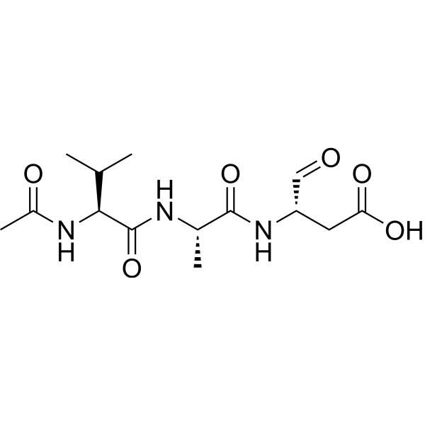
Ac-VAD-CHO
Ac-VAD-CHO (Ac-Val-Ala-Asp-CHO) is a pan-caspase inhibitor. Ac-VAD-CHO inhibits dissipation of MMP and cytochrome c release in hypoxia-exposed cells.
商品は「研究用試薬」です。人や動物の医療用・臨床診断用・食品用の製品ではありません。
研究用途以外に使用した場合、当社は一切の責任を負いかねます。
- CAS 番号: 147837-52-3
- 分子式: C14H23N3O6
- 分子量:329.35
-
保管条件:
Please store the product under the recommended conditions in the Certificate of Analysis.

To place orders, for customer services and technical support, please contact: MedChemExpress USA
Tel: 609-228-6898 E-mail: [email protected] [email protected]


-
 生物活性
生物活性
-
化学情報
- プロトコル
- 純度とドキュメンテーション
- 参考文献
-
 Help & FAQs


-
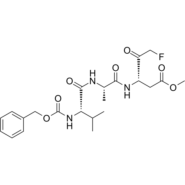
HY-16658
Z-VAD(OMe)-FMKPan-caspase Inhibitor -
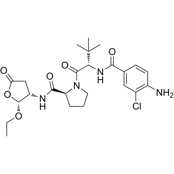
HY-13205
BelnacasanCaspase-1 Inhibitor -
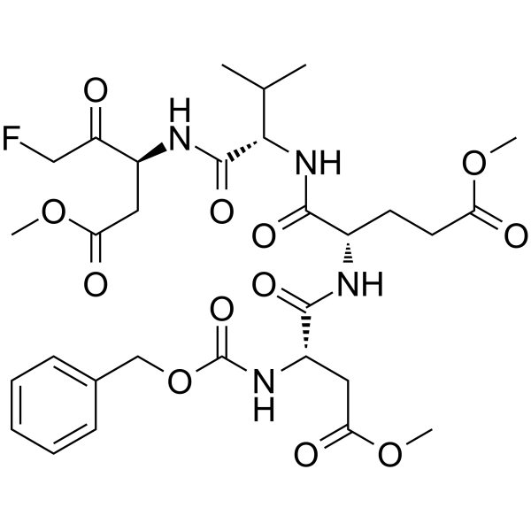
HY-12466
Z-DEVD-FMKCaspase-3 Inhibitor -
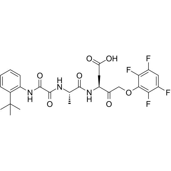
HY-10396
Emricasanpan-Caspase Inhibitor -
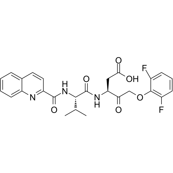
HY-12305
Q-VD-OPhPan-caspase Inhibitor -
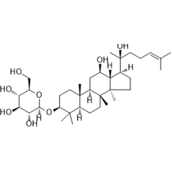
HY-N0605
Ginsenoside Rh2Caspase Activator -
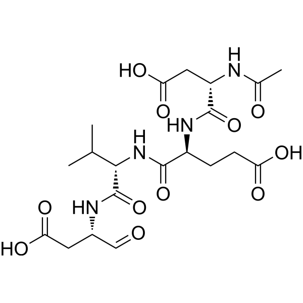
HY-P1001
Ac-DEVD-CHOCaspase-3 Inhibitor -
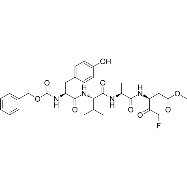
HY-P1009
Z-YVAD-FMKCaspase 1/4 Inhibitor -
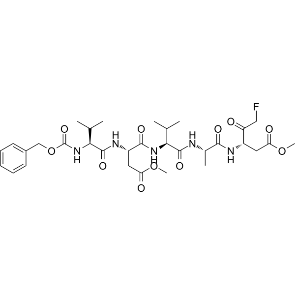
HY-P1008
Z-VDVAD-FMKCaspase-2 Inhibitor -
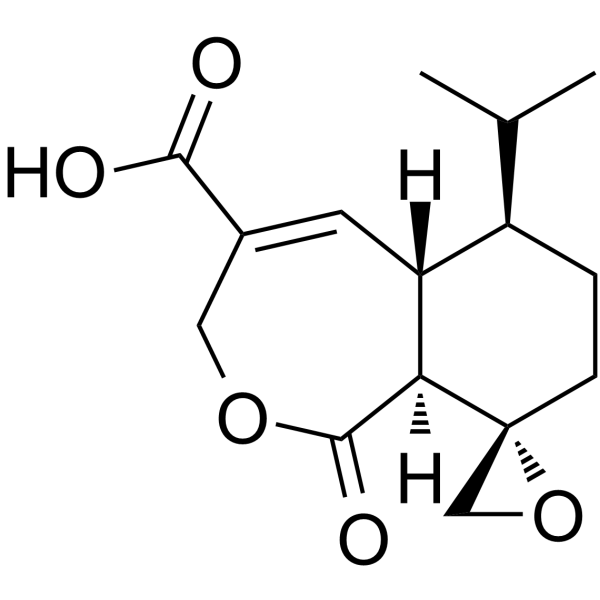
HY-120838
Heptelidic acidGAPDH Inhibitor -
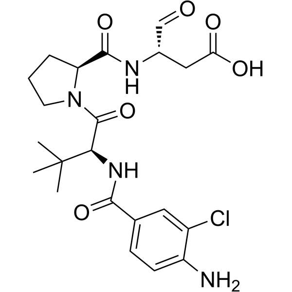
HY-112226
VRT-043198Caspase-1/4 Inhibitor -
HY-N0745
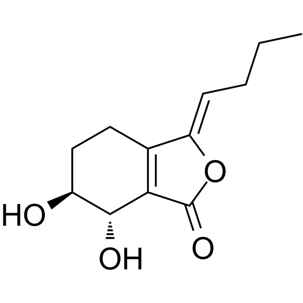
Senkyunolide IMetabolite of Z-ligustilide
Caspase アイソフォーム固有の製品をすべて表示
More

生物活性
製品説明


化学情報
-
CAS 番号 147837-52-3
-
分子量 329.35
-
分子式 C14H23N3O6
-
別名
Ac-Val-Ala-Asp-CHO
-
配列
Ac-Val-Ala-Asp-CHO
-
シーケンスの短縮
Ac-VAD-CHO
-
輸送条件
Room temperature in continental US; may vary elsewhere.
-
保管条件
Please store the product under the recommended conditions in the Certificate of Analysis.

プロトコル
-
Mitochondrial membrane-potential fluorescent assay
Mitochondrial membrane potential fluorescent assays estimate ΔΨm in living cells using lipophilic cationic dyes such as TMRM, TMRE, rhodamine 123, and JC-1, which accumulate in mitochondria according to membrane polarization; loss of signal after FCCP or CCCP treatment is interpreted as mitochondrial depolarization. TMRM/TMRE and rhodamine 123 are commonly used for semi-quantitative live-cell microscopy or flow cytometry, while JC-1 can report a shift from red aggregate fluorescence to green monomer fluorescence during depolarization; interpretation requires controls because dye concentration, quenching mode, cell type, dye efflux, and mitochondrial mass can affect fluorescence independently of ΔΨm.
-
Mitochondrial membrane-potential and mitochondrial mass staining
Mitochondrial membrane potential staining measures the electrochemical polarization across the mitochondrial inner membrane in live cells using lipophilic cationic fluorescent probes; early rhodamine-based work showed that selective mitochondrial dye accumulation is lost when the mitochondrial transmembrane potential is dissipated. JC-1 reports mitochondrial polarization by shifting from green monomer fluorescence to red J-aggregate fluorescence as dye concentration increases within energized mitochondria; therefore, the red/green fluorescence ratio is used as a relative readout of mitochondrial membrane potential. TMRE or TMRM staining provides a single-channel relative readout because these cationic rhodamine esters accumulate in polarized mitochondria, and lower fluorescence indicates reduced mitochondrial polarization when acquisition and dye-loading conditions are controlled. Mitochondrial mass staining is commonly performed with MitoTracker Green FM or related MitoTracker dyes as
-
Apoptosis Solutions
Apoptosis is a regulated, generally non-lytic cell-death pathway that removes unwanted, damaged, infected, or abnormal cells through coordinated morphological changes, caspase activation, DNA fragmentation, and membrane remodeling. The intrinsic apoptosis pathway is controlled mainly by mitochondrial outer membrane permeabilization, BCL-2 family proteins, cytochrome c release, apoptosome formation, caspase-9 activation, and downstream executioner caspase-3/7 activation. The extrinsic apoptosis pathway is initiated by death receptors such as Fas, TNFR, and TRAIL receptors, which recruit adaptor proteins and activate caspase-8 before engaging executioner caspases or mitochondrial amplification through BID cleavage. Apoptosis is linked to many phenotypes, including cancer cell killing, tissue homeostasis, immune regulation, neurodegeneration, infection response, and treatment-induced cytotoxicity; unresolved questions include how apoptosis interacts with necroptosis, pyroptosis, ferroptos


純度とドキュメンテーション
参考文献

Calculators
濃度 (開始) × 体積 (開始) = 濃度 (終了) × 体積 (終了)


Ac-VAD-CHO 関連分類
HY-W011254 関連分類
 Powered by Bioz